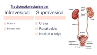
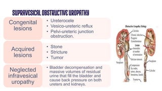
OBSTRUCTIVE
UROPATHY
By : Abdelrahman Elkhiat
WhatisObstructiveUropathy?
● Pathological changes in the urinary
tract and kidney that result from
chronic partial obstruction to the flow
of urine
The obstructive lesion is either
Infravesical
Urethra
Bladder neck
Supravesical
Ureter
Renal pelvis
Neck of a calyx
INFRAVESICALOBSTRUCTIVEUROPATHY
• Urethral meatal stenosis
• Posterior urethral valves
Congenital
lesions
• Urethral stone
• Urethral stricture
• Benign prostatic hyperplasia
Acquired
lesions
Pathophysiology
High resistance caused by an obstructive
lesion in the urethra or bladder neck,
Detrusor muscle contractions become
stronger to overcome the high resistance
Detrusor muscle contractions become
stronger to overcome the high resistance
Progressive hypertrophy of the
detrusor muscle bundles
Hypertrophied muscle bundles protrude
significantly and are called trabecula
Strong detrusor contractions
Increased
intravesical pressure
Herniation of the bladder
mucosa at these weak points
A diverticulum is a bigger herniation that
extends beyond the bladder wall
A “cellule” is a small herniation
within the bladder wal
Small diverticulum is harmless and
does not require to be removed.
Huge diverticulum is surgically
removed to avoid complications.
Cont.Pathophysiology
● A bladder that completely empties its urine content, in spite of
high infravesical resistance, is known as a “Compensated
bladder”.
● A bladder that is chronically fatigued from sustained strong
detrusor contractions against high resistance is known as a
“Decompensated bladder”.
● With incomplete bladder emptying in a decompensated bladder,
“residual urine” starts to collect in the bladder.
.
Cont.Pathophysiology
● As the detrusor contractions get weaker, the volume of residual
urine increases progressively.
● In an advanced stage, the volume of residual urine becomes
equal to the bladder capacity. At this stage, urine entering the
full bladder from the ureters escapes involuntarily through the
urethra: “Overflow incontinence”.
● High volumes of residual urine will hinder urine flow from the
ureters to the bladder causing Bilateral hydroureters and
bilateral hydronephroses. If neglected, renal failure and
Uremia occur
Diagnosis
● Palpation: a fully distended bladder is felt as a
midline suprapubic swelling
● Cystoscopy: shows bladder trabecula and necks
of bladder diverticula.
● Detection of residual urine:
 post-voiding ultrasound of the bladder
 post-voiding catheterization
 post-voiding cystogram
● Cystogram: shows bladder diverticula.
● Detection of a lesion that caused obstructive uropathy:
e.g. urethral stricture, BPH.
SymptomsandTreatment
● Partial obstruction:
the patient can micturate (void), but will experience obstructive urinary
symptoms (hesitancy, difficulty, straining, weak and narrow urine
stream, interruption and bifurcation of the urine stream, terminal
dribbling or sense of incomplete bladder emptying).
The lesion causing the obstruction should be treated to correct the
urine stream.
SymptomsandTreatment
● Complete obstruction:
 The patient cannot micturate (acute retention).
 A filled bladder causes severe suprapubic pain.
 Urethral catheterization is required to relieve the retention.
 If urethral catheterization fails (e.g. if there is urethral stricture), an
emergency suprapubic cystostomy is done.
 At a later stage, the lesion causing the obstruction should be treated to
correct the urine stream.
SUPRAVESICALOBSTRUCTIVEUROPATHY
• Ureterocele
• Vesico-ureteric reflux
• Pelvi-ureteric junction
obstruction.
Congenital
lesions
• Stone
• Stricture
• Tumor
Acquired
lesions
• Bladder decompensation and
massive volumes of residual
urine that fill the bladder and
cause back pressure on both
ureters and kidneys.
Neglected
infravesical
uropathy
Pathophysiology
● Complete obstruction
 Caused by:
o A small stone impacted in the ureter from muscle spasm and edema
o Iatrogenic ligation of the ureter during a difficult hysterectomy or colonic
surgery.
 The completely obstructed kidney stops producing urine.
 Bilateral complete obstruction of the ureters leads to “obstructive anuria”
Pathophysiology
● Partial obstruction
 Hydroureter: dilated, elongated and tortuous ureter.
 Hydronephrosis: progressive aseptic dilatation of the pelvicalyceal
system with ischemic atrophy of the renal parenchyma. The normal
“cupping” of the calyces turns to “clubbing” (ballooning of the calyces).
 In advanced cases, the completely atrophied renal parenchyma becomes
as thin as a paper, and even transparent, with the renal pelvis and calyces
hugely dilated
Symptoms
● Renal colic with acute complete obstruction:
The colic is caused by reflex ureteral muscle spasm around the stone.
It is usually severe and may require hospitalization.
● Dull aching renal pain with chronic partial obstruction:
The pain is caused by irritation of the sensory nerves in the renal capsule.
The pain is persistent, and is usually mild to moderate in intensity.
Diagnosis
● Detection of the lesion that caused the obstructive uropathy; e.g. stone, tumor or
stricture in the ureter are seen by
○ Ultrasound
○ IVU
○ CT scan.
Complications
● In case of acute complete bilateral obstruction:
 Obstructive anuria: both kidneys stop producing urine. The patient has no
desire to void and the bladder is empty for more than 24 hours. Acute
progressive uremia develops. It is fatal if the obstruction is not rapidly
relieved
● In case of neglected partial chronic obstruction:
 progressive hydroureter and hydronephrosis, up to complete parenchymal
atrophy.
 Uremia occurs if partial obstruction is bilateral and neglected.
Treatment
● Complete obstruction is an emergency that requires
immediate kidney drainage by introduction of a
ureteric catheter.
● If ureteric catheterization is not technically possible,
percutaneous nephrostomy is done.
● The lesion causing obstruction must be removed to
avoid complications.
● Advanced unilateral hydronephrosis with complete parenchymal
atrophy and renal pain is managed by nephrectomy (only if the
kidney is not functioning and symptomatic)
● Advanced bilateral hydronephroses with complete parenchymal
atrophy and uremia is managed by hemodialysis or renal
transplantation.

obstructive uropathy.pptx

  • 1.
  • 2.
    WhatisObstructiveUropathy? ● Pathological changesin the urinary tract and kidney that result from chronic partial obstruction to the flow of urine
  • 3.
    The obstructive lesionis either Infravesical Urethra Bladder neck Supravesical Ureter Renal pelvis Neck of a calyx
  • 4.
    INFRAVESICALOBSTRUCTIVEUROPATHY • Urethral meatalstenosis • Posterior urethral valves Congenital lesions • Urethral stone • Urethral stricture • Benign prostatic hyperplasia Acquired lesions
  • 5.
    Pathophysiology High resistance causedby an obstructive lesion in the urethra or bladder neck, Detrusor muscle contractions become stronger to overcome the high resistance Detrusor muscle contractions become stronger to overcome the high resistance Progressive hypertrophy of the detrusor muscle bundles Hypertrophied muscle bundles protrude significantly and are called trabecula Strong detrusor contractions Increased intravesical pressure Herniation of the bladder mucosa at these weak points A diverticulum is a bigger herniation that extends beyond the bladder wall A “cellule” is a small herniation within the bladder wal Small diverticulum is harmless and does not require to be removed. Huge diverticulum is surgically removed to avoid complications.
  • 6.
    Cont.Pathophysiology ● A bladderthat completely empties its urine content, in spite of high infravesical resistance, is known as a “Compensated bladder”. ● A bladder that is chronically fatigued from sustained strong detrusor contractions against high resistance is known as a “Decompensated bladder”. ● With incomplete bladder emptying in a decompensated bladder, “residual urine” starts to collect in the bladder. .
  • 7.
    Cont.Pathophysiology ● As thedetrusor contractions get weaker, the volume of residual urine increases progressively. ● In an advanced stage, the volume of residual urine becomes equal to the bladder capacity. At this stage, urine entering the full bladder from the ureters escapes involuntarily through the urethra: “Overflow incontinence”. ● High volumes of residual urine will hinder urine flow from the ureters to the bladder causing Bilateral hydroureters and bilateral hydronephroses. If neglected, renal failure and Uremia occur
  • 8.
    Diagnosis ● Palpation: afully distended bladder is felt as a midline suprapubic swelling ● Cystoscopy: shows bladder trabecula and necks of bladder diverticula. ● Detection of residual urine:  post-voiding ultrasound of the bladder  post-voiding catheterization  post-voiding cystogram
  • 9.
    ● Cystogram: showsbladder diverticula. ● Detection of a lesion that caused obstructive uropathy: e.g. urethral stricture, BPH.
  • 10.
    SymptomsandTreatment ● Partial obstruction: thepatient can micturate (void), but will experience obstructive urinary symptoms (hesitancy, difficulty, straining, weak and narrow urine stream, interruption and bifurcation of the urine stream, terminal dribbling or sense of incomplete bladder emptying). The lesion causing the obstruction should be treated to correct the urine stream.
  • 11.
    SymptomsandTreatment ● Complete obstruction: The patient cannot micturate (acute retention).  A filled bladder causes severe suprapubic pain.  Urethral catheterization is required to relieve the retention.  If urethral catheterization fails (e.g. if there is urethral stricture), an emergency suprapubic cystostomy is done.  At a later stage, the lesion causing the obstruction should be treated to correct the urine stream.
  • 12.
    SUPRAVESICALOBSTRUCTIVEUROPATHY • Ureterocele • Vesico-uretericreflux • Pelvi-ureteric junction obstruction. Congenital lesions • Stone • Stricture • Tumor Acquired lesions • Bladder decompensation and massive volumes of residual urine that fill the bladder and cause back pressure on both ureters and kidneys. Neglected infravesical uropathy
  • 13.
    Pathophysiology ● Complete obstruction Caused by: o A small stone impacted in the ureter from muscle spasm and edema o Iatrogenic ligation of the ureter during a difficult hysterectomy or colonic surgery.  The completely obstructed kidney stops producing urine.  Bilateral complete obstruction of the ureters leads to “obstructive anuria”
  • 14.
    Pathophysiology ● Partial obstruction Hydroureter: dilated, elongated and tortuous ureter.  Hydronephrosis: progressive aseptic dilatation of the pelvicalyceal system with ischemic atrophy of the renal parenchyma. The normal “cupping” of the calyces turns to “clubbing” (ballooning of the calyces).  In advanced cases, the completely atrophied renal parenchyma becomes as thin as a paper, and even transparent, with the renal pelvis and calyces hugely dilated
  • 15.
    Symptoms ● Renal colicwith acute complete obstruction: The colic is caused by reflex ureteral muscle spasm around the stone. It is usually severe and may require hospitalization. ● Dull aching renal pain with chronic partial obstruction: The pain is caused by irritation of the sensory nerves in the renal capsule. The pain is persistent, and is usually mild to moderate in intensity.
  • 16.
    Diagnosis ● Detection ofthe lesion that caused the obstructive uropathy; e.g. stone, tumor or stricture in the ureter are seen by ○ Ultrasound ○ IVU ○ CT scan.
  • 17.
    Complications ● In caseof acute complete bilateral obstruction:  Obstructive anuria: both kidneys stop producing urine. The patient has no desire to void and the bladder is empty for more than 24 hours. Acute progressive uremia develops. It is fatal if the obstruction is not rapidly relieved ● In case of neglected partial chronic obstruction:  progressive hydroureter and hydronephrosis, up to complete parenchymal atrophy.  Uremia occurs if partial obstruction is bilateral and neglected.
  • 18.
    Treatment ● Complete obstructionis an emergency that requires immediate kidney drainage by introduction of a ureteric catheter. ● If ureteric catheterization is not technically possible, percutaneous nephrostomy is done. ● The lesion causing obstruction must be removed to avoid complications.
  • 19.
    ● Advanced unilateralhydronephrosis with complete parenchymal atrophy and renal pain is managed by nephrectomy (only if the kidney is not functioning and symptomatic) ● Advanced bilateral hydronephroses with complete parenchymal atrophy and uremia is managed by hemodialysis or renal transplantation.