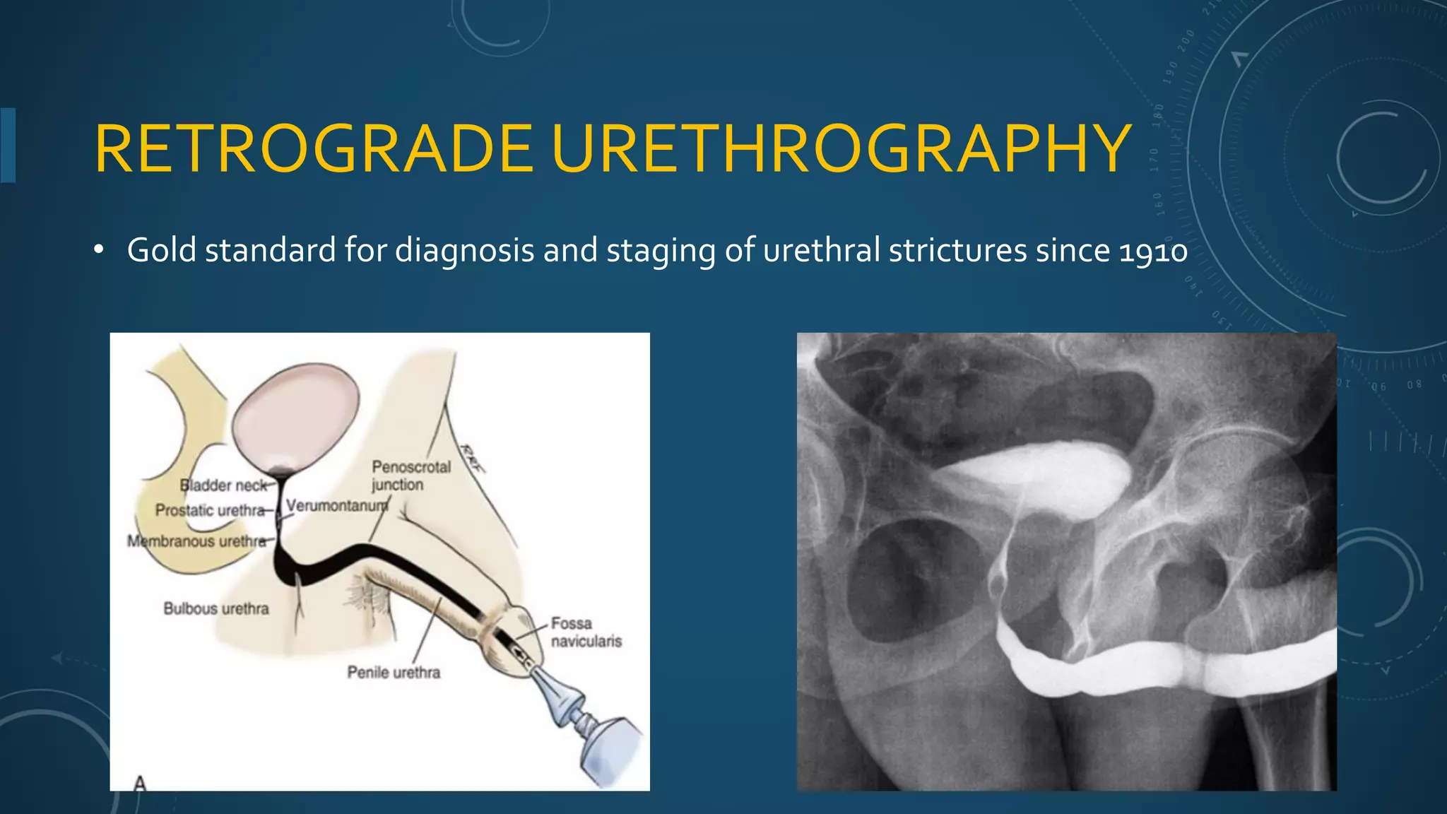
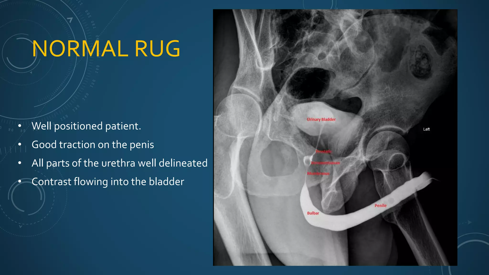
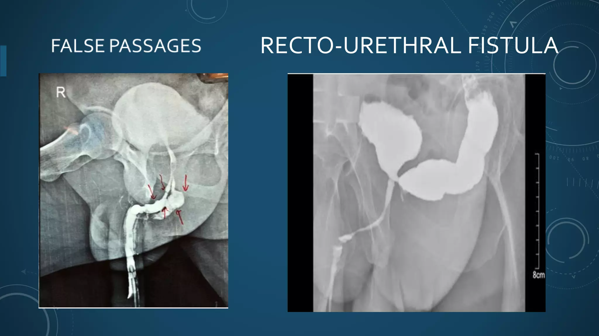
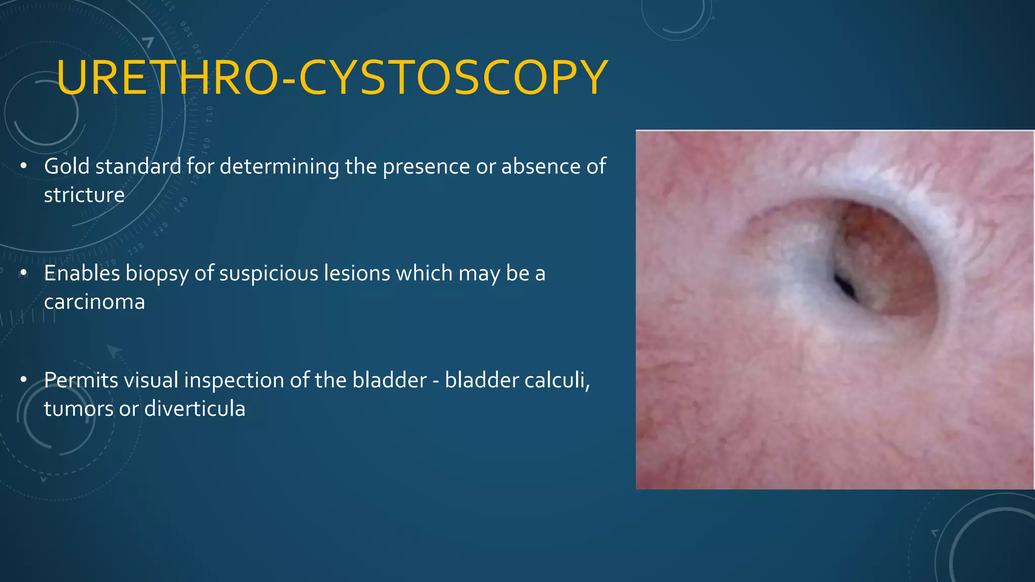
URETHRAL STRICTURES
DR. M.YIGAH
INTRODUCTION
• A urethral stricture is a narrowing of the urethra caused by scarring of the epithelium and
its surrounding corpus spongiosum.
• 2nd commonest cause of Lower UrinaryTract Obstruction in Sub Saharan Africa.
(Mbibu et al 2002)
ANATOMY OFTHE URETHRA
ANATOMYOFTHE
PENIS
• Glans penis is the expanded
part of the Corpus
spongiosum
• Corpus spongiosum is
attached to perineal body
• The corpus cavernosum is
attached to Ischiopubic rami
EPIDEMIOLOGY
• Prevalence in Sub Saharan Africa
• 2nd commonest cause of lower urinary tract obstruction (Mbibu et al, 2002)
• Hospital Prevalence – 4.2% in Ouagadougou (Yameogo et al, 2017)
• Accounts for 14.3% ofAcute Urinary Retention in SouthAfrica– ( Stephan et al, 2011 )
• Gender Differences
• Female Urethral strictures < 3% of all strictures (Mugalo et al 2013)
• Age
• Peak age range – (20 and 39) years
EPIDEMIOLOGY
• Prevalence in Ghana
• Out of the 11,084 OPD visits to the urology clinic of KBTH in 2011, it accounted for
1015 (9%) of all diagnosis (Mensah et al 2013)
• Cape CoastTeaching Hospital
• Diagnosed 94 times out 173,507 OPD visits to the hospital
• Formal Cystostomy – 5 over the past 2 months
• 1 Urethroplasty over the past 3 months
• Urethroplasty – GH ₵4000
IATROGENIC CAUSES
• Urethral Instrumentation for diagnostic or therapeutic purposes
• Transurethral Catheterization
• Urethroscopy
• Transurethral Resection of the Prostate (TURP) or BladderTumors (TURBT)
• Urethral dilatation
• Internal Urethrotomy
• Genitourinary Surgery
• Radical Prostatectomy
• Hypospadia repairs
• Circumcision
• External beam radiotherapy or brachytherapy
TRAUMATIC CAUSES
• Blunt Injury
• Straddle injuries – e.g. falling onto a bicycle crossbar.
• RoadTraffic Accidents and Industrial Accidents
• Penile fractures
• Constriction penile bands or rings
• Penetrating injury
• Gunshot wounds
• Stab wounds
• Penile amputation injuries
SOME CAUSESOF URETHRAL STRICTURES
INFLAMMATORY CAUSES
• Post-infectious inflammatory causes
• Gonococcal urethritis
• Non-specific Urethritis (e.g Chlamydia sp.)
• Schistosomiasis
• Tuberculosis
• Non-infective inflammatory causes
• Lichen Sclerosis
• Allergic reactions – latex catheters
• Reiter’s syndrome
IDIOPATHIC & CONGENITAL
• Idiopathic strictures – Urethral strictures of unknown origin
• Absence of urethral inflammation, trauma, infection, and urethral manipulation.
• Congenital strictures
• Associated with other conditions
• Amniotic band syndrome
• Prune-belly syndrome
EPIDEMIOLOGY
Developed Nations
• Idiopathic urethral strictures
• Iatrogenic stricture
• External trauma
• Post-infectious strictures
Developing Nations
• Post-infectious strictures
• External trauma
• Iatrogenic urethral strictures
• Idiopathic strictures
(Irekpita & Udefiagbon, 2019)
COMMON SITE OF URETHRAL STRICTURES
• Bulbar urethra – 40.2%
• Membranous urethra – 35.57%
• Pendulous urethra – 14.43%
• Prostatic – 4.63%
• Female Urethra – 3.10%
• Fossa navicularis – 2.07%
PATHOGENESIS
• Strictures are the consequence of epithelial damage and spongiofibrosis
• Noxious stimulus (bacterial, chemical, physical)
• Squamous metaplasia
• Fissures develop in epithelium
• Extravasation of urine into corpus spongiosum
• Fibrosis develops in the corpus spongiosum
• Fibrotic plaques coalesce
• Contraction of this scar reduces the urethral lumen.
NATURAL HISTORY
• A stricture narrows the lumen to (10–12 French) before significantly impairing urine flow
• Compensatory hypertrophy of the detrusor muscles
• Decreases compliance to filling of bladder
• Development of hydroureters and hydronephrosis due to vesico-ureteric reflux
• Urinary stasis can predispose infections and bladder stones
• Development of periurethral abscess leading to fistulation (Watering Can Perineum)
COMPLICATIONS
• Infections - Urinary tract infections, prostatitis, epididymo-orchitis
• Development of fistula
• Bladder calculi
• Diverticula
• Urinary Retention
• Renal failure
CLINICAL PRESENTATION
• Progressive worsening of symptoms of lower urinary tract obstruction.
• Frequency and Nocturia
• Feeling of incomplete emptying of urine
• Poor urine stream
• Straining
• Hesitancy
• Terminal dribbling
• .
CLINICAL PRESENTATION
• Complications especially in long standing cases or neglected cases
• Acute or Chronic Urinary Retention
• Infections – cystitis, prostatitis, epididymitis and pyelonephritis
• Urethrocutaneous fistulae – scrotal, perineal fistulae (Watering can perineum)
• Bladder stones
• Renal failure
FURTHER ENQUIRY ON HISTORY
• Onset, severity and rapid progression of the LUTS
• Post gonococcal strictures – can take up to 20 years to develop
• History of confirmed STD or past symptoms suggestive of STD
• Trauma – within 2 months
• Preceding trauma to the perineum or penis, RTA
• Lower abdominal pain, blood at the urethral meatus, inability to pass urine - PFUI
CLINICAL HISTORY
• Past Medical History
• History of a stricture and subsequent management – urethral dilatation, internal urethrotomy
• History of urologic surgeries – Hypospadia repairs, prostatectomy etc
• History of difficult urethral catheterization
• TB, Schistosomiasis, BPH, Diabetes mellitus
• Social History
• Smoking or chewing of tobacco.
PHYSICAL EXAMINATION
• General
• Ill-looking and febrile - infection
• Examining the oral mucosa for suitability for harvest of buccal or lingual mucosal graft
• Abdomen
• Lower abdominal pain in UTI, retention
• Bladder may be palpable in patient with urinary retention.
• Examining the hernia orifices
• DRE – for patient >40yrs and patients with PFUI
PHYSICAL EXAMINATION
• Genitalia and Perineum
• Palpation of the penis, scrotum, perineum and urethra for thickenings or Indurations
• Assess the urethral meatus for stenosis and bleeding
• Scrotal, Penile or Perineal hematoma
• Presence or absence of a fistula
• Presence of Chordie, Hypospadia and whether the patient has been circumcised
EXAMINATION OFTHE GENITALS & PERINEUM
DIAGNOSTIC INVESTIGATIONS
1. Imaging Studies
• Retrograde Urethrography (RUG)
• Voiding Cysto-Urethrography (VCUG)
• Urethrosonography
• MRI & CT Scan
2. Urethrocystoscopy
3. Urethral Instrumentation
RETROGRADE URETHROGRAPHY
• Gold standard for diagnosis and staging of urethral strictures since 1910
RETROGRADE URETHROGRAPHY
• A scout film should be taken before the contrast is injected
RETROGRADE URETHROGRAPHY
• Positioning of the patients
• Should be done (or supervised) by the treating Urologist
• Lateral Oblique (45 degrees)
• Gentle traction on the penis
RETROGRADE URETHROGRAPHY
• Contrast should be seen proximal to the stricture.
NORMAL RUG
• Well positioned patient.
• Good traction on the penis
• All parts of the urethra well delineated
• Contrast flowing into the bladder
POST-TRAUMATIC BULBAR
URETHRAL STRICTURE
POST INFLAMMATORY PENILE
URETHRAL STRICTURE
RECTO-URETHRAL FISTULA
LIMITATIONS OF RUG
• Difficult to assess distal urethral strictures (urethral meatus and fossa navicularis)
• Difficult assessing the proximal part of the urethra e.g Bladder neck
• Injecting of contrast can be very uncomfortable and predispose to UTI
• Reaction to contrast agent.
VOIDING CYSTO-URETHROGRAM (VCUG/MCUG)
BULBAR STRICTURE
RUG
BULBAR STRICTURE
ON MCUG
URETHRO-CYSTOSCOPY
• Gold standard for determining the presence or absence of
stricture
• Enables biopsy of suspicious lesions which may be a
carcinoma
• Permits visual inspection of the bladder - bladder calculi,
tumors or diverticula
URETHROSONOGRAPHY
Used to stage the stricture - location, length
and caliber.
Best used for penile urethral strictures
compared to the bulbar portion.
Limited by the stricture location and its is
operator dependent
MRI & CT SCAN
• Best ancillary imaging modality for assessing post-traumatic pelvic anatomy.
• Evaluate the configuration of any pelvic fractures
• 3 Dimension Spiral CT Cystourethrography (CTUG) – Novel technique
SUPPORTIVE INVESTIGATIONS
• PostVoid Residual Urine Measurement
• Uroflowmetry
• urethral obstruction – PFR < 15ml/sec and plateau curve profile
• American Urological Association Symptom Score (or IPSS)
• Questionnaire used to quantify the severity of the subjective voiding symptoms in both sexes
• Urinalysis
• BUE and Cr
FINAL DIANOSIS
• Confirm the stricture and its characteristic
• Cause of the stricture
• Site of the stricture
• Length of the stricture
• Complete or Incomplete (Caliber of the stricture)
• Density and depth of the spongiofibrosis
• Complication of the stricture e.g. Bladder calculi, Renal failure
• Associated urological pathology that might affect LUTS
TREATMENTOPTIONS
• Temporary Measures
• Supra-pubic catheterization – Formal or Stab Cystostomy
• Definitive Treatment
• Dilation
• Endoscopic DirectVision Internal Urethrotomy
• Urethroplasty
INDICATIONS FORTEMPORARYTREATMENT
1. Complications of Urethral strictures –
• Urinary retention,
• Fistula,
• Uraemia etc.
2. Unfit for surgery – Old age, Bleeding disorders
3. Lack of funds for definitive treatment
4. Lack of expertise and facilities for open or endoscopic surgery
DEFINITIVE
TREATMENT
URETHRAL DILATION
• Goal: Stretch the scar without injuring the urethra to produce more scars
• A palliative treatment for most patients
• A potential curative treatment for well selected patients
• Short (< 2 cm) incomplete epithelial stricture with no (or minimal) spongiofibrosis
• Frequency of dilation Increasing intervals
• e.g. 2 weeks, 1 month, 3months and 6months and continued indefinitely
FLEXIBLE FILIFORM
BOUGIES & FOLLOWERS
CURVED STEEL BOUGIES
FLEXIBLE FILIFORM BOUGIES AND FOLLOWERS
COMPLICATIONS OF URETHRAL DILATION
• Bleeding,
• Clot Retention,
• Urethral rupture,
• Infections - Prostatitis, Cystitis, Epididymo-orchitis, Pyelonephritis, Bacteraemia
• Rupture of prostate (BPH)
ENDOSCOPIC DIRECTVISION INTERNAL URETHROTOMY (DVIU)
• Internal Urethrotomy is any procedure that opens the stricture by incising through the scar
transurethrally to release the contraction to allow the lumen to heal enlarged.
• DirectVision Internal Urethrotomy (DVIU) was introduced in 1974 by Sachse.
• The urethra is then splinted with an indwelling catheter for about 2 – 7 days
• (14 – 21 days for difficult strictures)
ENDOSCOPIC DIRECTVISION INTERNAL
URETHROTOMY
1st line therapy
• Short < 1 cm bulbar stricture > 15F in calibre in the absence
of dense and deep spongiofibrosis.
• A stricture-free rate (SFR) of up to 50%–70%
THE CASE FOR PATIENT SELECTION
PANSADORO AND EMILIOZZI 1996
Stricture Location Bulbar Penile Peno-scrotal
Success Rate (SFR) 42% 16% 11%
Stricture Length < 1cm > 1cm
Success Rate (SFR) 71% 18%
Diameter > 15 FR <15 FR
Success Rate (SFR) 69% 34%
No of Strictures Single Multiple
Success Rate (SFR) 50% 16%
Primary or RepeatTreatment Primary Repeat
Success Rate (SFR) 47% 0%
ATTEMPTSTO IMPROVINGTHE OUTCOME OF DVIU
• Long term continuous catheterization – i.e for 6 weeks
• Intermittent self or office catheterization
• Urethral dilation
• Pharmacological management
• Colchicine
• MitomycinC
• Triamcinolone
• Laser therapy
CONTRAINDICATIONS TO DVIU
1. Stricture Characteristics
• Long strictures (> 4cm)
• Complete stricture
• Strictures with dense spongiofibrosis
2. 2 previous failed DVIU
3. Recurrent strictures that occur <3 months after DVIU
4. Complications of strictures: Untreated urinary tract infection, Fistula etc
5. Coagulation disorders
OUTCOME OF DVIU/DILATION
• Best Outcome
• A stricture-free rate (SFR) - 50%–70% (for well selected patients).
• Recurrence of stricture
• Recurrence at < 3 months – SFR is 0 % after 2 years for the 2nd attempt
• Recurrence at > 6 months - SFR is 40% after 2 years for the 2nd attempt
• Repeated DVIU/dilation
• Patients undergoing a third incision or dilation have a 100% recurrence rate (palliative)
COMPLICATIONS OF INTERNAL URETHROTOMY
1. Recurrence of stricture,
2. Urethral haemorrhage
3. Clot retention
4. Erectile dysfunction
5. UTI
URETHRAL STENTING
• The concept of urethral stenting dates back to at least 1969
• Championed by Milroy et al. in 1988.
• Criteria for using Permanent Stent
• Short, incomplete and recurrent bulbar stricture who are
• medically unfit for urethroplasty
• cannot tolerate intermittent self-catheterization/dilation
URETHROPLASTY
• Gold standard for management of urethral stricture and stenosis.
• Indications
• All forms of stricture and stenosis
• Failed Urethral Dilation or DVIU
• Complex strictures – Long and Multiple, Pelvic Fractures
• Associated urologic conditions – e.g. Hypospadia
• Associated complication – False passages, Fistulae, Calculus, urethral diverticula
.
TYPES OF OPEN URETHROPLASTY
1. Excision and Primary Anastomosis
2. Substitution (Augmentation) UrethroplastyTechniques
• Augmented anastomotic procedure.
• Onlay Augmentation Procedure
• Tube Augmentation
EXCISION AND PRIMARY ANASTOMOSIS
• The most dependable technique of short anterior urethral reconstruction
• High success rate – 90 – 95%
• (G. Barbagli et al 2008; Santucci et al 2007)
• Patient selection
• Short (<2cm) especially in the anterior bulbar urethral
• Dense bulbar strictures (obliterative and near obliterative)
EXCISIONAND PRIMARY ANASTOMOSIS
• The best results are achieved when the following technical points are observed:
• The area of fibrosis is totally excised;
• the urethral anastomosis is widely spatulated
• creating a large ovoid anastomosis
• anastomosis is tension free
• Techniques
• Vessel‐sparing excision and primary anastomosis
• Non-vessel sparing technique
VESSEL‐SPARING EXCISION AND PRIMARY
ANASTOMOSIS
VESSEL‐SPARING EXCISION AND PRIMARY
ANASTOMOSIS
VESSEL‐SPARING EXCISION AND PRIMARY
ANASTOMOSIS
VESSEL‐SPARING EXCISION AND PRIMARY
ANASTOMOSIS
EXCISION AND PRIMARY ANASTOMOSIS
• Complications
• Ejaculatory dysfunction
• Erectile dysfunction
• Chordie
• Penile shortening
• Stricture recurrence
SUBSTITUTION URETHROPLASTY
• Strictures too lengthy for tension free EPA
• Substitutes – Grafts and Flaps
• Various grafts have been used for reconstructing the Urethra.
• STGF or FTSG – Penis, Post-auricular region
• Oral Mucosal Grafts - buccal, lingual, labial
• Bladder Epithelial Graft,
• Rectal Mucosal Graft.
• Acellular collagen matrix
DONOR SITES FOR GRAFT
BUCCAL POST-AURICULARLINGUAL
SUBSTITUTION URETHROPLASTY
• One-stage GraftTechniques
1. An augmented anastomotic procedure
• Excision of stricture and restoration of roof or floor strip of native urethra with a graft
2. On-lay Augmentation Procedure
• To incise the stricture and carry out a patch augmentation
3. Tube augmentation.
• To excise the stricture and put in a circumferential patch
THE AUGMENTED ANASTOMOTIC URETHROPLASTY
• Combination Excision and Substitutional Urethroplasty
• Bulbar strictures deemed too long for straight forward primary anastomosis.
DORSAL ONLAY
AUGMENTED ANASTOMOSIS
For short distal Bulbar strictures ≤ 2cm
Superior spread-fixation (reduces graft
shrinkage)
Enhances graft take due to vascular corporal
body,
VENTRAL ONLAY
AUGMENTED ANASTOMOSIS
• Short proximal bulbar strictures ≤ 2cm
• A risk of graft shrinkage
• Prone to diverticulum
ON-LAY (NON-TRANSECTING)
AUGMENTATION URETHROPLASTY
VENTRAL ONLAY
NON-TRANSECTED
AUGMENTATION URETHROPLASTY
• For Proximal Bulbar Stricture that are > 2 cm.
• Inferior graft take compared to dorsal version
• Prone to graft contraction
• Prone to diverticulum
DORSAL ONLAY
NON-TRANSECTED
AUGMENTATION URETHROPLASTY
• For longer (> 2cm) middle and distal bulbar
strictures.
• Superior graft coverage
• Superior graft take.
• Less prone to graft sacculation or diverticulum
ASOPATECHNIQUE
NON-TRANSECTING
AUGMENTED URETHROPLASTY
Variant of dorsal onlay for penile and bulbar
strictures
Urethra plate > 1 cm
No circumferential mobilization of the urethra
Preserves the perforating neurovascular structures
KULKARNI
NON-TRANSECTING
AUGMENTED URETHROPLASTY
For Penile or Peno-bulbar stricture
Suitable for pan urethral strictures
Eliminates the need for full circumferential
mobilization
Preserves the perforating neurovascular structures
and Bulbospongiosus muscle
PALMINTERI
NON-TRANSECTING
AUGMENTED URETHROPLASTY
• For very narrow strictures where a single
graft would not be sufficient to obtain
adequate lumen width
• The strictured urethra is incised in the
midline and augmented dorsally and
ventrally using two oral grafts
• Preserves sexual function
MULTI-STAGED URETHROPLASTY
• Staged reconstruction is planned repair strategy characterized by more than one
operation and inherent free tissue transfer.
• INDICATIONS ,
• Multiple prior urethroplasty failures
• Long obliterative strictures
• Failed Hypospadia repairs
• Urethral reconstruction after failed urethral stent placement
• Presence of diverticulum or fistula
• Strictures caused by lichen sclerosis
MULTI-STAGED URETHROPLASTY
• 1st Stage of urethroplasty
• Marsupialization of the urethra
• Placement of FTSG, STSG or buccal grafts over the dartos fascia
• 2nd stage of Urethroplasty
• Tubularization of the graft.
Time between the 1st and 2nd surgery --- 6 -12 months.
TECHNIQUES OF MULTI-STAGED
URETHROPLASTY
• Staged oral mucosa graft urethroplasty
• Johanson’s technique
• Mesh graft urethroplasty
FLAP URETHROPLASTY
• The flap can be harvested from the penis or scrotum
• Techniques of flap urethroplasty
• Quartey’s penile flap – bulbar or pendulous strictures
• Jordan’s ventral transverse skin island flap procedure – fossa navicularis strictures
• Ventral longitudinal island flap (Orandi) – strictures of the pendulous urethra
• Dorsal transverse preputial island flap (Duckett) – pendulous and distal bulbous urethra
• Hairless scrotal island flap (Jordan) for bulbo-membranous stricture.
QUARTEY’S PENILE FLAP
URETHROPLASTY
ORANDI FLAP
Ventral longitudinal island flap for
long penile strictures
FLAPVS GRAFT
• There is no advantage of a flap over a graft in terms of stricture recurrence rate.
• Flap are technically more challenging and have more complications
• Wound infections
• Penile hematoma and Seroma
• Skin necrosis
• Urethrocutaneous fistula
• Higher risk of sacculation (diverticulum formation )
POST OPERATIVE EVALUATION
• There is not even a standard definition of what constitutes post-operative success or
a recurrent urethral stricture.
• Commonly Used AssessmentTools
• RUG,VCUG,
• Urethro -cystoscopy
• urethral calibration,
• AUA-SI, uroflowmetry & PVR,
COST EFFECTIVE WAY OF MANAGING STRICTURES
• For the management of short bulbar urethral strictures is to reserve urethroplasty
for patients in whom a single endoscopic attempt fails .
• For longer strictures, in which the success rate of DVIU is expected to be < 35%,
urethroplasty as primary therapy is cost effective.
• (Wright et al, 2006)
TAKE HOME MESSAGE
The BEST WAY to treat a stricture is to PREVENT IT
Gentle urethral instrumentation
Public education on STD and the use of condoms
Complying with all industrial and road safety measures
REFERENCES
• Campbell-Walsh UrologyTextbook 11th Edition.
• An InternationalConsultation on Urethral Strictures - Marrakech, Morocco,October 13-16, 2010.
• Anthony R. Mundy and Daniela E.Andrich, 2010, Urethral strictures
• BAJA’ Principles and Practice od SurgeryVol II
• Onyeanunam N Ekeke 2017, African Journal of Urology
• Eshiobo I, Ernest U. A Review of the epidemiology and management of urethral stricture disease in Sub-
SaharanAfrica. Curr Med Issues 2019;17:118-24.
• Mensah et al.-2013-Contemporary Evaluation andTreatment of Male Urethral Stricture Disease inWest
Africa
THANK YOU

Urethral strictures

  • 1.
  • 2.
    INTRODUCTION • A urethralstricture is a narrowing of the urethra caused by scarring of the epithelium and its surrounding corpus spongiosum. • 2nd commonest cause of Lower UrinaryTract Obstruction in Sub Saharan Africa. (Mbibu et al 2002)
  • 3.
  • 4.
    ANATOMYOFTHE PENIS • Glans penisis the expanded part of the Corpus spongiosum • Corpus spongiosum is attached to perineal body • The corpus cavernosum is attached to Ischiopubic rami
  • 5.
    EPIDEMIOLOGY • Prevalence inSub Saharan Africa • 2nd commonest cause of lower urinary tract obstruction (Mbibu et al, 2002) • Hospital Prevalence – 4.2% in Ouagadougou (Yameogo et al, 2017) • Accounts for 14.3% ofAcute Urinary Retention in SouthAfrica– ( Stephan et al, 2011 ) • Gender Differences • Female Urethral strictures < 3% of all strictures (Mugalo et al 2013) • Age • Peak age range – (20 and 39) years
  • 6.
    EPIDEMIOLOGY • Prevalence inGhana • Out of the 11,084 OPD visits to the urology clinic of KBTH in 2011, it accounted for 1015 (9%) of all diagnosis (Mensah et al 2013) • Cape CoastTeaching Hospital • Diagnosed 94 times out 173,507 OPD visits to the hospital • Formal Cystostomy – 5 over the past 2 months • 1 Urethroplasty over the past 3 months • Urethroplasty – GH ₵4000
  • 7.
    IATROGENIC CAUSES • UrethralInstrumentation for diagnostic or therapeutic purposes • Transurethral Catheterization • Urethroscopy • Transurethral Resection of the Prostate (TURP) or BladderTumors (TURBT) • Urethral dilatation • Internal Urethrotomy • Genitourinary Surgery • Radical Prostatectomy • Hypospadia repairs • Circumcision • External beam radiotherapy or brachytherapy
  • 8.
    TRAUMATIC CAUSES • BluntInjury • Straddle injuries – e.g. falling onto a bicycle crossbar. • RoadTraffic Accidents and Industrial Accidents • Penile fractures • Constriction penile bands or rings • Penetrating injury • Gunshot wounds • Stab wounds • Penile amputation injuries
  • 9.
  • 10.
    INFLAMMATORY CAUSES • Post-infectiousinflammatory causes • Gonococcal urethritis • Non-specific Urethritis (e.g Chlamydia sp.) • Schistosomiasis • Tuberculosis • Non-infective inflammatory causes • Lichen Sclerosis • Allergic reactions – latex catheters • Reiter’s syndrome
  • 11.
    IDIOPATHIC & CONGENITAL •Idiopathic strictures – Urethral strictures of unknown origin • Absence of urethral inflammation, trauma, infection, and urethral manipulation. • Congenital strictures • Associated with other conditions • Amniotic band syndrome • Prune-belly syndrome
  • 12.
    EPIDEMIOLOGY Developed Nations • Idiopathicurethral strictures • Iatrogenic stricture • External trauma • Post-infectious strictures Developing Nations • Post-infectious strictures • External trauma • Iatrogenic urethral strictures • Idiopathic strictures (Irekpita & Udefiagbon, 2019)
  • 13.
    COMMON SITE OFURETHRAL STRICTURES • Bulbar urethra – 40.2% • Membranous urethra – 35.57% • Pendulous urethra – 14.43% • Prostatic – 4.63% • Female Urethra – 3.10% • Fossa navicularis – 2.07%
  • 14.
    PATHOGENESIS • Strictures arethe consequence of epithelial damage and spongiofibrosis • Noxious stimulus (bacterial, chemical, physical) • Squamous metaplasia • Fissures develop in epithelium • Extravasation of urine into corpus spongiosum • Fibrosis develops in the corpus spongiosum • Fibrotic plaques coalesce • Contraction of this scar reduces the urethral lumen.
  • 15.
    NATURAL HISTORY • Astricture narrows the lumen to (10–12 French) before significantly impairing urine flow • Compensatory hypertrophy of the detrusor muscles • Decreases compliance to filling of bladder • Development of hydroureters and hydronephrosis due to vesico-ureteric reflux • Urinary stasis can predispose infections and bladder stones • Development of periurethral abscess leading to fistulation (Watering Can Perineum)
  • 16.
    COMPLICATIONS • Infections -Urinary tract infections, prostatitis, epididymo-orchitis • Development of fistula • Bladder calculi • Diverticula • Urinary Retention • Renal failure
  • 17.
    CLINICAL PRESENTATION • Progressiveworsening of symptoms of lower urinary tract obstruction. • Frequency and Nocturia • Feeling of incomplete emptying of urine • Poor urine stream • Straining • Hesitancy • Terminal dribbling • .
  • 18.
    CLINICAL PRESENTATION • Complicationsespecially in long standing cases or neglected cases • Acute or Chronic Urinary Retention • Infections – cystitis, prostatitis, epididymitis and pyelonephritis • Urethrocutaneous fistulae – scrotal, perineal fistulae (Watering can perineum) • Bladder stones • Renal failure
  • 19.
    FURTHER ENQUIRY ONHISTORY • Onset, severity and rapid progression of the LUTS • Post gonococcal strictures – can take up to 20 years to develop • History of confirmed STD or past symptoms suggestive of STD • Trauma – within 2 months • Preceding trauma to the perineum or penis, RTA • Lower abdominal pain, blood at the urethral meatus, inability to pass urine - PFUI
  • 20.
    CLINICAL HISTORY • PastMedical History • History of a stricture and subsequent management – urethral dilatation, internal urethrotomy • History of urologic surgeries – Hypospadia repairs, prostatectomy etc • History of difficult urethral catheterization • TB, Schistosomiasis, BPH, Diabetes mellitus • Social History • Smoking or chewing of tobacco.
  • 21.
    PHYSICAL EXAMINATION • General •Ill-looking and febrile - infection • Examining the oral mucosa for suitability for harvest of buccal or lingual mucosal graft • Abdomen • Lower abdominal pain in UTI, retention • Bladder may be palpable in patient with urinary retention. • Examining the hernia orifices • DRE – for patient >40yrs and patients with PFUI
  • 22.
    PHYSICAL EXAMINATION • Genitaliaand Perineum • Palpation of the penis, scrotum, perineum and urethra for thickenings or Indurations • Assess the urethral meatus for stenosis and bleeding • Scrotal, Penile or Perineal hematoma • Presence or absence of a fistula • Presence of Chordie, Hypospadia and whether the patient has been circumcised
  • 23.
  • 24.
    DIAGNOSTIC INVESTIGATIONS 1. ImagingStudies • Retrograde Urethrography (RUG) • Voiding Cysto-Urethrography (VCUG) • Urethrosonography • MRI & CT Scan 2. Urethrocystoscopy 3. Urethral Instrumentation
  • 25.
    RETROGRADE URETHROGRAPHY • Goldstandard for diagnosis and staging of urethral strictures since 1910
  • 26.
    RETROGRADE URETHROGRAPHY • Ascout film should be taken before the contrast is injected
  • 27.
    RETROGRADE URETHROGRAPHY • Positioningof the patients • Should be done (or supervised) by the treating Urologist • Lateral Oblique (45 degrees) • Gentle traction on the penis
  • 28.
    RETROGRADE URETHROGRAPHY • Contrastshould be seen proximal to the stricture.
  • 29.
    NORMAL RUG • Wellpositioned patient. • Good traction on the penis • All parts of the urethra well delineated • Contrast flowing into the bladder
  • 30.
  • 31.
  • 32.
  • 33.
    LIMITATIONS OF RUG •Difficult to assess distal urethral strictures (urethral meatus and fossa navicularis) • Difficult assessing the proximal part of the urethra e.g Bladder neck • Injecting of contrast can be very uncomfortable and predispose to UTI • Reaction to contrast agent.
  • 34.
  • 35.
  • 36.
    URETHRO-CYSTOSCOPY • Gold standardfor determining the presence or absence of stricture • Enables biopsy of suspicious lesions which may be a carcinoma • Permits visual inspection of the bladder - bladder calculi, tumors or diverticula
  • 37.
    URETHROSONOGRAPHY Used to stagethe stricture - location, length and caliber. Best used for penile urethral strictures compared to the bulbar portion. Limited by the stricture location and its is operator dependent
  • 38.
    MRI & CTSCAN • Best ancillary imaging modality for assessing post-traumatic pelvic anatomy. • Evaluate the configuration of any pelvic fractures • 3 Dimension Spiral CT Cystourethrography (CTUG) – Novel technique
  • 39.
    SUPPORTIVE INVESTIGATIONS • PostVoidResidual Urine Measurement • Uroflowmetry • urethral obstruction – PFR < 15ml/sec and plateau curve profile • American Urological Association Symptom Score (or IPSS) • Questionnaire used to quantify the severity of the subjective voiding symptoms in both sexes • Urinalysis • BUE and Cr
  • 40.
    FINAL DIANOSIS • Confirmthe stricture and its characteristic • Cause of the stricture • Site of the stricture • Length of the stricture • Complete or Incomplete (Caliber of the stricture) • Density and depth of the spongiofibrosis • Complication of the stricture e.g. Bladder calculi, Renal failure • Associated urological pathology that might affect LUTS
  • 41.
    TREATMENTOPTIONS • Temporary Measures •Supra-pubic catheterization – Formal or Stab Cystostomy • Definitive Treatment • Dilation • Endoscopic DirectVision Internal Urethrotomy • Urethroplasty
  • 42.
    INDICATIONS FORTEMPORARYTREATMENT 1. Complicationsof Urethral strictures – • Urinary retention, • Fistula, • Uraemia etc. 2. Unfit for surgery – Old age, Bleeding disorders 3. Lack of funds for definitive treatment 4. Lack of expertise and facilities for open or endoscopic surgery
  • 43.
  • 44.
    URETHRAL DILATION • Goal:Stretch the scar without injuring the urethra to produce more scars • A palliative treatment for most patients • A potential curative treatment for well selected patients • Short (< 2 cm) incomplete epithelial stricture with no (or minimal) spongiofibrosis • Frequency of dilation Increasing intervals • e.g. 2 weeks, 1 month, 3months and 6months and continued indefinitely
  • 45.
    FLEXIBLE FILIFORM BOUGIES &FOLLOWERS CURVED STEEL BOUGIES
  • 46.
  • 47.
    COMPLICATIONS OF URETHRALDILATION • Bleeding, • Clot Retention, • Urethral rupture, • Infections - Prostatitis, Cystitis, Epididymo-orchitis, Pyelonephritis, Bacteraemia • Rupture of prostate (BPH)
  • 48.
    ENDOSCOPIC DIRECTVISION INTERNALURETHROTOMY (DVIU) • Internal Urethrotomy is any procedure that opens the stricture by incising through the scar transurethrally to release the contraction to allow the lumen to heal enlarged. • DirectVision Internal Urethrotomy (DVIU) was introduced in 1974 by Sachse. • The urethra is then splinted with an indwelling catheter for about 2 – 7 days • (14 – 21 days for difficult strictures)
  • 49.
    ENDOSCOPIC DIRECTVISION INTERNAL URETHROTOMY 1stline therapy • Short < 1 cm bulbar stricture > 15F in calibre in the absence of dense and deep spongiofibrosis. • A stricture-free rate (SFR) of up to 50%–70%
  • 50.
    THE CASE FORPATIENT SELECTION PANSADORO AND EMILIOZZI 1996 Stricture Location Bulbar Penile Peno-scrotal Success Rate (SFR) 42% 16% 11% Stricture Length < 1cm > 1cm Success Rate (SFR) 71% 18% Diameter > 15 FR <15 FR Success Rate (SFR) 69% 34% No of Strictures Single Multiple Success Rate (SFR) 50% 16% Primary or RepeatTreatment Primary Repeat Success Rate (SFR) 47% 0%
  • 51.
    ATTEMPTSTO IMPROVINGTHE OUTCOMEOF DVIU • Long term continuous catheterization – i.e for 6 weeks • Intermittent self or office catheterization • Urethral dilation • Pharmacological management • Colchicine • MitomycinC • Triamcinolone • Laser therapy
  • 52.
    CONTRAINDICATIONS TO DVIU 1.Stricture Characteristics • Long strictures (> 4cm) • Complete stricture • Strictures with dense spongiofibrosis 2. 2 previous failed DVIU 3. Recurrent strictures that occur <3 months after DVIU 4. Complications of strictures: Untreated urinary tract infection, Fistula etc 5. Coagulation disorders
  • 53.
    OUTCOME OF DVIU/DILATION •Best Outcome • A stricture-free rate (SFR) - 50%–70% (for well selected patients). • Recurrence of stricture • Recurrence at < 3 months – SFR is 0 % after 2 years for the 2nd attempt • Recurrence at > 6 months - SFR is 40% after 2 years for the 2nd attempt • Repeated DVIU/dilation • Patients undergoing a third incision or dilation have a 100% recurrence rate (palliative)
  • 54.
    COMPLICATIONS OF INTERNALURETHROTOMY 1. Recurrence of stricture, 2. Urethral haemorrhage 3. Clot retention 4. Erectile dysfunction 5. UTI
  • 55.
    URETHRAL STENTING • Theconcept of urethral stenting dates back to at least 1969 • Championed by Milroy et al. in 1988. • Criteria for using Permanent Stent • Short, incomplete and recurrent bulbar stricture who are • medically unfit for urethroplasty • cannot tolerate intermittent self-catheterization/dilation
  • 56.
    URETHROPLASTY • Gold standardfor management of urethral stricture and stenosis. • Indications • All forms of stricture and stenosis • Failed Urethral Dilation or DVIU • Complex strictures – Long and Multiple, Pelvic Fractures • Associated urologic conditions – e.g. Hypospadia • Associated complication – False passages, Fistulae, Calculus, urethral diverticula .
  • 57.
    TYPES OF OPENURETHROPLASTY 1. Excision and Primary Anastomosis 2. Substitution (Augmentation) UrethroplastyTechniques • Augmented anastomotic procedure. • Onlay Augmentation Procedure • Tube Augmentation
  • 58.
    EXCISION AND PRIMARYANASTOMOSIS • The most dependable technique of short anterior urethral reconstruction • High success rate – 90 – 95% • (G. Barbagli et al 2008; Santucci et al 2007) • Patient selection • Short (<2cm) especially in the anterior bulbar urethral • Dense bulbar strictures (obliterative and near obliterative)
  • 59.
    EXCISIONAND PRIMARY ANASTOMOSIS •The best results are achieved when the following technical points are observed: • The area of fibrosis is totally excised; • the urethral anastomosis is widely spatulated • creating a large ovoid anastomosis • anastomosis is tension free • Techniques • Vessel‐sparing excision and primary anastomosis • Non-vessel sparing technique
  • 60.
    VESSEL‐SPARING EXCISION ANDPRIMARY ANASTOMOSIS
  • 61.
    VESSEL‐SPARING EXCISION ANDPRIMARY ANASTOMOSIS
  • 62.
    VESSEL‐SPARING EXCISION ANDPRIMARY ANASTOMOSIS
  • 63.
    VESSEL‐SPARING EXCISION ANDPRIMARY ANASTOMOSIS
  • 64.
    EXCISION AND PRIMARYANASTOMOSIS • Complications • Ejaculatory dysfunction • Erectile dysfunction • Chordie • Penile shortening • Stricture recurrence
  • 65.
    SUBSTITUTION URETHROPLASTY • Stricturestoo lengthy for tension free EPA • Substitutes – Grafts and Flaps • Various grafts have been used for reconstructing the Urethra. • STGF or FTSG – Penis, Post-auricular region • Oral Mucosal Grafts - buccal, lingual, labial • Bladder Epithelial Graft, • Rectal Mucosal Graft. • Acellular collagen matrix
  • 66.
    DONOR SITES FORGRAFT BUCCAL POST-AURICULARLINGUAL
  • 67.
    SUBSTITUTION URETHROPLASTY • One-stageGraftTechniques 1. An augmented anastomotic procedure • Excision of stricture and restoration of roof or floor strip of native urethra with a graft 2. On-lay Augmentation Procedure • To incise the stricture and carry out a patch augmentation 3. Tube augmentation. • To excise the stricture and put in a circumferential patch
  • 68.
    THE AUGMENTED ANASTOMOTICURETHROPLASTY • Combination Excision and Substitutional Urethroplasty • Bulbar strictures deemed too long for straight forward primary anastomosis.
  • 69.
    DORSAL ONLAY AUGMENTED ANASTOMOSIS Forshort distal Bulbar strictures ≤ 2cm Superior spread-fixation (reduces graft shrinkage) Enhances graft take due to vascular corporal body,
  • 70.
    VENTRAL ONLAY AUGMENTED ANASTOMOSIS •Short proximal bulbar strictures ≤ 2cm • A risk of graft shrinkage • Prone to diverticulum
  • 71.
  • 72.
    VENTRAL ONLAY NON-TRANSECTED AUGMENTATION URETHROPLASTY •For Proximal Bulbar Stricture that are > 2 cm. • Inferior graft take compared to dorsal version • Prone to graft contraction • Prone to diverticulum
  • 73.
    DORSAL ONLAY NON-TRANSECTED AUGMENTATION URETHROPLASTY •For longer (> 2cm) middle and distal bulbar strictures. • Superior graft coverage • Superior graft take. • Less prone to graft sacculation or diverticulum
  • 74.
    ASOPATECHNIQUE NON-TRANSECTING AUGMENTED URETHROPLASTY Variant ofdorsal onlay for penile and bulbar strictures Urethra plate > 1 cm No circumferential mobilization of the urethra Preserves the perforating neurovascular structures
  • 75.
    KULKARNI NON-TRANSECTING AUGMENTED URETHROPLASTY For Penileor Peno-bulbar stricture Suitable for pan urethral strictures Eliminates the need for full circumferential mobilization Preserves the perforating neurovascular structures and Bulbospongiosus muscle
  • 76.
    PALMINTERI NON-TRANSECTING AUGMENTED URETHROPLASTY • Forvery narrow strictures where a single graft would not be sufficient to obtain adequate lumen width • The strictured urethra is incised in the midline and augmented dorsally and ventrally using two oral grafts • Preserves sexual function
  • 77.
    MULTI-STAGED URETHROPLASTY • Stagedreconstruction is planned repair strategy characterized by more than one operation and inherent free tissue transfer. • INDICATIONS , • Multiple prior urethroplasty failures • Long obliterative strictures • Failed Hypospadia repairs • Urethral reconstruction after failed urethral stent placement • Presence of diverticulum or fistula • Strictures caused by lichen sclerosis
  • 78.
    MULTI-STAGED URETHROPLASTY • 1stStage of urethroplasty • Marsupialization of the urethra • Placement of FTSG, STSG or buccal grafts over the dartos fascia • 2nd stage of Urethroplasty • Tubularization of the graft. Time between the 1st and 2nd surgery --- 6 -12 months.
  • 79.
    TECHNIQUES OF MULTI-STAGED URETHROPLASTY •Staged oral mucosa graft urethroplasty • Johanson’s technique • Mesh graft urethroplasty
  • 80.
    FLAP URETHROPLASTY • Theflap can be harvested from the penis or scrotum • Techniques of flap urethroplasty • Quartey’s penile flap – bulbar or pendulous strictures • Jordan’s ventral transverse skin island flap procedure – fossa navicularis strictures • Ventral longitudinal island flap (Orandi) – strictures of the pendulous urethra • Dorsal transverse preputial island flap (Duckett) – pendulous and distal bulbous urethra • Hairless scrotal island flap (Jordan) for bulbo-membranous stricture.
  • 81.
  • 82.
    ORANDI FLAP Ventral longitudinalisland flap for long penile strictures
  • 83.
    FLAPVS GRAFT • Thereis no advantage of a flap over a graft in terms of stricture recurrence rate. • Flap are technically more challenging and have more complications • Wound infections • Penile hematoma and Seroma • Skin necrosis • Urethrocutaneous fistula • Higher risk of sacculation (diverticulum formation )
  • 85.
    POST OPERATIVE EVALUATION •There is not even a standard definition of what constitutes post-operative success or a recurrent urethral stricture. • Commonly Used AssessmentTools • RUG,VCUG, • Urethro -cystoscopy • urethral calibration, • AUA-SI, uroflowmetry & PVR,
  • 86.
    COST EFFECTIVE WAYOF MANAGING STRICTURES • For the management of short bulbar urethral strictures is to reserve urethroplasty for patients in whom a single endoscopic attempt fails . • For longer strictures, in which the success rate of DVIU is expected to be < 35%, urethroplasty as primary therapy is cost effective. • (Wright et al, 2006)
  • 87.
    TAKE HOME MESSAGE TheBEST WAY to treat a stricture is to PREVENT IT Gentle urethral instrumentation Public education on STD and the use of condoms Complying with all industrial and road safety measures
  • 88.
    REFERENCES • Campbell-Walsh UrologyTextbook11th Edition. • An InternationalConsultation on Urethral Strictures - Marrakech, Morocco,October 13-16, 2010. • Anthony R. Mundy and Daniela E.Andrich, 2010, Urethral strictures • BAJA’ Principles and Practice od SurgeryVol II • Onyeanunam N Ekeke 2017, African Journal of Urology • Eshiobo I, Ernest U. A Review of the epidemiology and management of urethral stricture disease in Sub- SaharanAfrica. Curr Med Issues 2019;17:118-24. • Mensah et al.-2013-Contemporary Evaluation andTreatment of Male Urethral Stricture Disease inWest Africa
  • 89.

Editor's Notes

  • #3 Male urethral stricture is one of the oldest known urologic diseases, and continues to be a common and challenging urologic condition. 2nd only to BPH Promise – At the end of this – identify the common causes of strictures, how evaluates patients and the various treatment modalities in our arsenal
  • #4 Length - (18 – 22) cm & Internal diameter – (8 – 9)mm >> [1mm of diameter – 3FR] Nomenclature – Strictures and Stenosis Penis – Dorsum, Ventrum, Roots, Body and Glans
  • #5 Coverings of the penis Orientation of erectile tissues
  • #6 True global data on Urethral stricture is lacking
  • #7 True global data on Urethral stricture is lacking
  • #8 Others have confirmed the law of diminishing returns with repeated dilation or internal urethrotomy for recurrent strictures.
  • #9 External trauma may be either blunt or penetrating in nature The posterior urethral stenosis secondary to trauma are associate with pelvic fractures unlike the post-traumatic anterior urethral strictures. Pelvic fractures account for most posterior urethral injuries
  • #11 Inflammatory cause – Long Urethral Strictures
  • #12 Rarest cause of strictures – 2 out of 36 paediatric strictures The entity known as a congenital stricture is difficult to understand. If a stricture is found at a natural place where a fusion of structures occurs (i.e., the posterior and anterior urethra)
  • #13 Historically urethral stricture were predominantly caused by urethritis with trauma causing a few. In the developed nations post-gonococcal urethritis is a rare cause of strictures because of better treatment and preventive measures. More developed nations and most active urban centres - Trauma and iatrogenic causes predominate Less developed nations and most rural areas – Urethritis > Trauma and Iatrogenic
  • #14 inflammatory etiologies - fossa navicularis and penile urethra Iatrogenic strictures were predominant in the bulbar urethra
  • #15 Any process that injures the urethral epithelium or the underlying corpus spongiosum to the point that healing results in a scar can cause a urethral stricture. Urethral stricture disease represents the final common pathway of a variety of different insults to the urethra
  • #16 Stricture increases the static urethral resistance Hydroureters and Hydronephrosis predisposes renal failure
  • #18 Presentation of patients depends on point in the natural history of disease that the patient sought medical attentions. Feeling of incomplete emptying of urine is the symptom with the strongest association with the stricture disease Further enquiry most of the patient are found to have tolerated notable voiding obstructive symptoms for a long time before progressing to complete obstruction
  • #19 An impression of urethral stricture is made when there is a failed transurethral catheterization in an attempt to
  • #20 Trauma - Motor vehicle accidents, pedestrian injuries, falls, and industrial work accidents
  • #21 Smoking – decreases the success rate of urethroplasty. Chewing tobacco - causes an abnormal buccal mucosa
  • #22 Clinical features are usually unrewarding unless the patient presents with complications or signs of fractures
  • #23 In the absence of lower abdominal pain, inability to void, bleeding from the urethra meatus, penile, scrotal or penile haematoma – PFUI are unlikey
  • #25 For an appropriate treatment plan to be devised, it is important to determine the location, length, depth, and density of the stricture (spongiofibrosis). The length and location of the stricture can be determined with radiography, urethroscopy, and ultrasonography. The depth and density of the scar in the spongy tissue can be deduced from the physical examination, the appearance of the urethra in contrast-enhanced studies, and the amount of elasticity noted on urethroscopy.
  • #26 Contrast-enhanced X-ray imaging study of the urethra. Retrograde. Dynamic RUG Cost - 315
  • #27 Helps to identify any radio-opaque pathology e.g. radio-opaque bladder calculi
  • #28 whether in supine of erect position What is the cost of RUG/MCUG
  • #29 Contrast should be seen proximal to the stricture to fully assess the extent of the stricture (except in cases of complete obstruction) Avoid introducing air bubbles during injection
  • #32 Long pendulous incomplete urethral stricture of inflammatory origin e.g Lichen Sclerosis
  • #34 Since it is difficult to fully assess the proximal part of the urethra (proximal to the urethra), the MCUG is used to complement the RUG to get a complete picture
  • #35 Costs - 415
  • #36 In well taken radiographs, the RUG and MCUG can be overlapped without distortion of images MCUG + RUG - 565
  • #37 Despite its widespread use, cystoscopy is a relatively under-reported modality for urethral stricture assessment in the current urological literature. The endoscope cannot be advanced beyond the stricture. The cystoscopy can be done after an internal urethrotomy or through a supra-pubic tract In some centres paediatric urethroscope are used to stage the stricture.
  • #38 Ancillary techniques for evaluating urethral strictures.
  • #39 3-D Spiral CT Cystourethrography for evaluating post-traumatic posterior urethral defects in 2003. Defines the pelvic anatomy, location and stricture length, degree and direction of alignment of urethral ends, relationship of bony fragments to the urethra and the presence of associated pathology (fistula, false passages, diverticula)
  • #40 PVR is the volume of urine left in the bladder after voiding It can be measured using an USG or a catheter. When the PVR > 100, it means the bladder is not emptying completely Anything less than the 15 is suggestive of the bladder dysfunction or increases urethral resistance Uroflowmetry Measurement of the urine speed, volume and time to complete voiding Patients urinates into a special toilet or funnel that has a container for collecting urine . Normal PFR (Qmax ) ≥ 15ml/sec American Urological Association Symptom Score (or IPSS) Not a diagnostic tool and non-specific for strictures Used to establish a baseline against which the outcome of urethroplasty can be measured
  • #42 Before any Surgeon makes a decision on the treatment modality, he or she Know the complete anatomy of the stricture through a thorough radiologic and endoscopic evaluation Should be familiar with the available techniques and their uses and limitations. Last, patient and surgeon should agree upon a desired outcome of any intervention and understand the long-term goals and expectations.
  • #45 Oldest and simplest treatment of urethral stricture disease A Bougie - thin, flexible surgical instrument for exploring or dilating a passage of the body. If bleeding occurs during dilation, the stricture has been torn rather than stretched, possibly further injuring the involved area. Dilation can be curative and, in the literature, in correctly selected patients, has short-term and midterm efficacy rates equal to internal urethrotomy.
  • #49 Goal: is for the resultant larger luminal caliber to be maintained after healing. It can be done purposefully or accidentally. It can also be done blindly or under direct vision using a urethroscope. One of the cold blade used to incise the scar is named after him (Saches Urethrome) There is no standard or proven optimal Length of drainage and with most contemporary series reporting 3–10 days of catheterization). Silicon catheter are preferred to latex catheters because risk of delevoping reaction to the latex catheter
  • #50 SFR varied significantly based on the stricture characteristics of location, length, diameter, primary versus recurrent, and single versus multiple strictures.
  • #51 Retrospective study of 224 patients who underwent DVIU and followed up for 98 months. SFR varied significantly based on the stricture characteristics of location, length, diameter, primary versus recurrent, and single versus multiple strictures.
  • #52 long-term catheterization after internal urethrotomy is similar to that seen with 3 to 7 days of catheterization, and even 6 weeks is insufficient time to oppose the forces of wound contraction. There is no solid large prospective trial to compare the long-term outcome with respect to these adjuvant therapies
  • #53 2 previous failed DVIU (unless necessitated by patient comorbidities or economic resources.)
  • #54 In addition, the literature is unclear regarding the goal of internal urethrotomy. For many, an internal urethrotomy is successful if it offers temporary relief. In many cases, internal urethrotomy has been reported as successful despite the fact that it has been associated with eventual stricture recurrence. (1997 by Steenkamp et al) also found that incremental increases in length resulted in higher failure rates and recommended initial dilation for strictures < 2 cm, urethroplasty for strictures > 4 cm, and a trial of DVIU or dilation for strictures 2–4 cm in length. Many studies have shown that the success of reconstruction is diminished by multiple prior urethral dilations and internal urethrotomy. The appeal of DVIU/dilation is its relative ease of performance, minimal resource requirements, and simplicity in not requiring expertise in urethral reconstruction. The procedure can be performed in the office (under local anesthesia), requires minimal recovery time, and has a low cost burden to the patient in terms of disability precluding work.
  • #56 when they reported “a new treatment for urethral strictures”. Temporary stenting is still a largely experimental and the appropriate circumstances for temporary urethral stenting have not been determined. Complications – Migration of stent, Infection, Persistent pain,
  • #57 Relegated to 2nd line because of initial expenses, lack of facilities, lack of expertise, long hospital stay It has been said that there is no tissue better than the urethra to replace the urethra. This is true for short stricture in the bulbar urethra amenable for excision and primary anastomosis.
  • #58 In determining the type of urethral reconstruction that is appropriate, one must consider the length of the stricture, its likely cause (in particular if lichen sclerosus is present), and what previous surgery has been carried out Effects on Urethroplasty The success rate for patient who have had a previous surgical intervention (e.g. DVIU or Dilation) decreases from 90% to ~60%. (Culty and Boccon-Gibod 2007)
  • #59 The most dependable technique of anterior urethral reconstruction is the complete excision of the area of fibrosis, with a primary anastomosis of the normal ends of the anterior urethra. Patient selection In general, longer strictures strictures (2–4 cm) versus the penile and distal bulbar urethra due to the favourable tissue characteristics of the proximal bulb region Both short-term and long-term success rates are highest for anastomotic urethroplasties, respectively, in one series, and 5–10% recurrence rates in another series. The long-term results of urethroplasty. Andrich DE, Dunglison N, Greenwell TJ, Mundy AR J Urol. 2003 Jul; 170(1):90-2. With vigorous mobilization, dissection of Buck fascia to improve compliance, development of the intracrural space, and detachment of the bulbospongiosus from the perineal body, significant lengths of stricture can be excised and reanastomosed. Strictures of 1 to 2 cm are generally easily excised with reanastomosis. It is often possible to carry out an anastomotic urethral reconstruction for strictures longer than 2–3 cm using techniques (covered elsewhere) to straighten the natural curve of the bulbar urethra Effects on Urethroplasty The success rate for patient who have had a previous surgical intervention (e.g. DVIU or Dilation) decreases from 90% to ~60%. (Culty and Boccon-Gibod 2007)
  • #60 Limiting the potential for using anastomotic urethral reconstruction are anatomical considerations and the length of the stricture
  • #66 There is no substitute for the urethra but in long urethral strictures For strictures too lengthy for EPA (because of the potential for causing chordee) Acellular Collagen Matrix – Biological Skin substitutes taken from foetal bovine
  • #67 Why are these places the donor sites
  • #68 Augmentation urethral reconstruction can be a one-stage or a two-stage procedure
  • #69 On lay - a graft applied to the surface of a tissue In this repair, up to two centimeters of afflicted urethra is excised, The obliterated urethra is completely excised, An anastomosis performed ventrally or dorsally The opposite urethral wall (urethrotomy defect) is filled with either a graft with an onlay graft or bulbar urethral strictures, where the thick spongiosum provides excellent support to the graf
  • #70 The dorsal spatulations are then sutured round a full-thickness graft of skin or buccal mucosa quilted onto the tunica albuginea
  • #73 The corpus spongiosum is opened along its ventral surface and The urethral lumen is fully exposed, extending the urethrotomy distally and proximally to the stenosis. The oral mucosa graft is sutured to the edge of the urethral mucosa plate. The spongiosum tissue is closed over the oral mucosa graft.
  • #75 The penile urethra is approached with a circular subcoronal incision and penile degloving The penile urethra is slit open ventrally and the stricture is laid open The full thickness of the dorsal urethra is then incised in the midline Free grafts are placed over the raw area of the incised dorsal urethra and sutured to the edge of the incised dorsal urethra he main drawbackof this technique is that the urethral plate needs to be morethan 1 cm wide, and the graft that can be inlayed is typi-cally narrower than the wide grafts that can be achievedwith an onlay procedure.1
  • #76 (a) The urethra is mobilized from the albuginea only along the left side. (b) The dorsolateral side of the urethra is incised longitudinally. (c) The oral mucosa graft is sutured to the underlying albuginea, and the right margin of the oral graft is sutured to the left margin of the urethral plate (d) and on the other side. eliminating the need for full circumferen-tial mobilization of the urethra and preserving the lateral vas-cular and nerve supply to the urethra
  • #77 the fibrotic urethral segment is not removed hence has a relatively higher failure rate than the augmented anastomotic urethroplasty
  • #78 Staged reconstructions are based on the marsupialization of the strictured urethra and involve a planned repair strategy characterized by more than one operation and inherent free tissue transfer. In all of these cases there is absence of enough healthy tissue to allow a successful one-stage reconstruction Alternatives to staged urethroplasty are definitive perineal urethrostomy, combined double face grafting Strictures caused by lichen sclerosis (complete removal of the native diseased urethra may be necessary)
  • #79 Time between the 1st and 2nd surgery - 6 -12 months (depending on the type of graft used) We wait 12 months between the first-stage and second stage surgeries if an STSG is used Patient may require more than 2 surgeries for the best outcome.
  • #81 Single staged reconstruction Penile skin has become a good urethral substitute because of ease of harvest, surgical handling characteristics, hairlessness, and compatibility in a wet environment. Ducketts – also used used to repair hypospadia
  • #83 The midline longitudinal incision and the penile skin island, based on the dartos fascia flap, are marked on the penile ventral surface (Fig. 3A,B). The penile urethra is fully dissected from the corpora cavernosa (Fig. 4A,B). The penile urethra is fully opened along its dorsal surface (Fig. 5A,B). The skin island is moved and sutured over the corpora cavernosa (Fig. 6A,B). The skin island is sutured to the left urethral mucosal margin and a Foley 14‐F silicone grooved catheter is inserted (Fig. 7A,B).
  • #86 As would be expected, each has its proponents based on availability, ease of use, sensitivity, and invasiveness. However, before deciding on the best procedure, one must consider what constitutes post-operative stricture recurrence and post-operative success. What constitutes Stricture Recurrence It has been stated that a stricture may stenose to a calibre of 10–12 French or a diameter of 3 mm before it significantly impairs the voiding flow rate flexible cystoscopy has been considered the most useful method to confirm the presence or absence of a stricture (It can be used to calibrate the lumen lumen of the urethra) A combination of the above factors