The pectoral girdle connects the upper limbs to the axial skeleton and consists of the clavicle and scapula in humans. The clavicle is an S-shaped bone that connects the sternum to the scapula. It has a lateral end that articulates with the acromion process of the scapula and a medial end that articulates with the manubrium of the sternum. The clavicle provides support for the shoulder joint and allows for a wide range of arm movement. It is attached to muscles like the deltoid, trapezius, pectoralis major, and sternocleidomastoid.
Slideshow: Clavicle
The Funky Professor videos can be viewed here;
http://publishing.rcseng.ac.uk/journal/video?doi=10.1308%2Fvideo.2016.1.10&videoTaxonomy=FUNK
Carpal Bone Anatomy Details PPT
Part-4 (UL Bone)
Carpal Bone names, attachments, clinical anatomy, General and specific points.
Carpal bones: 8
Like, share and comment.
Thank you
Slideshow: Clavicle
The Funky Professor videos can be viewed here;
http://publishing.rcseng.ac.uk/journal/video?doi=10.1308%2Fvideo.2016.1.10&videoTaxonomy=FUNK
Carpal Bone Anatomy Details PPT
Part-4 (UL Bone)
Carpal Bone names, attachments, clinical anatomy, General and specific points.
Carpal bones: 8
Like, share and comment.
Thank you
The scapula, also known as the shoulder blade, is a flat triangular bone located at the back of the trunk and resides over the posterior surface of ribs two to seven. ... It also articulates with the humerus and clavicle, forming the glenohumeral (shoulder) joint and acromioclavicular joint respectively.
The scapula, also known as the shoulder blade, is a flat triangular bone located at the back of the trunk and resides over the posterior surface of ribs two to seven. ... It also articulates with the humerus and clavicle, forming the glenohumeral (shoulder) joint and acromioclavicular joint respectively.
CLAVICLE Bone Osteology: Appendicular skeletonPriyanka Pundir
Clavicle bone: Osteology, Clavicle bone anatomy, Features of Clavicle bone, Side determination, Function of Clavicle, Characteristics of Clavicle Bone, Muscle Attachments, Ossification, Clinical Anatomy.
Biological screening of herbal drugs: Introduction and Need for
Phyto-Pharmacological Screening, New Strategies for evaluating
Natural Products, In vitro evaluation techniques for Antioxidants, Antimicrobial and Anticancer drugs. In vivo evaluation techniques
for Anti-inflammatory, Antiulcer, Anticancer, Wound healing, Antidiabetic, Hepatoprotective, Cardio protective, Diuretics and
Antifertility, Toxicity studies as per OECD guidelines
Macroeconomics- Movie Location
This will be used as part of your Personal Professional Portfolio once graded.
Objective:
Prepare a presentation or a paper using research, basic comparative analysis, data organization and application of economic information. You will make an informed assessment of an economic climate outside of the United States to accomplish an entertainment industry objective.
June 3, 2024 Anti-Semitism Letter Sent to MIT President Kornbluth and MIT Cor...Levi Shapiro
Letter from the Congress of the United States regarding Anti-Semitism sent June 3rd to MIT President Sally Kornbluth, MIT Corp Chair, Mark Gorenberg
Dear Dr. Kornbluth and Mr. Gorenberg,
The US House of Representatives is deeply concerned by ongoing and pervasive acts of antisemitic
harassment and intimidation at the Massachusetts Institute of Technology (MIT). Failing to act decisively to ensure a safe learning environment for all students would be a grave dereliction of your responsibilities as President of MIT and Chair of the MIT Corporation.
This Congress will not stand idly by and allow an environment hostile to Jewish students to persist. The House believes that your institution is in violation of Title VI of the Civil Rights Act, and the inability or
unwillingness to rectify this violation through action requires accountability.
Postsecondary education is a unique opportunity for students to learn and have their ideas and beliefs challenged. However, universities receiving hundreds of millions of federal funds annually have denied
students that opportunity and have been hijacked to become venues for the promotion of terrorism, antisemitic harassment and intimidation, unlawful encampments, and in some cases, assaults and riots.
The House of Representatives will not countenance the use of federal funds to indoctrinate students into hateful, antisemitic, anti-American supporters of terrorism. Investigations into campus antisemitism by the Committee on Education and the Workforce and the Committee on Ways and Means have been expanded into a Congress-wide probe across all relevant jurisdictions to address this national crisis. The undersigned Committees will conduct oversight into the use of federal funds at MIT and its learning environment under authorities granted to each Committee.
• The Committee on Education and the Workforce has been investigating your institution since December 7, 2023. The Committee has broad jurisdiction over postsecondary education, including its compliance with Title VI of the Civil Rights Act, campus safety concerns over disruptions to the learning environment, and the awarding of federal student aid under the Higher Education Act.
• The Committee on Oversight and Accountability is investigating the sources of funding and other support flowing to groups espousing pro-Hamas propaganda and engaged in antisemitic harassment and intimidation of students. The Committee on Oversight and Accountability is the principal oversight committee of the US House of Representatives and has broad authority to investigate “any matter” at “any time” under House Rule X.
• The Committee on Ways and Means has been investigating several universities since November 15, 2023, when the Committee held a hearing entitled From Ivory Towers to Dark Corners: Investigating the Nexus Between Antisemitism, Tax-Exempt Universities, and Terror Financing. The Committee followed the hearing with letters to those institutions on January 10, 202
Model Attribute Check Company Auto PropertyCeline George
In Odoo, the multi-company feature allows you to manage multiple companies within a single Odoo database instance. Each company can have its own configurations while still sharing common resources such as products, customers, and suppliers.
Acetabularia Information For Class 9 .docxvaibhavrinwa19
Acetabularia acetabulum is a single-celled green alga that in its vegetative state is morphologically differentiated into a basal rhizoid and an axially elongated stalk, which bears whorls of branching hairs. The single diploid nucleus resides in the rhizoid.
A Strategic Approach: GenAI in EducationPeter Windle
Artificial Intelligence (AI) technologies such as Generative AI, Image Generators and Large Language Models have had a dramatic impact on teaching, learning and assessment over the past 18 months. The most immediate threat AI posed was to Academic Integrity with Higher Education Institutes (HEIs) focusing their efforts on combating the use of GenAI in assessment. Guidelines were developed for staff and students, policies put in place too. Innovative educators have forged paths in the use of Generative AI for teaching, learning and assessments leading to pockets of transformation springing up across HEIs, often with little or no top-down guidance, support or direction.
This Gasta posits a strategic approach to integrating AI into HEIs to prepare staff, students and the curriculum for an evolving world and workplace. We will highlight the advantages of working with these technologies beyond the realm of teaching, learning and assessment by considering prompt engineering skills, industry impact, curriculum changes, and the need for staff upskilling. In contrast, not engaging strategically with Generative AI poses risks, including falling behind peers, missed opportunities and failing to ensure our graduates remain employable. The rapid evolution of AI technologies necessitates a proactive and strategic approach if we are to remain relevant.
Embracing GenAI - A Strategic ImperativePeter Windle
Artificial Intelligence (AI) technologies such as Generative AI, Image Generators and Large Language Models have had a dramatic impact on teaching, learning and assessment over the past 18 months. The most immediate threat AI posed was to Academic Integrity with Higher Education Institutes (HEIs) focusing their efforts on combating the use of GenAI in assessment. Guidelines were developed for staff and students, policies put in place too. Innovative educators have forged paths in the use of Generative AI for teaching, learning and assessments leading to pockets of transformation springing up across HEIs, often with little or no top-down guidance, support or direction.
This Gasta posits a strategic approach to integrating AI into HEIs to prepare staff, students and the curriculum for an evolving world and workplace. We will highlight the advantages of working with these technologies beyond the realm of teaching, learning and assessment by considering prompt engineering skills, industry impact, curriculum changes, and the need for staff upskilling. In contrast, not engaging strategically with Generative AI poses risks, including falling behind peers, missed opportunities and failing to ensure our graduates remain employable. The rapid evolution of AI technologies necessitates a proactive and strategic approach if we are to remain relevant.
Francesca Gottschalk - How can education support child empowerment.pptxEduSkills OECD
Francesca Gottschalk from the OECD’s Centre for Educational Research and Innovation presents at the Ask an Expert Webinar: How can education support child empowerment?
2024.06.01 Introducing a competency framework for languag learning materials ...Sandy Millin
http://sandymillin.wordpress.com/iateflwebinar2024
Published classroom materials form the basis of syllabuses, drive teacher professional development, and have a potentially huge influence on learners, teachers and education systems. All teachers also create their own materials, whether a few sentences on a blackboard, a highly-structured fully-realised online course, or anything in between. Despite this, the knowledge and skills needed to create effective language learning materials are rarely part of teacher training, and are mostly learnt by trial and error.
Knowledge and skills frameworks, generally called competency frameworks, for ELT teachers, trainers and managers have existed for a few years now. However, until I created one for my MA dissertation, there wasn’t one drawing together what we need to know and do to be able to effectively produce language learning materials.
This webinar will introduce you to my framework, highlighting the key competencies I identified from my research. It will also show how anybody involved in language teaching (any language, not just English!), teacher training, managing schools or developing language learning materials can benefit from using the framework.
How to Make a Field invisible in Odoo 17Celine George
It is possible to hide or invisible some fields in odoo. Commonly using “invisible” attribute in the field definition to invisible the fields. This slide will show how to make a field invisible in odoo 17.
The French Revolution, which began in 1789, was a period of radical social and political upheaval in France. It marked the decline of absolute monarchies, the rise of secular and democratic republics, and the eventual rise of Napoleon Bonaparte. This revolutionary period is crucial in understanding the transition from feudalism to modernity in Europe.
For more information, visit-www.vavaclasses.com
1. Pectoral Girdle:
The shoulder girdle or pectoral girdle is the set of bones in the appendicular skeleton which
connects to the arm on each side.
In humans it consists of the clavicle and scapula; in those species with three bones in the
shoulder, it consists of the clavicle and scapula
Clavicle:
The clavicle, or collarbone, is a long bone that serves as a strut between the shoulder blade and
the sternum (breastbone).
There are two clavicles, one on the left and one on the right.
The clavicle is the only long bone in the body that lies horizontally.
Type of Bone:
Even though it is classified as a long bone, the collarbone has no medullary (bone marrow)
cavity like other long bones
It is made up of spongy bone with a shell of compact bone.
Anatomical position:
The clavicle is an elongated, S-shaped bone that rests horizontally at the sternum across the
upper part of the ribcage, and the acromial end of the scapula.
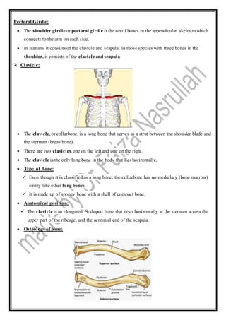
Osteologyof bone:
2. The bone has a cylindrical part called the shaft, and two ends: lateral and medial.
Shaft:
1. Lateral 1/3: flattened in sectionand thinner
consists of 2 borders : anterior and posterior and 2 surfaces: superior and inferior
BORDERS:
i. anterior border: concave forwards and gives origin to deltoid muscle
ii. posterior border: convex backwards and gives attachment to trapezius muscle
SURFACE:
i. superior surface: is subcutaneous
ii. inferior surface: shows conoid tubercle and trapezoid ridge which gives
attachment to conoid ligament (medial part of coracoclavicular ligament) and
trapezoid ligament (lateral part of coracoclavicular ligament) respectively
2. Medial 2/3: circular insectionand thicker
consists of 4 surfaces
i. anterior surface: convex forwards and gives origin to pectorals major
ii. posterior surface: concave backwards and gives origin to sternohyoid muscle
iii. superior surface: is rough on its medial part
iv. inferior surface: has rough oval impression at the medial end for costoclavicular
ligament and shows subclavian groove which gives attachment to subclavian muscle
Ends:
1. Lateral (Acromial) End: articulates with with the acromian process of the scapula to form
the Acromioclavicular joint
2. Medial (Sternal) End: articulates with the clavicular notch of the manubrium sterni to
form Sternoclavicular joint.
Side Determination:
Lateral end is flat and thinner and medial end is round and thicker.
There is a groove on inferior surface.
Medial part of shaft is convex anteriorly while lateral part is concave anteriorly.
3. Functions of clavicle:
It serves as a rigid support from which the scapula and free limb suspended; an arrangement
that keeps the upper limb away from the thorax so that the arm has maximum range of
movement.
Articulationof Clavicle:
The clavicle is an anterior bone whose sternal end articulates with the manubrium of the
sternum at the Sternoclavicular joint. The sternal end is also anchored to the first rib by the
costoclavicular ligament.
The acromial end of the clavicle articulates with the acromion of the scapula at the
Acromioclavicular joint.
Muscles attached to clavicle:
A total of five muscles are attached to the clavicle, found distributed at either the lateral third
or medial two thirds of the bone.
Two muscles are attached to the lateral thirdofthe clavicle:
4. i. The trapezius muscle, which is attached along the posterior surface of the bone.
ii. The deltoidmuscle, where the anterior portionof the muscle is attached to the
periosteum at the anterior surface of the bone.
Three muscles are attached to the medial thirdof the clavicle:
i. The sternocleidomastoidmuscle, where the muscular clavicular head of the muscle is
attached to the superior surface of the bone.
ii. The pectoralis major muscle, which is attached to the anterior surface of the bone.
iii. The subclavius muscle, which is attached to a groove found in the middle of the bone’s
inferior surface. From there, the muscle extends into both lateral and medial areas of the
clavicle.
Ligaments attached to clavicle:
Acromioclavicular ligaments (several) and coracoclavicular ligaments (there are
two: trapezoid and conoid). These ligaments stabilize the Acromioclavicular (AC) joint.
Sternoclavicular ligaments (many) stabilize the Sternoclavicular (SC) joint.
Bloodsupply:
Three arteries were found to supply the clavicle:
1. the suprascapular a.;
2. the thoracoacromial a.; and
3. the internal thoracic (mammary)
The main supply was primarily periosteal.
No nutrient artery was found.
Nerve Supply:
the common trunk of the supraclavicular nerves divides into three groups of descending
branches:
1. the medial supraclavicular nerve,
2. intermediate supraclavicular nerve and
3. Lateral supraclavicular nerve.
Clinical notes:
Clavicle Fracture:
Clavicle fractures are fairly common and occur in people of all ages. Most fractures occur in
the middle portion, or shaft, of the bone. Occasionally, the bone will break where it attaches
at the ribcage or shoulder blade.
5. Clavicle fractures vary. The bone can crack just slightly or break into many pieces
(comminuted fracture). The broken pieces of bone may line up straight or may be far out of
place (displaced fracture).
Comminuted
Causes:
Clavicle fractures are most often caused by a direct blow to the shoulder. This can happen
during a fall onto the shoulder or a car collision. A fall onto an outstretched arm can also
cause a clavicle fracture. In a baby, a clavicle fracture can occur during the passage through
the birth canal.