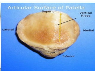
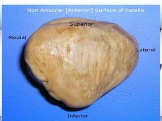
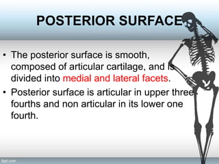
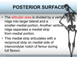
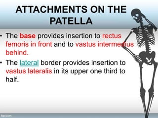
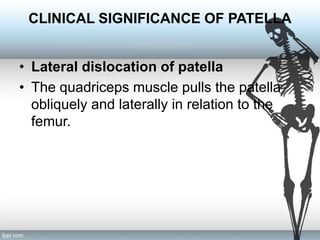
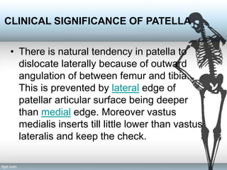
Lecture 4 PATELLA
Dr. EIMAAN SUMAYYAH
MSPT (Neuro), DPT (KMU)
PATELLA
• A sesamoid bone that lies within the quadriceps
tendon/patella ligament and forms part of the
knee joint.
• Situated in front of lower end of femur appx 1 cm
above the knee joint.
• It is the largest sesamoid bone of the body.
DETERMINATION OF SIDE OF
PATELLA
• Patella is triangular with apex of the
triangle directed downwards.
• Anterior surface is rough and non articular.
The upper three fourths of the posterior
surface are smooth and articular.
• The bone rests on broad lateral area when
placed on table as a vertical ridge divides
it into large lateral area and a
smaller medial area.
PATELLA
• The patella is triangular in shape with a superior
base and inferior apex.
• The apex lies about 1 cm from knee joint.
• It has three borders – superior, lateral and
medial, and two surfaces – anterior and
posterior.
ANTERIOR SURFACE
• Anterior surface is convex, rough and
ridged vertically.
• Covered by expansion from tendons of
rectus femoris and is separated from the
skin by prepatellar bursa.
POSTERIOR SURFACE
• The posterior surface is smooth,
composed of articular cartilage, and is
divided into medial and lateral facets.
• Posterior surface is articular in upper three
fourths and non articular in its lower one
fourth.
POSTERIOR SURFACE
• The articular area is divided by a vertical
ridge into larger lateral and a
smaller medial portion. Another vertical
ridge separates a medial strip
from medial portion.
• This medial strip articulates with a
reciprocal strip on medial side of
intercondylar notch of femur during
full flexion.
POSTERIOR SURFACE
• The rest of medial and lateral portion of
the articular surface are divided by two
transverse lines into three pairs of facets.
• The medial and lateral facets of the patella
articulate with the medial and lateral
condyles of the femur, respectively, to form
the patellofemoral component of the knee
joint.
ATTACHMENTS ON THE
PATELLA
• The base provides insertion to rectus
femoris in front and to vastus intermedius
behind.
• The lateral border provides insertion to
vastus lateralis in its upper one third to
half.
ATTACHMENTS ON THE
PATELLA
• The medial border provides insertion to
vastus lateralis in its upper third to half.
• Behind the apex, on the posterior aspect,
there is nonarticular area which provides
attachment to patellar ligament or
ligamentum patellae which gets attached
to the tibial tubercle, inferiorly.
PATELLOFEMORAL
ARTICULATION
• During different phases of movements of
the knee, different portions of the patella
articulate with femur. The lower pair of
articular facets articulates
during extension, middle
during flexion and upper pair during
mid flexion. Medial strip articulates during
full flexion.
CLINICAL SIGNIFICANCE OF PATELLA
• Lateral dislocation of patella
• The quadriceps muscle pulls the patella
obliquely and laterally in relation to the
femur.
CLINICAL SIGNIFICANCE OF PATELLA
• There is natural tendency in patella to
dislocate laterally because of outward
angulation of between femur and tibia.
This is prevented by lateral edge of
patellar articular surface being deeper
than medial edge. Moreover vastus
medialis inserts till little lower than vastus
lateralis and keep the check.
CLINICAL SIGNIFICANCE OF PATELLA
• Patellofemoral syndrome, or chondromalacia
patella, is a painful condition in which the
cartilage on the articular surfaces of the patella
becomes soft and breaks down. The condition
occurs when the patella rubs against the femur
due to problems with patellar alignment or
excessive physical activity involving the knee.
Patellofemoral syndrome is most commonly
seen in adolescent and young adult females.
CLINICAL SIGNIFICANCE OF PATELLA
• If patella lies higher, it is called patella alta
and if it is lower, it is called patella baja.
Next…
• Tibia and its attachments!!!!

Lec 4 patella

  • 1.
    Lecture 4 PATELLA Dr.EIMAAN SUMAYYAH MSPT (Neuro), DPT (KMU)
  • 3.
    PATELLA • A sesamoidbone that lies within the quadriceps tendon/patella ligament and forms part of the knee joint. • Situated in front of lower end of femur appx 1 cm above the knee joint. • It is the largest sesamoid bone of the body.
  • 4.
    DETERMINATION OF SIDEOF PATELLA • Patella is triangular with apex of the triangle directed downwards. • Anterior surface is rough and non articular. The upper three fourths of the posterior surface are smooth and articular. • The bone rests on broad lateral area when placed on table as a vertical ridge divides it into large lateral area and a smaller medial area.
  • 7.
    PATELLA • The patellais triangular in shape with a superior base and inferior apex. • The apex lies about 1 cm from knee joint. • It has three borders – superior, lateral and medial, and two surfaces – anterior and posterior.
  • 8.
    ANTERIOR SURFACE • Anteriorsurface is convex, rough and ridged vertically. • Covered by expansion from tendons of rectus femoris and is separated from the skin by prepatellar bursa.
  • 10.
    POSTERIOR SURFACE • Theposterior surface is smooth, composed of articular cartilage, and is divided into medial and lateral facets. • Posterior surface is articular in upper three fourths and non articular in its lower one fourth.
  • 12.
    POSTERIOR SURFACE • Thearticular area is divided by a vertical ridge into larger lateral and a smaller medial portion. Another vertical ridge separates a medial strip from medial portion. • This medial strip articulates with a reciprocal strip on medial side of intercondylar notch of femur during full flexion.
  • 13.
    POSTERIOR SURFACE • Therest of medial and lateral portion of the articular surface are divided by two transverse lines into three pairs of facets. • The medial and lateral facets of the patella articulate with the medial and lateral condyles of the femur, respectively, to form the patellofemoral component of the knee joint.
  • 14.
    ATTACHMENTS ON THE PATELLA •The base provides insertion to rectus femoris in front and to vastus intermedius behind. • The lateral border provides insertion to vastus lateralis in its upper one third to half.
  • 16.
    ATTACHMENTS ON THE PATELLA •The medial border provides insertion to vastus lateralis in its upper third to half. • Behind the apex, on the posterior aspect, there is nonarticular area which provides attachment to patellar ligament or ligamentum patellae which gets attached to the tibial tubercle, inferiorly.
  • 17.
    PATELLOFEMORAL ARTICULATION • During differentphases of movements of the knee, different portions of the patella articulate with femur. The lower pair of articular facets articulates during extension, middle during flexion and upper pair during mid flexion. Medial strip articulates during full flexion.
  • 18.
    CLINICAL SIGNIFICANCE OFPATELLA • Lateral dislocation of patella • The quadriceps muscle pulls the patella obliquely and laterally in relation to the femur.
  • 19.
    CLINICAL SIGNIFICANCE OFPATELLA • There is natural tendency in patella to dislocate laterally because of outward angulation of between femur and tibia. This is prevented by lateral edge of patellar articular surface being deeper than medial edge. Moreover vastus medialis inserts till little lower than vastus lateralis and keep the check.
  • 20.
    CLINICAL SIGNIFICANCE OFPATELLA • Patellofemoral syndrome, or chondromalacia patella, is a painful condition in which the cartilage on the articular surfaces of the patella becomes soft and breaks down. The condition occurs when the patella rubs against the femur due to problems with patellar alignment or excessive physical activity involving the knee. Patellofemoral syndrome is most commonly seen in adolescent and young adult females.
  • 21.
    CLINICAL SIGNIFICANCE OFPATELLA • If patella lies higher, it is called patella alta and if it is lower, it is called patella baja.
  • 23.
    Next… • Tibia andits attachments!!!!