SlideShare a Scribd company logo
SKELETON OF THE UPPER LIMB
The skeleton of the upper limb is divided into two parts -
the pectoral girdle and the freely movable part of the upper
limb: pectoral girdle, includes the clavicle and the scapula,
with the help of which the free part of the upper limb
articulates with the torso. free part of the upper limb,includes
bones of the arm (ossa brachii), bones of the forearm (ossa
antebrachii) and bones of the hand(ossa manus).
PECTORAL GIRDLE
The scapula:The scapula (“shoulder blade”) is a large,
triangular flat bone on the posterior side of the rib cage,
overlying ribs 2 through 7. The spine of the scapula,spina
scapulae is a prominent diagonal bony ridge seen on the
posterior surface. The spine strengthens the scapula, making it
more resistant to bending. Above the spine is
the supraspinous fossa,fossa supraspinata and below the
spine is the infraspinous fossa,fossa infraspinata. The spine
broadens toward the shoulder as the acromion. This process
serves for the attachment of several muscles, as well as for
articulation with the clavicle. Inferior to the acromion is a
shallow depression, the glenoid cavity,cavitas
glenoidalis into which the head of the humerus fits.
The coracoid process,processus coracoideus is a thick
upward projection lying superior and anterior to the glenoid
cavity. The scapula has anterior(costal surface)
and posterior surfaces.The anterior surface faces the ribs.On
the anterior surface of the scapula is a slightly concave area
known as the subscapular fossa. The posterior surface faces
the skin of the back.
Angles of the scapula
 Acromial angle,angulus acromialis; sharp bend at the
site where the spine of the scapula becomes continuous
with the lateral margin of the acromion.
SKELETON OF THE UPPER LIMB
 Inferior angle,angulus inferior; lower angle of the
scapula.
 Lateral angle,angulus lateralis; lateral angle of the
scapula bearing the glenoid cavity.
 Superior angle,angulus superior ;upper medial angle of
the scapula .
Margins of the scapula
 Medial margin,margo medialis; border of the scapula
facing the vertebral column.
 Lateral margin,margo lateralis; border of the scapula
facing the humerus.
 Superior margin,margo superior; upper border of the
scapula.
SKELETON OF THE UPPER LIMB
DonNMU.
Clinical application: The scapula has numerous surface
features because 15 muscles attach to it. Clinically, the
pectoral girdle is significant becausethe clavicle and acromion
of the scapula are frequentlybroken in trying to break a fall.
The acromion is used as a landmark for identifying the site for
an injection in the arm. This site is chosen because the
musculature of the shoulder is quite thick and contains few
nerves.
HOW TO KNOW LEFT OR RIGHT
SCAPULA
 Identify the glenoid cavity and place it laterally.
 Place the spine of the scapula posteriorly.In this way
you can be able to identify the position of the scapula
easily.
The clavicle:The slender S-shaped
clavicle(“collarbone”) connects the upper extremity to the
axial skeleton and holds the shoulder joint away from the
trunk to permit freedom of movement. The articulation of the
medial sternal extremity of the clavicle to the manubrium of
the sternum is referred to as the sternoclavicular joint. The
lateral acromial extremity of the clavicle articulates with the
acromion of the scapula.This articulation is referred to as the
acromioclavicular joint. A conoid tubercle is present on the
acromial extremity of the clavicle, and a costal tuberosity is
present on the inferior surface of the sternal extremity. Both
processes serve as attachments for ligaments.
SKELETON OF THE UPPER LIMB
Clinical application:The long, delicate clavicle is the most
commonly broken bone in the body. When a person receives a
blow to the shoulder, or attempts to break a fall with an
outstretched hand, the force is transmitted to the clavicle,
possibly causing it to fracture. The most vulnerable part of
this bone is through its center, immediately proximal to the
conoid tubercle. Because the clavicle is directly beneath the
skin and is not covered with muscle, a fracture can easily be
palpated, and frequently seen.
SKELETON OF THE UPPER LIMB
FREE PART OF THE UPPER LIMB
The humerus:The humerus is the longest bone of the upper
extremity.It consists of a proximal head(proximal epiphyses),
which articulates with the glenoidcavity of the scapula;
a body (“shaft”); and a distal end(distal epiphysis), which is
modified to articulate with the two bones of the forearm.
Surrounding the margin of the head is a slightly indented
groove denoting the anatomical neck. The surgical neck, the
constriction just below the head, is a frequent fracture
site. The greater tubercle is a large knob on the lateral
proximal portion of the humerus. The lesser tubercle is
slightly anterior to the greater tubercle and is separated from
the greater by an intertubercular groove. The tendon of the
long head of the biceps brachii muscle passes through this
groove. Along the lateral midregion of the body of the
humerus is a roughened area, the deltoid tuberosity, for the
attachment of the deltoid muscle. Small openings in the body
are called nutrient foramina. The humeral condyle on the
distal end of the humerus has two articular surfaces.
The capitulum is the lateral rounded part that articulates with
the radius. The trochlea is the pulleylike medial part that
articulates with the ulna. On either side above the condyle are
the lateral and medial epicondyles. The large medial
epicondyle protects the ulnar nerve that passes posteriorly
through the ulnar sulcus. It is popularly known as the “funny
bone” because striking the elbow on the edge of a table, for
example, stimulates the ulnar nerve and produces a tingling
sensation. The coronoid fossa is a depression above the
trochlea on the anterior surface. The olecranon fossa is a
depression on the distal posterior surface. Both fossae are
adapted to work with the ulna during movement of the
forearm.
Margins(borders):
SKELETON OF THE UPPER LIMB
 Medial margin,margo medialis: inner margin of the
humerus continuous distally with the medial
supracondylar ridge.
 Lateral margin,margo lateralis: outer margin of the
humerus continuous distally with the lateral
supracondylar ridge.
Surfaces:
 Anteromedial surface,facies anteromedialis: surface of
the humerus lying medial to the prolongation of the crest
of the greater tubercle.
 Anterolateral surface,facies anterolateralis: surface of
the humerus located lateral to the prolongation of the
crest of the greater tubercle.
 posterior surface,facies posterior.
clinical application:The medical term for tennis elbow is
lateral epicondylitis, which means inflammation of the tissues
surrounding the lateral epicondyle of the humerus. At least six
SKELETON OF THE UPPER LIMB
muscles that control backward (extension) movement of the
wrist and finger joints originate on the lateral epicondyle.
Repeated strenuous contractions of these muscles, as in
stroking with a tennis racket, may strain the periosteum and
muscle attachments, resulting in swelling, tenderness, and
pain around the epicondyle. Binding usually eases the pain,
but only rest can eliminate the causative factor, and recovery
generally follows.
HOW TO KNOW IF A HUMERUS IS LEFT OR
RIGHT.
clue I
1. Identify the head of the humerus and the medial
epicondyle. These two should be medially placed.
2.Identify the coronoid and radial fossae. These two should
be anteriorly placed with the olecranon fossa placed
posteriorly. In this position, we can determine if its left or
right.
clue II
1. place the head of the humerus and the medial
epicondyle at the medial position.
2.the olecranon fossa should be posteriorly placed.
3.If the head of the humerus faces left,it means it is a
right humerus and vice versa.
THE RADIUS:The radius consists of a body,corpus
radii with a small proximal end, and a large distal end. A
proximal disc-shaped head articulates with the capitulum of
the humerus and the radial notch of the ulna. The prominent
tuberosity of radius (radial tuberosity), for attachment of the
biceps brachii muscle, is located on the medial side of the
body, just below the head. On the distal end of the radius is a
double-faceted surface for articulation with the proximal
SKELETON OF THE UPPER LIMB
carpal bones. The distal end of the radius also has
a styloid process on the lateral tip and an ulnar notch on
the medial side that receives the distal end of the ulna. The
styloid processes on the ulna and radius provide lateral and
medial stability for articulation at the wrist.
Surfaces:
 Anterior surface,facies anterior.
 Posterior surface,facies posterior.
 Lateral surface, facies lateralis.
Borders:
 Anterior border,margo anterior.
 Posterior border,margo posterior.
 Interosseous border,margo interosseous.
Clinical application: When a person falls, the natural
tendency is to extend the hand to break the fall. This reflexive
movement frequently results in fractured bones. Common
fractures of the radius include a fracture of the head, as it is
driven forcefully against the capitulum; a fracture of the neck;
or a fracture of the distal end (Colles’ fracture), caused by
landing on an outstretched hand.
When falling, it is less traumatic to the body to withdraw the
appendages,bend the knees, and let the entire body hit the
surface.
Athletes learn that this is the safe way to fall.
how to know left and right radius
 The proximal end should face up and the distal end,
down.
SKELETON OF THE UPPER LIMB
 Place the radial tuberosity medially.
 Identify the nutrient foramen and place it anteriorly.i.e
away from you.
 The radial styloid process should also be at the lateral
position.
ULNA:The proximal end of the ulna articulates with the
humerus and radius. A distinct depression, the trochlear
notch, articulates with the trochlea of the humerus.
The coronoid process forms the anterior lip of the trochlear
notch, and the olecranon forms the posterior portion. Lateral
and inferior to the coronoid process is the radial notch, which
accommodates the head of the radius.
On the tapered distal end of the ulna is a knobbed
portion, the head, and a knoblike projection, the styloid
process. The ulna articulates at both ends with the radius.In
the forearm, it is located medially.
SKELETON OF THE UPPER LIMB
DonNMU.
how to know left and right ulna
 The proximal and distal epiphysis should first of all be placed at the right
position;proximal up, distal down.
 face the trochlear notch away from you and look at the olecranon.
 Check to see which side the radial notch is.
 If it is on the right, it a right ulna and vice versa.
BONES OF THE HAND
The hand contains 27 bones, grouped into the carpus,
metacarpus,
and phalanges.
SKELETON OF THE UPPER LIMB
Carpus:The carpus, or wrist, or carpal bone contains eight
bones arranged in two transverse rows of four bones each.
The proximal row, naming from the lateral (thumb) to the
medial side, consists of the:
 scaphoid (navicular),os scaphoideum
 lunate,os lunatum
 triquetrum,os triquetrum and
 pisiform,os pisiforme.The pisiform forms in a tendon as a
sesamoid bone.
The distal row, from lateral to medial, consists of the
 trapezium,os trapezium (greater multangular),
 trapezoid,os trapezoideum (lesser multangular),
 capitate,os capitatum and
 hamate,os hamatum. The scaphoid and lunate of the
proximal row articulate with the distal end of the radius.
Metacarpus:The metacarpus, or palm of the hand,
contains five metacarpal bones. Each metacarpal bone
consists of a proximal base, a body, and a distal head that is
rounded for articulation with the base of each proximal
phalanx. The heads of the metacarpal bones are distally
located and form the knuckles of a clenched fist.
The phalanges:The 14 phalanges are the bones of the
digits. A single finger bone is called a phalanx (fa'langks).
The phalanges of the fingers are arranged in a proximal row,
a middle row, and a distal row.
The thumb, or pollex (adjective, pollicis), lacks a middle
phalanx.
The digits are sequentially numbered I to V starting with the
SKELETON OF THE UPPER LIMB
thumb—the lateral side, in reference to anatomical position.
Clinical application: The hand is a marvel
of structural complexity that can withstand
considerable abuse. Other than sprained
ligaments of the fingers and joint dislocations,
the most common bone injury is a fracture to
the scaphoid—a wrist bone that accounts for
about 70% of carpal fractures. When immobilizing the
wrist joint, the wrist is positioned in the plane of
relaxed function. This is the position in which
SKELETON OF THE UPPER LIMB
the hand is about to grasp an object between
the thumb and index finger.
Kamal Umar Labaran(Med)
Donetsk National Medical University.

More Related Content

What's hot

Bone and Muscles of the Hand
Bone and Muscles of the HandBone and Muscles of the Hand
Bone and Muscles of the Hand
Sado Anatomist
 
Elbow joint anatomy and examination
Elbow joint anatomy and examinationElbow joint anatomy and examination
Elbow joint anatomy and examination
RamanGhimire3
 
Elbow joint
Elbow jointElbow joint
Elbow joint
Idris Siddiqui
 
Anatomy of upper limb
Anatomy of upper limbAnatomy of upper limb
Anatomy of upper limb
galibraihan
 
Surface anatomy of upper limb
Surface anatomy of upper limbSurface anatomy of upper limb
Surface anatomy of upper limb
Mathew Joseph
 
Joints of upper limb
Joints of upper limb Joints of upper limb
Joints of upper limb
sadhamhussain52
 
Seminar clinical anatomy of upper limb joints and muscles
Seminar clinical anatomy of upper limb joints and musclesSeminar clinical anatomy of upper limb joints and muscles
Seminar clinical anatomy of upper limb joints and muscles
Quan Fu Gan
 
Radiographic anatomy upper limb
Radiographic anatomy upper limbRadiographic anatomy upper limb
Radiographic anatomy upper limb
donishajohnson
 
Anatomy revision course pt1
Anatomy revision course pt1Anatomy revision course pt1
Anatomy revision course pt1
Murad Alshehry
 
Radius, Ulna, Elbow and Radioulnar Joint
Radius, Ulna, Elbow and Radioulnar JointRadius, Ulna, Elbow and Radioulnar Joint
Radius, Ulna, Elbow and Radioulnar JointSado Anatomist
 
Anatomy and Biomechanics of the Elbow Joint
Anatomy and Biomechanics of the Elbow JointAnatomy and Biomechanics of the Elbow Joint
Anatomy and Biomechanics of the Elbow Jointorthoprince
 
upper limb joints.
upper limb joints.upper limb joints.
upper limb joints.
mohammed Qazzaz
 
Anatomy revision 2
Anatomy revision 2Anatomy revision 2
Anatomy revision 2
Murad Alshehry
 
Anatomy (anatomy of bone, joint & muscles of upper limb)
Anatomy (anatomy of bone, joint & muscles of upper limb)Anatomy (anatomy of bone, joint & muscles of upper limb)
Anatomy (anatomy of bone, joint & muscles of upper limb)
Osama Al-Zahrani
 
Kin 191 B Elbow And Forearm Anatomy And Evaluation
Kin 191 B   Elbow And Forearm Anatomy And EvaluationKin 191 B   Elbow And Forearm Anatomy And Evaluation
Kin 191 B Elbow And Forearm Anatomy And EvaluationJLS10
 
Anatomy bones of upper limbs
Anatomy bones of upper limbsAnatomy bones of upper limbs
Anatomy bones of upper limbs
Shifa medicine collage
 
Radiology of the Elbow Joint. Dr. Sumit Sharma
Radiology of the Elbow Joint. Dr. Sumit SharmaRadiology of the Elbow Joint. Dr. Sumit Sharma
Radiology of the Elbow Joint. Dr. Sumit Sharma
Sumit Sharma
 
Pectoral Girdle and Shoulder
Pectoral Girdle and ShoulderPectoral Girdle and Shoulder
Pectoral Girdle and ShoulderSado Anatomist
 
Forearm bones
Forearm bonesForearm bones
Forearm bones
sameer abdallah
 
Anatomy of upper limb rehan
Anatomy of upper limb rehanAnatomy of upper limb rehan
Anatomy of upper limb rehan
Rehan Asad
 

What's hot (20)

Bone and Muscles of the Hand
Bone and Muscles of the HandBone and Muscles of the Hand
Bone and Muscles of the Hand
 
Elbow joint anatomy and examination
Elbow joint anatomy and examinationElbow joint anatomy and examination
Elbow joint anatomy and examination
 
Elbow joint
Elbow jointElbow joint
Elbow joint
 
Anatomy of upper limb
Anatomy of upper limbAnatomy of upper limb
Anatomy of upper limb
 
Surface anatomy of upper limb
Surface anatomy of upper limbSurface anatomy of upper limb
Surface anatomy of upper limb
 
Joints of upper limb
Joints of upper limb Joints of upper limb
Joints of upper limb
 
Seminar clinical anatomy of upper limb joints and muscles
Seminar clinical anatomy of upper limb joints and musclesSeminar clinical anatomy of upper limb joints and muscles
Seminar clinical anatomy of upper limb joints and muscles
 
Radiographic anatomy upper limb
Radiographic anatomy upper limbRadiographic anatomy upper limb
Radiographic anatomy upper limb
 
Anatomy revision course pt1
Anatomy revision course pt1Anatomy revision course pt1
Anatomy revision course pt1
 
Radius, Ulna, Elbow and Radioulnar Joint
Radius, Ulna, Elbow and Radioulnar JointRadius, Ulna, Elbow and Radioulnar Joint
Radius, Ulna, Elbow and Radioulnar Joint
 
Anatomy and Biomechanics of the Elbow Joint
Anatomy and Biomechanics of the Elbow JointAnatomy and Biomechanics of the Elbow Joint
Anatomy and Biomechanics of the Elbow Joint
 
upper limb joints.
upper limb joints.upper limb joints.
upper limb joints.
 
Anatomy revision 2
Anatomy revision 2Anatomy revision 2
Anatomy revision 2
 
Anatomy (anatomy of bone, joint & muscles of upper limb)
Anatomy (anatomy of bone, joint & muscles of upper limb)Anatomy (anatomy of bone, joint & muscles of upper limb)
Anatomy (anatomy of bone, joint & muscles of upper limb)
 
Kin 191 B Elbow And Forearm Anatomy And Evaluation
Kin 191 B   Elbow And Forearm Anatomy And EvaluationKin 191 B   Elbow And Forearm Anatomy And Evaluation
Kin 191 B Elbow And Forearm Anatomy And Evaluation
 
Anatomy bones of upper limbs
Anatomy bones of upper limbsAnatomy bones of upper limbs
Anatomy bones of upper limbs
 
Radiology of the Elbow Joint. Dr. Sumit Sharma
Radiology of the Elbow Joint. Dr. Sumit SharmaRadiology of the Elbow Joint. Dr. Sumit Sharma
Radiology of the Elbow Joint. Dr. Sumit Sharma
 
Pectoral Girdle and Shoulder
Pectoral Girdle and ShoulderPectoral Girdle and Shoulder
Pectoral Girdle and Shoulder
 
Forearm bones
Forearm bonesForearm bones
Forearm bones
 
Anatomy of upper limb rehan
Anatomy of upper limb rehanAnatomy of upper limb rehan
Anatomy of upper limb rehan
 

Similar to Skeleton of the upper limb

Presentation1.pptx, radiological anatomy of the upper limb joint.
Presentation1.pptx, radiological anatomy of the upper limb joint.Presentation1.pptx, radiological anatomy of the upper limb joint.
Presentation1.pptx, radiological anatomy of the upper limb joint.Abdellah Nazeer
 
01 Bones of upper limb.ppt111111111111111
01 Bones of upper limb.ppt11111111111111101 Bones of upper limb.ppt111111111111111
01 Bones of upper limb.ppt111111111111111
JamesAmaduKamara
 
Clinical examination in orthopedics
Clinical examination in orthopedicsClinical examination in orthopedics
Clinical examination in orthopedicsSpringer
 
The skeleton of the lower limb
The skeleton of the lower limbThe skeleton of the lower limb
The skeleton of the lower limb
Kamal Deen
 
06 Appendicular Skeleton Pectoral Girdle And Upper Limbs
06 Appendicular Skeleton   Pectoral Girdle And Upper Limbs06 Appendicular Skeleton   Pectoral Girdle And Upper Limbs
06 Appendicular Skeleton Pectoral Girdle And Upper Limbsguest334add
 
06 Appendicular Skeleton Pectoral Girdle And Upper Limbs
06 Appendicular Skeleton   Pectoral Girdle And Upper Limbs06 Appendicular Skeleton   Pectoral Girdle And Upper Limbs
06 Appendicular Skeleton Pectoral Girdle And Upper Limbs
Kevin Young
 
06 Appendicular Skeleton Pectoral Girdle And Upper Limbs
06 Appendicular Skeleton   Pectoral Girdle And Upper Limbs06 Appendicular Skeleton   Pectoral Girdle And Upper Limbs
06 Appendicular Skeleton Pectoral Girdle And Upper Limbs
guest295165
 
UPPER LIMB 4 ppt of human body..........
UPPER LIMB 4 ppt of human body..........UPPER LIMB 4 ppt of human body..........
UPPER LIMB 4 ppt of human body..........
aahanak787
 
Anatomy of Upper extremity
Anatomy of Upper extremityAnatomy of Upper extremity
Anatomy of Upper extremity
Sunil Pahari
 
Skeletal System 2
Skeletal System 2Skeletal System 2
Skeletal System 2Marc Potter
 
Joints upper limb 2nd lecture10122010
Joints upper limb 2nd lecture10122010Joints upper limb 2nd lecture10122010
Joints upper limb 2nd lecture10122010Lawrence James
 
Lesson 9 Elbow Joint (Elbow and Forearm).pptx
Lesson 9 Elbow Joint (Elbow and Forearm).pptxLesson 9 Elbow Joint (Elbow and Forearm).pptx
Lesson 9 Elbow Joint (Elbow and Forearm).pptx
JohnneErikaLarosa
 
Lesson 8 (The Shoulder).pptx
Lesson 8 (The Shoulder).pptxLesson 8 (The Shoulder).pptx
Lesson 8 (The Shoulder).pptx
JohnneErikaLarosa
 
1.Bones of The Upper Limbs.pdf
1.Bones of The Upper Limbs.pdf1.Bones of The Upper Limbs.pdf
1.Bones of The Upper Limbs.pdf
lamba christian
 
UPPERLIMB bs1.pptx
UPPERLIMB bs1.pptxUPPERLIMB bs1.pptx
UPPERLIMB bs1.pptx
THIRUVAZAHAN
 
Elbowinjuries
ElbowinjuriesElbowinjuries
Elbowinjuries
bandarindupriya08
 
elbow injuries and disorders presentation
elbow injuries and disorders  presentationelbow injuries and disorders  presentation
elbow injuries and disorders presentation
SalmaAzeem3
 
Anatomy of Humerus
Anatomy of HumerusAnatomy of Humerus
Anatomy of Humerus
Dr Adnan Sami
 
shoulder assessment (full)
shoulder assessment (full)shoulder assessment (full)
shoulder assessment (full)
SYED MASOOD
 
Elbow Region.pptx
Elbow Region.pptxElbow Region.pptx
Elbow Region.pptx
Chiibeneme
 

Similar to Skeleton of the upper limb (20)

Presentation1.pptx, radiological anatomy of the upper limb joint.
Presentation1.pptx, radiological anatomy of the upper limb joint.Presentation1.pptx, radiological anatomy of the upper limb joint.
Presentation1.pptx, radiological anatomy of the upper limb joint.
 
01 Bones of upper limb.ppt111111111111111
01 Bones of upper limb.ppt11111111111111101 Bones of upper limb.ppt111111111111111
01 Bones of upper limb.ppt111111111111111
 
Clinical examination in orthopedics
Clinical examination in orthopedicsClinical examination in orthopedics
Clinical examination in orthopedics
 
The skeleton of the lower limb
The skeleton of the lower limbThe skeleton of the lower limb
The skeleton of the lower limb
 
06 Appendicular Skeleton Pectoral Girdle And Upper Limbs
06 Appendicular Skeleton   Pectoral Girdle And Upper Limbs06 Appendicular Skeleton   Pectoral Girdle And Upper Limbs
06 Appendicular Skeleton Pectoral Girdle And Upper Limbs
 
06 Appendicular Skeleton Pectoral Girdle And Upper Limbs
06 Appendicular Skeleton   Pectoral Girdle And Upper Limbs06 Appendicular Skeleton   Pectoral Girdle And Upper Limbs
06 Appendicular Skeleton Pectoral Girdle And Upper Limbs
 
06 Appendicular Skeleton Pectoral Girdle And Upper Limbs
06 Appendicular Skeleton   Pectoral Girdle And Upper Limbs06 Appendicular Skeleton   Pectoral Girdle And Upper Limbs
06 Appendicular Skeleton Pectoral Girdle And Upper Limbs
 
UPPER LIMB 4 ppt of human body..........
UPPER LIMB 4 ppt of human body..........UPPER LIMB 4 ppt of human body..........
UPPER LIMB 4 ppt of human body..........
 
Anatomy of Upper extremity
Anatomy of Upper extremityAnatomy of Upper extremity
Anatomy of Upper extremity
 
Skeletal System 2
Skeletal System 2Skeletal System 2
Skeletal System 2
 
Joints upper limb 2nd lecture10122010
Joints upper limb 2nd lecture10122010Joints upper limb 2nd lecture10122010
Joints upper limb 2nd lecture10122010
 
Lesson 9 Elbow Joint (Elbow and Forearm).pptx
Lesson 9 Elbow Joint (Elbow and Forearm).pptxLesson 9 Elbow Joint (Elbow and Forearm).pptx
Lesson 9 Elbow Joint (Elbow and Forearm).pptx
 
Lesson 8 (The Shoulder).pptx
Lesson 8 (The Shoulder).pptxLesson 8 (The Shoulder).pptx
Lesson 8 (The Shoulder).pptx
 
1.Bones of The Upper Limbs.pdf
1.Bones of The Upper Limbs.pdf1.Bones of The Upper Limbs.pdf
1.Bones of The Upper Limbs.pdf
 
UPPERLIMB bs1.pptx
UPPERLIMB bs1.pptxUPPERLIMB bs1.pptx
UPPERLIMB bs1.pptx
 
Elbowinjuries
ElbowinjuriesElbowinjuries
Elbowinjuries
 
elbow injuries and disorders presentation
elbow injuries and disorders  presentationelbow injuries and disorders  presentation
elbow injuries and disorders presentation
 
Anatomy of Humerus
Anatomy of HumerusAnatomy of Humerus
Anatomy of Humerus
 
shoulder assessment (full)
shoulder assessment (full)shoulder assessment (full)
shoulder assessment (full)
 
Elbow Region.pptx
Elbow Region.pptxElbow Region.pptx
Elbow Region.pptx
 

More from Kamal Deen

Breast cancer
Breast cancerBreast cancer
Breast cancer
Kamal Deen
 
Radiation diseases
Radiation diseasesRadiation diseases
Radiation diseases
Kamal Deen
 
The arthrosyndesmology
The arthrosyndesmologyThe arthrosyndesmology
The arthrosyndesmology
Kamal Deen
 
Anatomy of the cranial bones
Anatomy of the cranial bonesAnatomy of the cranial bones
Anatomy of the cranial bones
Kamal Deen
 
Bone classification
Bone classificationBone classification
Bone classification
Kamal Deen
 
Locomotor system
Locomotor systemLocomotor system
Locomotor system
Kamal Deen
 
OSTEOLOGY OF THE VERTEBRAE
OSTEOLOGY OF THE VERTEBRAE OSTEOLOGY OF THE VERTEBRAE
OSTEOLOGY OF THE VERTEBRAE
Kamal Deen
 

More from Kamal Deen (7)

Breast cancer
Breast cancerBreast cancer
Breast cancer
 
Radiation diseases
Radiation diseasesRadiation diseases
Radiation diseases
 
The arthrosyndesmology
The arthrosyndesmologyThe arthrosyndesmology
The arthrosyndesmology
 
Anatomy of the cranial bones
Anatomy of the cranial bonesAnatomy of the cranial bones
Anatomy of the cranial bones
 
Bone classification
Bone classificationBone classification
Bone classification
 
Locomotor system
Locomotor systemLocomotor system
Locomotor system
 
OSTEOLOGY OF THE VERTEBRAE
OSTEOLOGY OF THE VERTEBRAE OSTEOLOGY OF THE VERTEBRAE
OSTEOLOGY OF THE VERTEBRAE
 

Recently uploaded

Maxilla, Mandible & Hyoid Bone & Clinical Correlations by Dr. RIG.pptx
Maxilla, Mandible & Hyoid Bone & Clinical Correlations by Dr. RIG.pptxMaxilla, Mandible & Hyoid Bone & Clinical Correlations by Dr. RIG.pptx
Maxilla, Mandible & Hyoid Bone & Clinical Correlations by Dr. RIG.pptx
Dr. Rabia Inam Gandapore
 
For Better Surat #ℂall #Girl Service ❤85270-49040❤ Surat #ℂall #Girls
For Better Surat #ℂall #Girl Service ❤85270-49040❤ Surat #ℂall #GirlsFor Better Surat #ℂall #Girl Service ❤85270-49040❤ Surat #ℂall #Girls
For Better Surat #ℂall #Girl Service ❤85270-49040❤ Surat #ℂall #Girls
Savita Shen $i11
 
basicmodesofventilation2022-220313203758.pdf
basicmodesofventilation2022-220313203758.pdfbasicmodesofventilation2022-220313203758.pdf
basicmodesofventilation2022-220313203758.pdf
aljamhori teaching hospital
 
POST OPERATIVE OLIGURIA and its management
POST OPERATIVE OLIGURIA and its managementPOST OPERATIVE OLIGURIA and its management
POST OPERATIVE OLIGURIA and its management
touseefaziz1
 
BRACHYTHERAPY OVERVIEW AND APPLICATORS
BRACHYTHERAPY OVERVIEW  AND  APPLICATORSBRACHYTHERAPY OVERVIEW  AND  APPLICATORS
BRACHYTHERAPY OVERVIEW AND APPLICATORS
Krishan Murari
 
ARTHROLOGY PPT NCISM SYLLABUS AYURVEDA STUDENTS
ARTHROLOGY PPT NCISM SYLLABUS AYURVEDA STUDENTSARTHROLOGY PPT NCISM SYLLABUS AYURVEDA STUDENTS
ARTHROLOGY PPT NCISM SYLLABUS AYURVEDA STUDENTS
Dr. Vinay Pareek
 
Ozempic: Preoperative Management of Patients on GLP-1 Receptor Agonists
Ozempic: Preoperative Management of Patients on GLP-1 Receptor Agonists  Ozempic: Preoperative Management of Patients on GLP-1 Receptor Agonists
Ozempic: Preoperative Management of Patients on GLP-1 Receptor Agonists
Saeid Safari
 
Physiology of Special Chemical Sensation of Taste
Physiology of Special Chemical Sensation of TastePhysiology of Special Chemical Sensation of Taste
Physiology of Special Chemical Sensation of Taste
MedicoseAcademics
 
263778731218 Abortion Clinic /Pills In Harare ,
263778731218 Abortion Clinic /Pills In Harare ,263778731218 Abortion Clinic /Pills In Harare ,
263778731218 Abortion Clinic /Pills In Harare ,
sisternakatoto
 
Cervical & Brachial Plexus By Dr. RIG.pptx
Cervical & Brachial Plexus By Dr. RIG.pptxCervical & Brachial Plexus By Dr. RIG.pptx
Cervical & Brachial Plexus By Dr. RIG.pptx
Dr. Rabia Inam Gandapore
 
heat stroke and heat exhaustion in children
heat stroke and heat exhaustion in childrenheat stroke and heat exhaustion in children
heat stroke and heat exhaustion in children
SumeraAhmad5
 
micro teaching on communication m.sc nursing.pdf
micro teaching on communication m.sc nursing.pdfmicro teaching on communication m.sc nursing.pdf
micro teaching on communication m.sc nursing.pdf
Anurag Sharma
 
Knee anatomy and clinical tests 2024.pdf
Knee anatomy and clinical tests 2024.pdfKnee anatomy and clinical tests 2024.pdf
Knee anatomy and clinical tests 2024.pdf
vimalpl1234
 
24 Upakrama.pptx class ppt useful in all
24 Upakrama.pptx class ppt useful in all24 Upakrama.pptx class ppt useful in all
24 Upakrama.pptx class ppt useful in all
DrSathishMS1
 
Are There Any Natural Remedies To Treat Syphilis.pdf
Are There Any Natural Remedies To Treat Syphilis.pdfAre There Any Natural Remedies To Treat Syphilis.pdf
Are There Any Natural Remedies To Treat Syphilis.pdf
Little Cross Family Clinic
 
Physiology of Chemical Sensation of smell.pdf
Physiology of Chemical Sensation of smell.pdfPhysiology of Chemical Sensation of smell.pdf
Physiology of Chemical Sensation of smell.pdf
MedicoseAcademics
 
Surat @ℂall @Girls ꧁❤8527049040❤꧂@ℂall @Girls Service Vip Top Model Safe
Surat @ℂall @Girls ꧁❤8527049040❤꧂@ℂall @Girls Service Vip Top Model SafeSurat @ℂall @Girls ꧁❤8527049040❤꧂@ℂall @Girls Service Vip Top Model Safe
Surat @ℂall @Girls ꧁❤8527049040❤꧂@ℂall @Girls Service Vip Top Model Safe
Savita Shen $i11
 
Phone Us ❤85270-49040❤ #ℂall #gIRLS In Surat By Surat @ℂall @Girls Hotel With...
Phone Us ❤85270-49040❤ #ℂall #gIRLS In Surat By Surat @ℂall @Girls Hotel With...Phone Us ❤85270-49040❤ #ℂall #gIRLS In Surat By Surat @ℂall @Girls Hotel With...
Phone Us ❤85270-49040❤ #ℂall #gIRLS In Surat By Surat @ℂall @Girls Hotel With...
Savita Shen $i11
 
How to Give Better Lectures: Some Tips for Doctors
How to Give Better Lectures: Some Tips for DoctorsHow to Give Better Lectures: Some Tips for Doctors
How to Give Better Lectures: Some Tips for Doctors
LanceCatedral
 
Couples presenting to the infertility clinic- Do they really have infertility...
Couples presenting to the infertility clinic- Do they really have infertility...Couples presenting to the infertility clinic- Do they really have infertility...
Couples presenting to the infertility clinic- Do they really have infertility...
Sujoy Dasgupta
 

Recently uploaded (20)

Maxilla, Mandible & Hyoid Bone & Clinical Correlations by Dr. RIG.pptx
Maxilla, Mandible & Hyoid Bone & Clinical Correlations by Dr. RIG.pptxMaxilla, Mandible & Hyoid Bone & Clinical Correlations by Dr. RIG.pptx
Maxilla, Mandible & Hyoid Bone & Clinical Correlations by Dr. RIG.pptx
 
For Better Surat #ℂall #Girl Service ❤85270-49040❤ Surat #ℂall #Girls
For Better Surat #ℂall #Girl Service ❤85270-49040❤ Surat #ℂall #GirlsFor Better Surat #ℂall #Girl Service ❤85270-49040❤ Surat #ℂall #Girls
For Better Surat #ℂall #Girl Service ❤85270-49040❤ Surat #ℂall #Girls
 
basicmodesofventilation2022-220313203758.pdf
basicmodesofventilation2022-220313203758.pdfbasicmodesofventilation2022-220313203758.pdf
basicmodesofventilation2022-220313203758.pdf
 
POST OPERATIVE OLIGURIA and its management
POST OPERATIVE OLIGURIA and its managementPOST OPERATIVE OLIGURIA and its management
POST OPERATIVE OLIGURIA and its management
 
BRACHYTHERAPY OVERVIEW AND APPLICATORS
BRACHYTHERAPY OVERVIEW  AND  APPLICATORSBRACHYTHERAPY OVERVIEW  AND  APPLICATORS
BRACHYTHERAPY OVERVIEW AND APPLICATORS
 
ARTHROLOGY PPT NCISM SYLLABUS AYURVEDA STUDENTS
ARTHROLOGY PPT NCISM SYLLABUS AYURVEDA STUDENTSARTHROLOGY PPT NCISM SYLLABUS AYURVEDA STUDENTS
ARTHROLOGY PPT NCISM SYLLABUS AYURVEDA STUDENTS
 
Ozempic: Preoperative Management of Patients on GLP-1 Receptor Agonists
Ozempic: Preoperative Management of Patients on GLP-1 Receptor Agonists  Ozempic: Preoperative Management of Patients on GLP-1 Receptor Agonists
Ozempic: Preoperative Management of Patients on GLP-1 Receptor Agonists
 
Physiology of Special Chemical Sensation of Taste
Physiology of Special Chemical Sensation of TastePhysiology of Special Chemical Sensation of Taste
Physiology of Special Chemical Sensation of Taste
 
263778731218 Abortion Clinic /Pills In Harare ,
263778731218 Abortion Clinic /Pills In Harare ,263778731218 Abortion Clinic /Pills In Harare ,
263778731218 Abortion Clinic /Pills In Harare ,
 
Cervical & Brachial Plexus By Dr. RIG.pptx
Cervical & Brachial Plexus By Dr. RIG.pptxCervical & Brachial Plexus By Dr. RIG.pptx
Cervical & Brachial Plexus By Dr. RIG.pptx
 
heat stroke and heat exhaustion in children
heat stroke and heat exhaustion in childrenheat stroke and heat exhaustion in children
heat stroke and heat exhaustion in children
 
micro teaching on communication m.sc nursing.pdf
micro teaching on communication m.sc nursing.pdfmicro teaching on communication m.sc nursing.pdf
micro teaching on communication m.sc nursing.pdf
 
Knee anatomy and clinical tests 2024.pdf
Knee anatomy and clinical tests 2024.pdfKnee anatomy and clinical tests 2024.pdf
Knee anatomy and clinical tests 2024.pdf
 
24 Upakrama.pptx class ppt useful in all
24 Upakrama.pptx class ppt useful in all24 Upakrama.pptx class ppt useful in all
24 Upakrama.pptx class ppt useful in all
 
Are There Any Natural Remedies To Treat Syphilis.pdf
Are There Any Natural Remedies To Treat Syphilis.pdfAre There Any Natural Remedies To Treat Syphilis.pdf
Are There Any Natural Remedies To Treat Syphilis.pdf
 
Physiology of Chemical Sensation of smell.pdf
Physiology of Chemical Sensation of smell.pdfPhysiology of Chemical Sensation of smell.pdf
Physiology of Chemical Sensation of smell.pdf
 
Surat @ℂall @Girls ꧁❤8527049040❤꧂@ℂall @Girls Service Vip Top Model Safe
Surat @ℂall @Girls ꧁❤8527049040❤꧂@ℂall @Girls Service Vip Top Model SafeSurat @ℂall @Girls ꧁❤8527049040❤꧂@ℂall @Girls Service Vip Top Model Safe
Surat @ℂall @Girls ꧁❤8527049040❤꧂@ℂall @Girls Service Vip Top Model Safe
 
Phone Us ❤85270-49040❤ #ℂall #gIRLS In Surat By Surat @ℂall @Girls Hotel With...
Phone Us ❤85270-49040❤ #ℂall #gIRLS In Surat By Surat @ℂall @Girls Hotel With...Phone Us ❤85270-49040❤ #ℂall #gIRLS In Surat By Surat @ℂall @Girls Hotel With...
Phone Us ❤85270-49040❤ #ℂall #gIRLS In Surat By Surat @ℂall @Girls Hotel With...
 
How to Give Better Lectures: Some Tips for Doctors
How to Give Better Lectures: Some Tips for DoctorsHow to Give Better Lectures: Some Tips for Doctors
How to Give Better Lectures: Some Tips for Doctors
 
Couples presenting to the infertility clinic- Do they really have infertility...
Couples presenting to the infertility clinic- Do they really have infertility...Couples presenting to the infertility clinic- Do they really have infertility...
Couples presenting to the infertility clinic- Do they really have infertility...
 

Skeleton of the upper limb

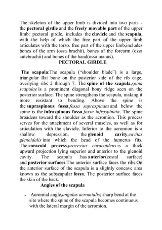
  • 1. SKELETON OF THE UPPER LIMB The skeleton of the upper limb is divided into two parts - the pectoral girdle and the freely movable part of the upper limb: pectoral girdle, includes the clavicle and the scapula, with the help of which the free part of the upper limb articulates with the torso. free part of the upper limb,includes bones of the arm (ossa brachii), bones of the forearm (ossa antebrachii) and bones of the hand(ossa manus). PECTORAL GIRDLE The scapula:The scapula (“shoulder blade”) is a large, triangular flat bone on the posterior side of the rib cage, overlying ribs 2 through 7. The spine of the scapula,spina scapulae is a prominent diagonal bony ridge seen on the posterior surface. The spine strengthens the scapula, making it more resistant to bending. Above the spine is the supraspinous fossa,fossa supraspinata and below the spine is the infraspinous fossa,fossa infraspinata. The spine broadens toward the shoulder as the acromion. This process serves for the attachment of several muscles, as well as for articulation with the clavicle. Inferior to the acromion is a shallow depression, the glenoid cavity,cavitas glenoidalis into which the head of the humerus fits. The coracoid process,processus coracoideus is a thick upward projection lying superior and anterior to the glenoid cavity. The scapula has anterior(costal surface) and posterior surfaces.The anterior surface faces the ribs.On the anterior surface of the scapula is a slightly concave area known as the subscapular fossa. The posterior surface faces the skin of the back. Angles of the scapula  Acromial angle,angulus acromialis; sharp bend at the site where the spine of the scapula becomes continuous with the lateral margin of the acromion.
  • 2. SKELETON OF THE UPPER LIMB  Inferior angle,angulus inferior; lower angle of the scapula.  Lateral angle,angulus lateralis; lateral angle of the scapula bearing the glenoid cavity.  Superior angle,angulus superior ;upper medial angle of the scapula . Margins of the scapula  Medial margin,margo medialis; border of the scapula facing the vertebral column.  Lateral margin,margo lateralis; border of the scapula facing the humerus.  Superior margin,margo superior; upper border of the scapula.
  • 3. SKELETON OF THE UPPER LIMB DonNMU. Clinical application: The scapula has numerous surface features because 15 muscles attach to it. Clinically, the pectoral girdle is significant becausethe clavicle and acromion of the scapula are frequentlybroken in trying to break a fall. The acromion is used as a landmark for identifying the site for an injection in the arm. This site is chosen because the musculature of the shoulder is quite thick and contains few nerves. HOW TO KNOW LEFT OR RIGHT SCAPULA  Identify the glenoid cavity and place it laterally.  Place the spine of the scapula posteriorly.In this way you can be able to identify the position of the scapula easily. The clavicle:The slender S-shaped clavicle(“collarbone”) connects the upper extremity to the axial skeleton and holds the shoulder joint away from the trunk to permit freedom of movement. The articulation of the medial sternal extremity of the clavicle to the manubrium of the sternum is referred to as the sternoclavicular joint. The lateral acromial extremity of the clavicle articulates with the acromion of the scapula.This articulation is referred to as the acromioclavicular joint. A conoid tubercle is present on the acromial extremity of the clavicle, and a costal tuberosity is present on the inferior surface of the sternal extremity. Both processes serve as attachments for ligaments.
  • 4. SKELETON OF THE UPPER LIMB Clinical application:The long, delicate clavicle is the most commonly broken bone in the body. When a person receives a blow to the shoulder, or attempts to break a fall with an outstretched hand, the force is transmitted to the clavicle, possibly causing it to fracture. The most vulnerable part of this bone is through its center, immediately proximal to the conoid tubercle. Because the clavicle is directly beneath the skin and is not covered with muscle, a fracture can easily be palpated, and frequently seen.
  • 5. SKELETON OF THE UPPER LIMB FREE PART OF THE UPPER LIMB The humerus:The humerus is the longest bone of the upper extremity.It consists of a proximal head(proximal epiphyses), which articulates with the glenoidcavity of the scapula; a body (“shaft”); and a distal end(distal epiphysis), which is modified to articulate with the two bones of the forearm. Surrounding the margin of the head is a slightly indented groove denoting the anatomical neck. The surgical neck, the constriction just below the head, is a frequent fracture site. The greater tubercle is a large knob on the lateral proximal portion of the humerus. The lesser tubercle is slightly anterior to the greater tubercle and is separated from the greater by an intertubercular groove. The tendon of the long head of the biceps brachii muscle passes through this groove. Along the lateral midregion of the body of the humerus is a roughened area, the deltoid tuberosity, for the attachment of the deltoid muscle. Small openings in the body are called nutrient foramina. The humeral condyle on the distal end of the humerus has two articular surfaces. The capitulum is the lateral rounded part that articulates with the radius. The trochlea is the pulleylike medial part that articulates with the ulna. On either side above the condyle are the lateral and medial epicondyles. The large medial epicondyle protects the ulnar nerve that passes posteriorly through the ulnar sulcus. It is popularly known as the “funny bone” because striking the elbow on the edge of a table, for example, stimulates the ulnar nerve and produces a tingling sensation. The coronoid fossa is a depression above the trochlea on the anterior surface. The olecranon fossa is a depression on the distal posterior surface. Both fossae are adapted to work with the ulna during movement of the forearm. Margins(borders):
  • 6. SKELETON OF THE UPPER LIMB  Medial margin,margo medialis: inner margin of the humerus continuous distally with the medial supracondylar ridge.  Lateral margin,margo lateralis: outer margin of the humerus continuous distally with the lateral supracondylar ridge. Surfaces:  Anteromedial surface,facies anteromedialis: surface of the humerus lying medial to the prolongation of the crest of the greater tubercle.  Anterolateral surface,facies anterolateralis: surface of the humerus located lateral to the prolongation of the crest of the greater tubercle.  posterior surface,facies posterior. clinical application:The medical term for tennis elbow is lateral epicondylitis, which means inflammation of the tissues surrounding the lateral epicondyle of the humerus. At least six
  • 7. SKELETON OF THE UPPER LIMB muscles that control backward (extension) movement of the wrist and finger joints originate on the lateral epicondyle. Repeated strenuous contractions of these muscles, as in stroking with a tennis racket, may strain the periosteum and muscle attachments, resulting in swelling, tenderness, and pain around the epicondyle. Binding usually eases the pain, but only rest can eliminate the causative factor, and recovery generally follows. HOW TO KNOW IF A HUMERUS IS LEFT OR RIGHT. clue I 1. Identify the head of the humerus and the medial epicondyle. These two should be medially placed. 2.Identify the coronoid and radial fossae. These two should be anteriorly placed with the olecranon fossa placed posteriorly. In this position, we can determine if its left or right. clue II 1. place the head of the humerus and the medial epicondyle at the medial position. 2.the olecranon fossa should be posteriorly placed. 3.If the head of the humerus faces left,it means it is a right humerus and vice versa. THE RADIUS:The radius consists of a body,corpus radii with a small proximal end, and a large distal end. A proximal disc-shaped head articulates with the capitulum of the humerus and the radial notch of the ulna. The prominent tuberosity of radius (radial tuberosity), for attachment of the biceps brachii muscle, is located on the medial side of the body, just below the head. On the distal end of the radius is a double-faceted surface for articulation with the proximal
  • 8. SKELETON OF THE UPPER LIMB carpal bones. The distal end of the radius also has a styloid process on the lateral tip and an ulnar notch on the medial side that receives the distal end of the ulna. The styloid processes on the ulna and radius provide lateral and medial stability for articulation at the wrist. Surfaces:  Anterior surface,facies anterior.  Posterior surface,facies posterior.  Lateral surface, facies lateralis. Borders:  Anterior border,margo anterior.  Posterior border,margo posterior.  Interosseous border,margo interosseous. Clinical application: When a person falls, the natural tendency is to extend the hand to break the fall. This reflexive movement frequently results in fractured bones. Common fractures of the radius include a fracture of the head, as it is driven forcefully against the capitulum; a fracture of the neck; or a fracture of the distal end (Colles’ fracture), caused by landing on an outstretched hand. When falling, it is less traumatic to the body to withdraw the appendages,bend the knees, and let the entire body hit the surface. Athletes learn that this is the safe way to fall. how to know left and right radius  The proximal end should face up and the distal end, down.
  • 9. SKELETON OF THE UPPER LIMB  Place the radial tuberosity medially.  Identify the nutrient foramen and place it anteriorly.i.e away from you.  The radial styloid process should also be at the lateral position. ULNA:The proximal end of the ulna articulates with the humerus and radius. A distinct depression, the trochlear notch, articulates with the trochlea of the humerus. The coronoid process forms the anterior lip of the trochlear notch, and the olecranon forms the posterior portion. Lateral and inferior to the coronoid process is the radial notch, which accommodates the head of the radius. On the tapered distal end of the ulna is a knobbed portion, the head, and a knoblike projection, the styloid process. The ulna articulates at both ends with the radius.In the forearm, it is located medially.
  • 10. SKELETON OF THE UPPER LIMB DonNMU. how to know left and right ulna  The proximal and distal epiphysis should first of all be placed at the right position;proximal up, distal down.  face the trochlear notch away from you and look at the olecranon.  Check to see which side the radial notch is.  If it is on the right, it a right ulna and vice versa. BONES OF THE HAND The hand contains 27 bones, grouped into the carpus, metacarpus, and phalanges.
  • 11. SKELETON OF THE UPPER LIMB Carpus:The carpus, or wrist, or carpal bone contains eight bones arranged in two transverse rows of four bones each. The proximal row, naming from the lateral (thumb) to the medial side, consists of the:  scaphoid (navicular),os scaphoideum  lunate,os lunatum  triquetrum,os triquetrum and  pisiform,os pisiforme.The pisiform forms in a tendon as a sesamoid bone. The distal row, from lateral to medial, consists of the  trapezium,os trapezium (greater multangular),  trapezoid,os trapezoideum (lesser multangular),  capitate,os capitatum and  hamate,os hamatum. The scaphoid and lunate of the proximal row articulate with the distal end of the radius. Metacarpus:The metacarpus, or palm of the hand, contains five metacarpal bones. Each metacarpal bone consists of a proximal base, a body, and a distal head that is rounded for articulation with the base of each proximal phalanx. The heads of the metacarpal bones are distally located and form the knuckles of a clenched fist. The phalanges:The 14 phalanges are the bones of the digits. A single finger bone is called a phalanx (fa'langks). The phalanges of the fingers are arranged in a proximal row, a middle row, and a distal row. The thumb, or pollex (adjective, pollicis), lacks a middle phalanx. The digits are sequentially numbered I to V starting with the
  • 12. SKELETON OF THE UPPER LIMB thumb—the lateral side, in reference to anatomical position. Clinical application: The hand is a marvel of structural complexity that can withstand considerable abuse. Other than sprained ligaments of the fingers and joint dislocations, the most common bone injury is a fracture to the scaphoid—a wrist bone that accounts for about 70% of carpal fractures. When immobilizing the wrist joint, the wrist is positioned in the plane of relaxed function. This is the position in which
  • 13. SKELETON OF THE UPPER LIMB the hand is about to grasp an object between the thumb and index finger. Kamal Umar Labaran(Med) Donetsk National Medical University.