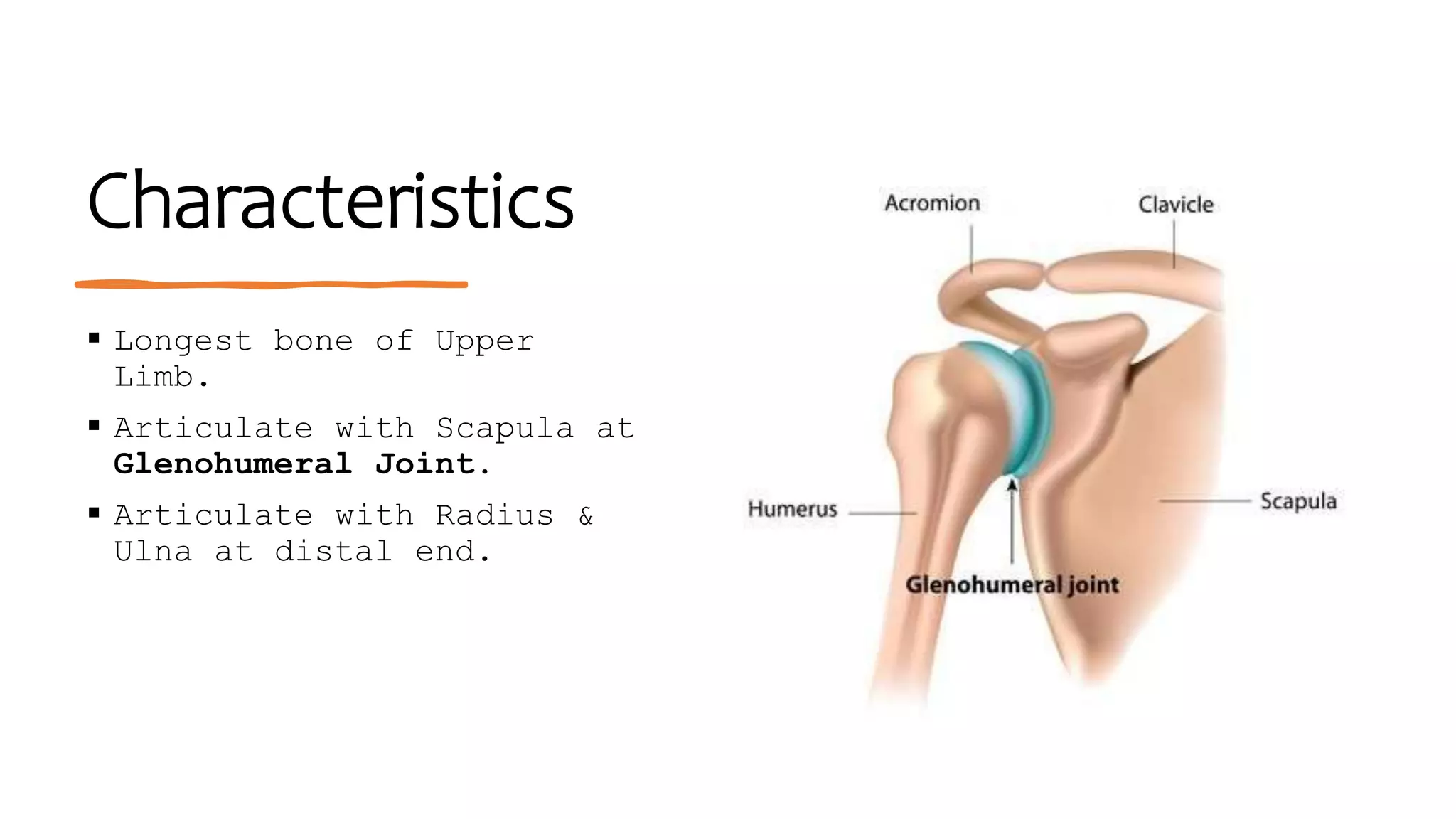
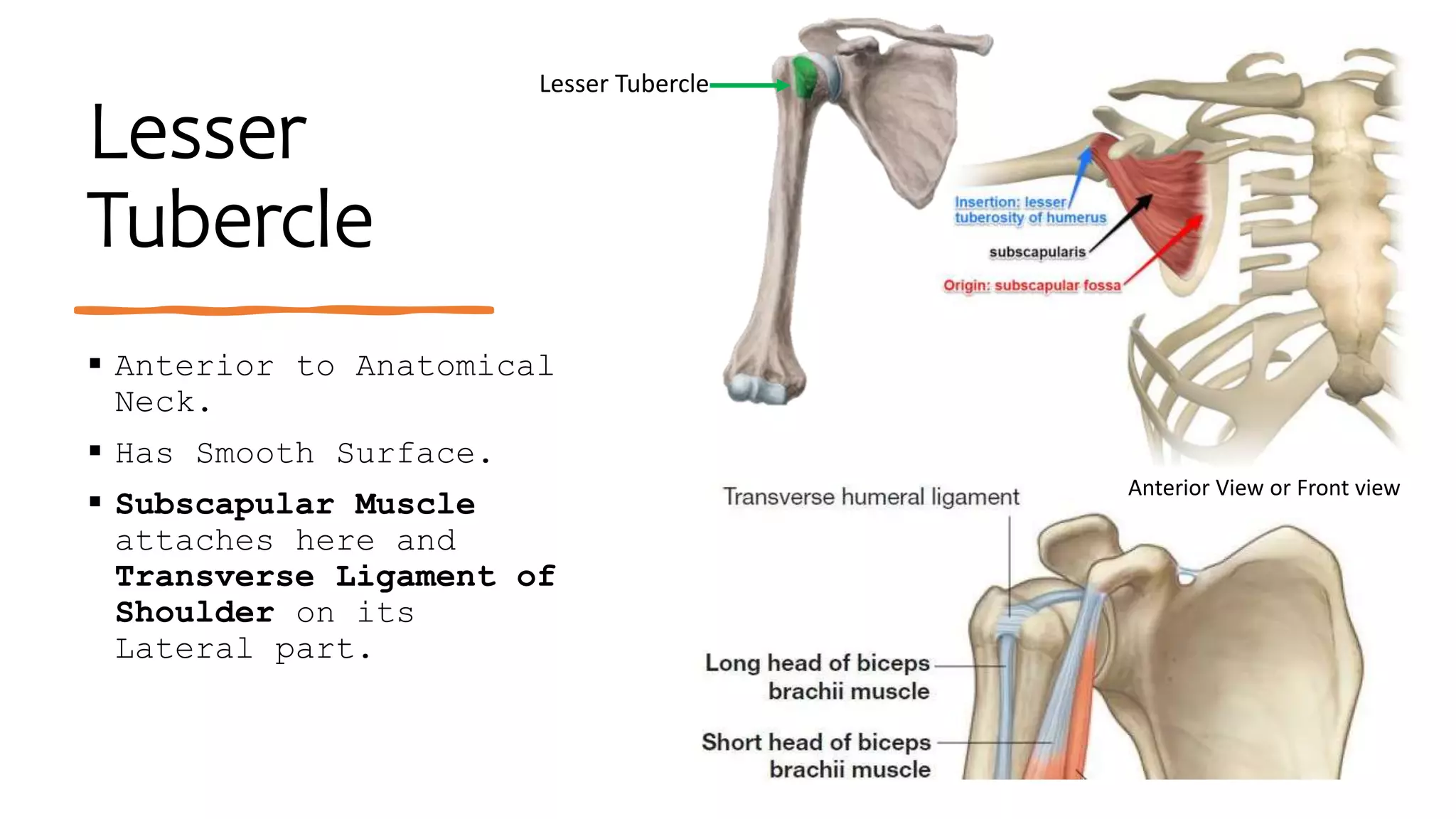
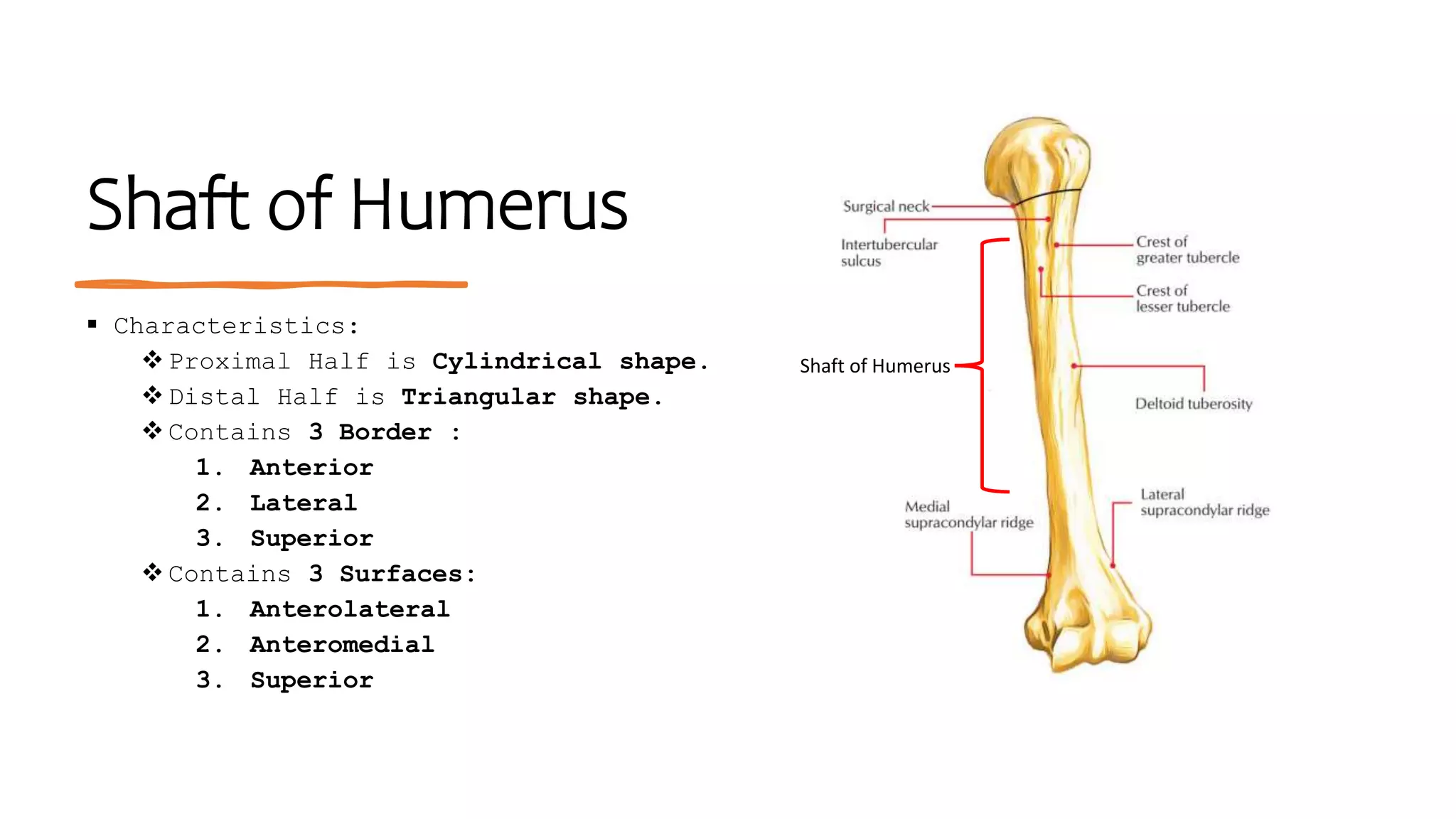
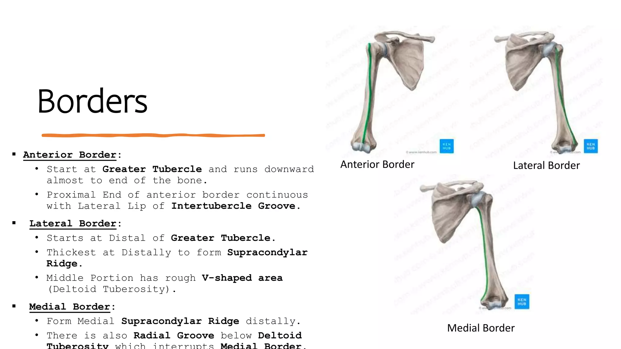
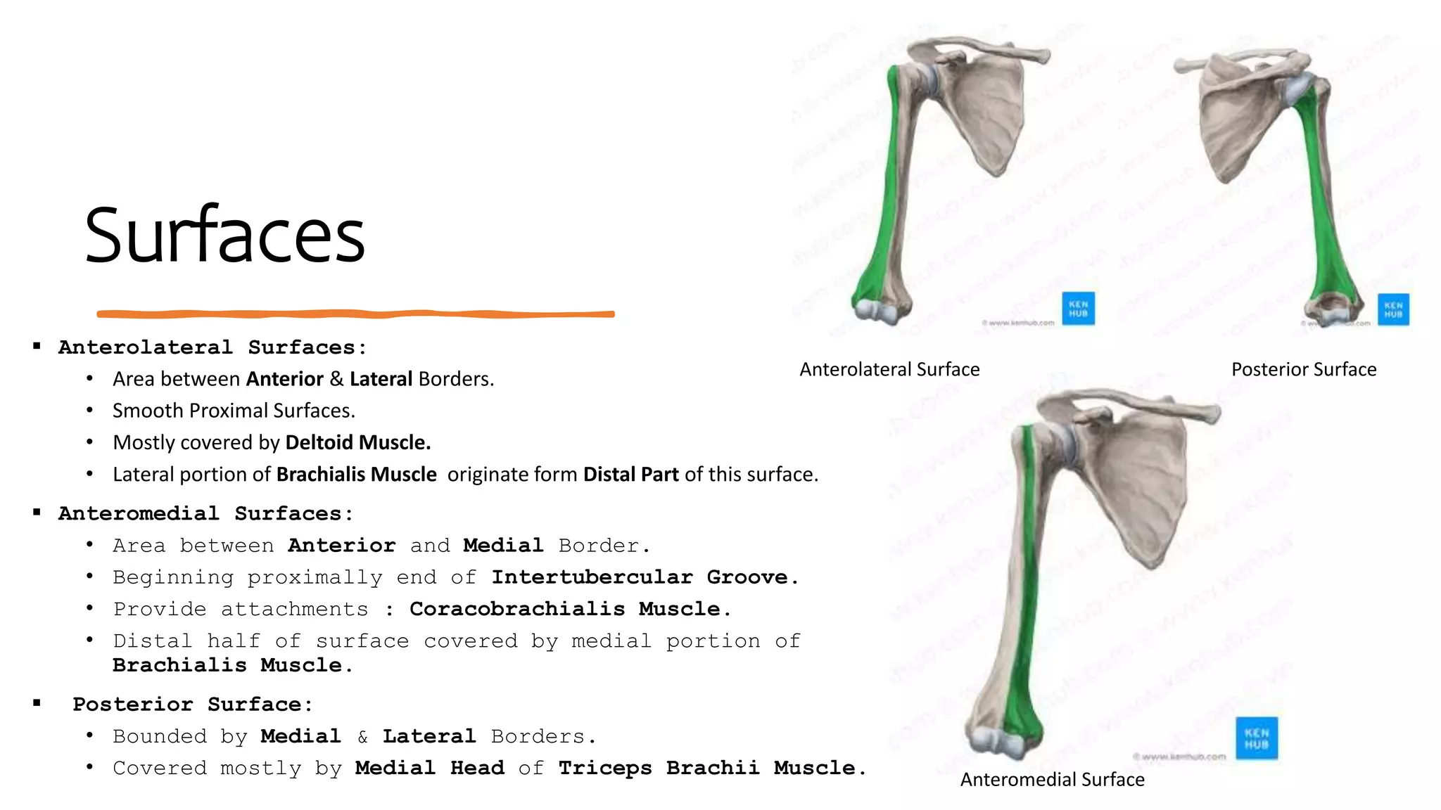
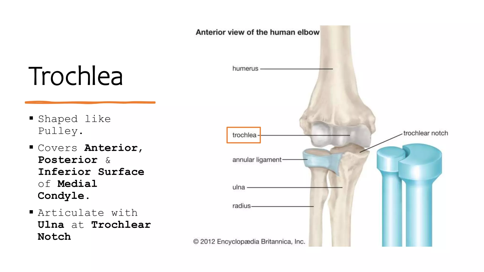
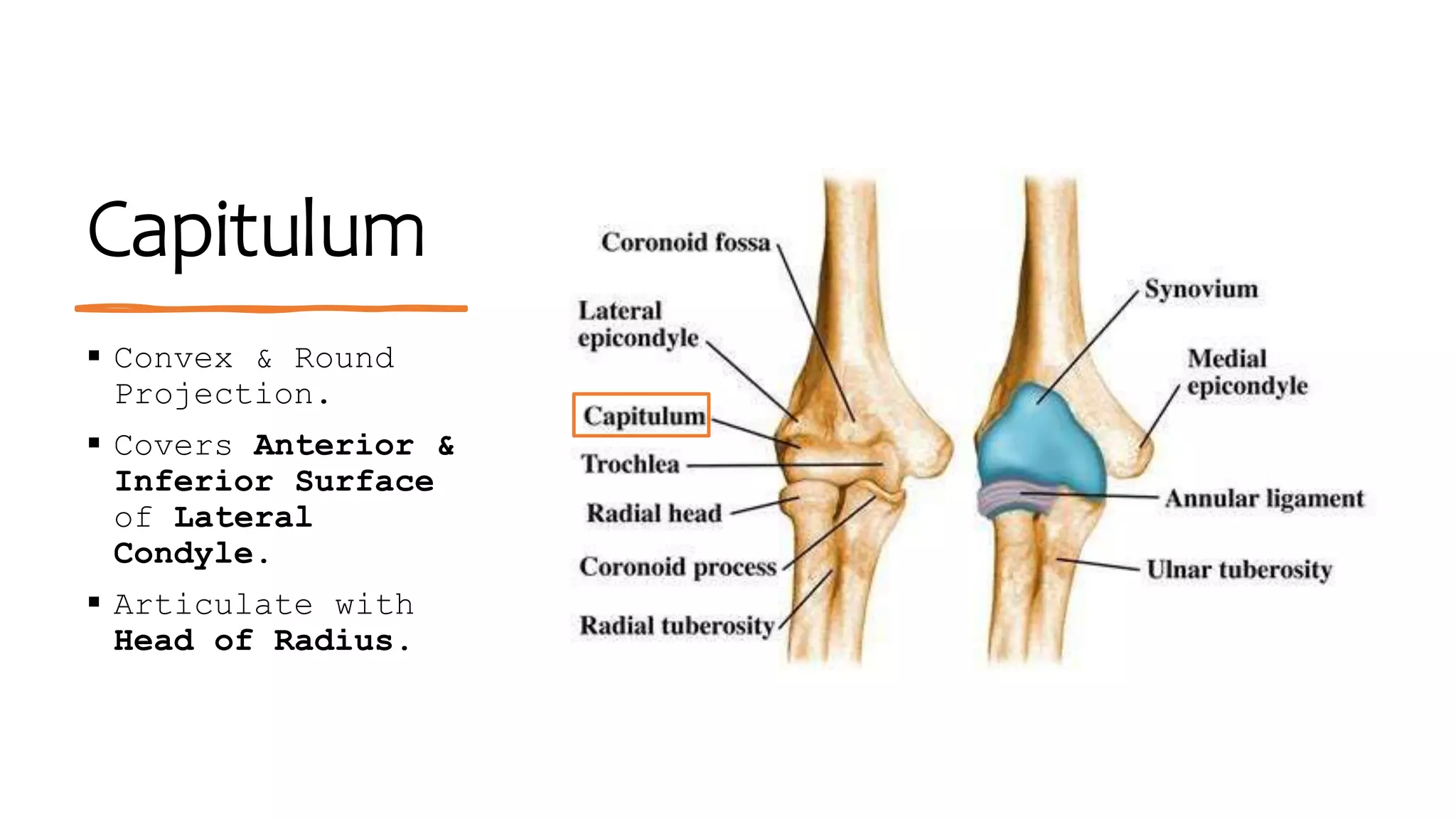
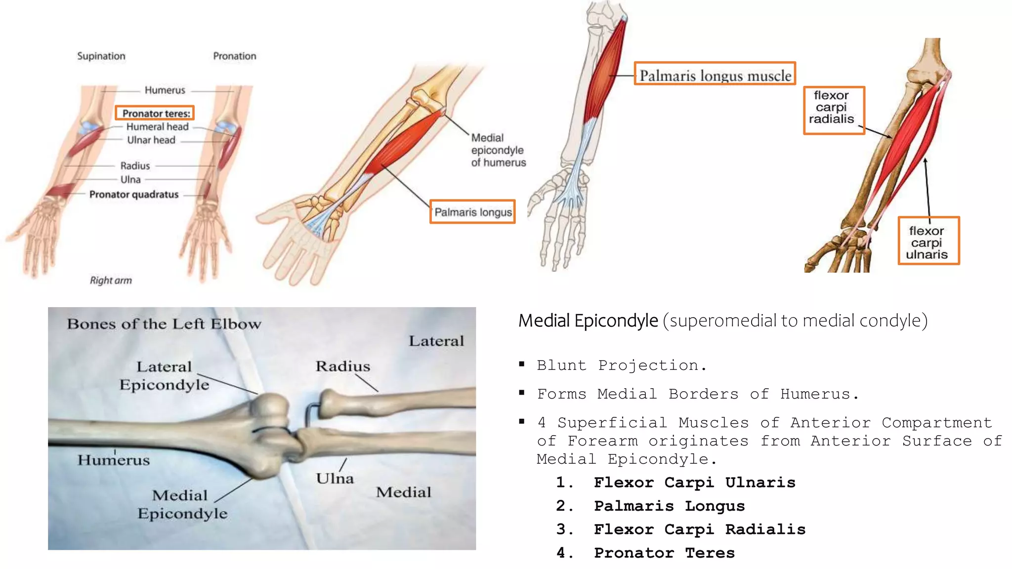
Humerus
Upper Limb
Characteristics
 Longest bone of Upper
Limb.
 Articulate with Scapula at
Glenohumeral Joint.
 Articulate with Radius &
Ulna at distal end.
Head of Humerus
 Hemispheroidal shape.
 Hyaline Cartilage covers its smooth
Articular Surface.
 Head faces in Medial, Superior &
Posterior direction.
 Articulate with Glenoid Fossa of
Scapula.
Anatomical Neck
 Below Articulating Head.
 The Joint Capsule of the Shoulder
Joint is attached.
Greater Tubercle
 It’s most Lateral portion of Proximal end
of Humerus.
 At Posterosuperior aspect, have 3 smooth
and flat surface for muscle attachment.
Muscles:
1. Supraspinatus
2. Infraspinatus
3. Teres Minor
• Deltoid muscle covers lateral aspect of
Greater Tubercle giving shoulder round
shape.
Greater Tubercle of Humerus
Posterior View or Back View
Lesser
Tubercle
 Anterior to Anatomical
Neck.
 Has Smooth Surface.
 Subscapular Muscle
attaches here and
Transverse Ligament of
Shoulder on its
Lateral part.
Lesser Tubercle
Anterior View or Front view
Intertubercular Groove/Sulcus or
Bicipital Groove
 Groove located between two Tubercles
 In this Groove :
Long tendon of Bicep Brachii
Muscle.
Branch of Anterior Circumflex
Humeral Artery.
 Groove consist of Lateral Lip &
Medial Lip.
Lateral Lip (aka crest of Greater
tubercle): Pectoralis Major
attaches to it.
Medial Lip: Teres Major Tendons &
Bicipital
groove
Shaft of Humerus
 Characteristics:
 Proximal Half is Cylindrical shape.
 Distal Half is Triangular shape.
 Contains 3 Border :
1. Anterior
2. Lateral
3. Superior
 Contains 3 Surfaces:
1. Anterolateral
2. Anteromedial
3. Superior
Shaft of Humerus
Borders
 Anterior Border:
• Start at Greater Tubercle and runs downward
almost to end of the bone.
• Proximal End of anterior border continuous
with Lateral Lip of Intertubercle Groove.
 Lateral Border:
• Starts at Distal of Greater Tubercle.
• Thickest at Distally to form Supracondylar
Ridge.
• Middle Portion has rough V-shaped area
(Deltoid Tuberosity).
 Medial Border:
• Form Medial Supracondylar Ridge distally.
• There is also Radial Groove below Deltoid
Medial Border
Lateral Border
Anterior Border
Surfaces
 Anterolateral Surfaces:
• Area between Anterior & Lateral Borders.
• Smooth Proximal Surfaces.
• Mostly covered by Deltoid Muscle.
• Lateral portion of Brachialis Muscle originate form Distal Part of this surface.
 Anteromedial Surfaces:
• Area between Anterior and Medial Border.
• Beginning proximally end of Intertubercular Groove.
• Provide attachments : Coracobrachialis Muscle.
• Distal half of surface covered by medial portion of
Brachialis Muscle.
 Posterior Surface:
• Bounded by Medial & Lateral Borders.
• Covered mostly by Medial Head of Triceps Brachii Muscle.
Posterior Surface
Anterolateral Surface
Anteromedial Surface
Distal End
 Consist Articular Parts
& Non-Articular Parts.
 Articular Parts:
Trochlea
Capitulum
 Non-Articular Parts:
Medial Epicondyle
Lateral Epicondyle
Posterior View Anterior View View
Trochlea
 Shaped like
Pulley.
 Covers Anterior,
Posterior &
Inferior Surface
of Medial
Condyle.
 Articulate with
Ulna at Trochlear
Notch
Capitulum
 Convex & Round
Projection.
 Covers Anterior &
Inferior Surface
of Lateral
Condyle.
 Articulate with
Head of Radius.
Medial Epicondyle (superomedial to medial condyle)
 Blunt Projection.
 Forms Medial Borders of Humerus.
 4 Superficial Muscles of Anterior Compartment
of Forearm originates from Anterior Surface of
Medial Epicondyle.
1. Flexor Carpi Ulnaris
2. Palmaris Longus
3. Flexor Carpi Radialis
4. Pronator Teres
Lateral Epicondyle
(superolateral to
lateral condyle)
 Impression on Lateral & Anterior
Surfaces. (for muscle
attachment)
 7 Muscles of Superficial Group
of Posterior Compartment of
Forearm originates.
1. Extensor Carpi Radialis
Longus
2. Extensor Carpi Radialis
Brevis
3. Extensor Digitorum
4. Extensor Digiti Minimi
5. Extensor Carpi Ulnaris
6. Brachioradialis
7. Anconeus
Fossa
 Olecranon Fossa:
 Hallowed area in Posterior Surface.
 Superior to Trochlea.
 Tip of Ulnar Olecranon Process lodges
into this Fossa.
 Coronoid Fossa:
 Smaller Hollow Area on Anterior
Surfaces.
 Superior to Trochlea.
 Coronoid Process of Ulna lodges here.
 Radial Fossa:
 Superior to Capitulum
 Head of Radius loges here.

Humerus.pptx

  • 1.
  • 2.
    Characteristics  Longest boneof Upper Limb.  Articulate with Scapula at Glenohumeral Joint.  Articulate with Radius & Ulna at distal end.
  • 3.
    Head of Humerus Hemispheroidal shape.  Hyaline Cartilage covers its smooth Articular Surface.  Head faces in Medial, Superior & Posterior direction.  Articulate with Glenoid Fossa of Scapula.
  • 4.
    Anatomical Neck  BelowArticulating Head.  The Joint Capsule of the Shoulder Joint is attached.
  • 5.
    Greater Tubercle  It’smost Lateral portion of Proximal end of Humerus.  At Posterosuperior aspect, have 3 smooth and flat surface for muscle attachment. Muscles: 1. Supraspinatus 2. Infraspinatus 3. Teres Minor • Deltoid muscle covers lateral aspect of Greater Tubercle giving shoulder round shape. Greater Tubercle of Humerus Posterior View or Back View
  • 6.
    Lesser Tubercle  Anterior toAnatomical Neck.  Has Smooth Surface.  Subscapular Muscle attaches here and Transverse Ligament of Shoulder on its Lateral part. Lesser Tubercle Anterior View or Front view
  • 7.
    Intertubercular Groove/Sulcus or BicipitalGroove  Groove located between two Tubercles  In this Groove : Long tendon of Bicep Brachii Muscle. Branch of Anterior Circumflex Humeral Artery.  Groove consist of Lateral Lip & Medial Lip. Lateral Lip (aka crest of Greater tubercle): Pectoralis Major attaches to it. Medial Lip: Teres Major Tendons & Bicipital groove
  • 8.
    Shaft of Humerus Characteristics:  Proximal Half is Cylindrical shape.  Distal Half is Triangular shape.  Contains 3 Border : 1. Anterior 2. Lateral 3. Superior  Contains 3 Surfaces: 1. Anterolateral 2. Anteromedial 3. Superior Shaft of Humerus
  • 9.
    Borders  Anterior Border: •Start at Greater Tubercle and runs downward almost to end of the bone. • Proximal End of anterior border continuous with Lateral Lip of Intertubercle Groove.  Lateral Border: • Starts at Distal of Greater Tubercle. • Thickest at Distally to form Supracondylar Ridge. • Middle Portion has rough V-shaped area (Deltoid Tuberosity).  Medial Border: • Form Medial Supracondylar Ridge distally. • There is also Radial Groove below Deltoid Medial Border Lateral Border Anterior Border
  • 10.
    Surfaces  Anterolateral Surfaces: •Area between Anterior & Lateral Borders. • Smooth Proximal Surfaces. • Mostly covered by Deltoid Muscle. • Lateral portion of Brachialis Muscle originate form Distal Part of this surface.  Anteromedial Surfaces: • Area between Anterior and Medial Border. • Beginning proximally end of Intertubercular Groove. • Provide attachments : Coracobrachialis Muscle. • Distal half of surface covered by medial portion of Brachialis Muscle.  Posterior Surface: • Bounded by Medial & Lateral Borders. • Covered mostly by Medial Head of Triceps Brachii Muscle. Posterior Surface Anterolateral Surface Anteromedial Surface
  • 11.
    Distal End  ConsistArticular Parts & Non-Articular Parts.  Articular Parts: Trochlea Capitulum  Non-Articular Parts: Medial Epicondyle Lateral Epicondyle Posterior View Anterior View View
  • 12.
    Trochlea  Shaped like Pulley. Covers Anterior, Posterior & Inferior Surface of Medial Condyle.  Articulate with Ulna at Trochlear Notch
  • 13.
    Capitulum  Convex &Round Projection.  Covers Anterior & Inferior Surface of Lateral Condyle.  Articulate with Head of Radius.
  • 14.
    Medial Epicondyle (superomedialto medial condyle)  Blunt Projection.  Forms Medial Borders of Humerus.  4 Superficial Muscles of Anterior Compartment of Forearm originates from Anterior Surface of Medial Epicondyle. 1. Flexor Carpi Ulnaris 2. Palmaris Longus 3. Flexor Carpi Radialis 4. Pronator Teres
  • 15.
    Lateral Epicondyle (superolateral to lateralcondyle)  Impression on Lateral & Anterior Surfaces. (for muscle attachment)  7 Muscles of Superficial Group of Posterior Compartment of Forearm originates. 1. Extensor Carpi Radialis Longus 2. Extensor Carpi Radialis Brevis 3. Extensor Digitorum 4. Extensor Digiti Minimi 5. Extensor Carpi Ulnaris 6. Brachioradialis 7. Anconeus
  • 16.
    Fossa  Olecranon Fossa: Hallowed area in Posterior Surface.  Superior to Trochlea.  Tip of Ulnar Olecranon Process lodges into this Fossa.  Coronoid Fossa:  Smaller Hollow Area on Anterior Surfaces.  Superior to Trochlea.  Coronoid Process of Ulna lodges here.  Radial Fossa:  Superior to Capitulum  Head of Radius loges here.