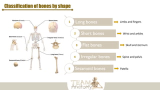
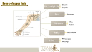
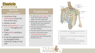
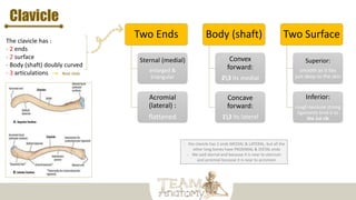
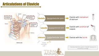
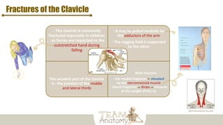
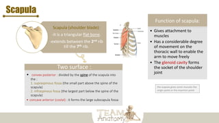
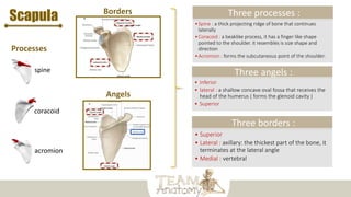
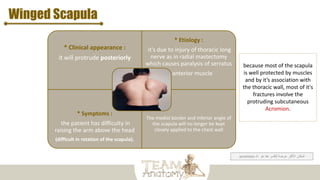
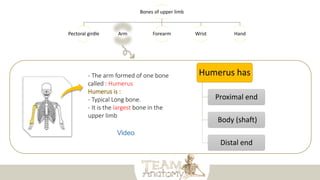
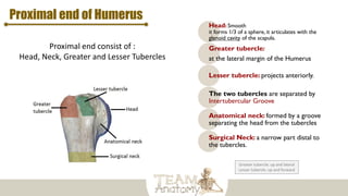
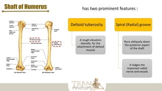
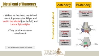
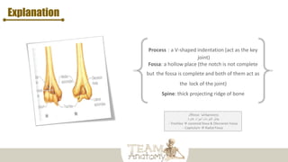
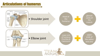
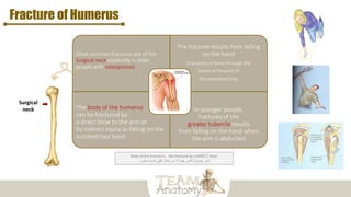
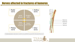
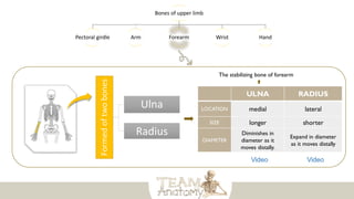
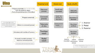
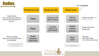
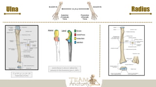
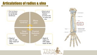
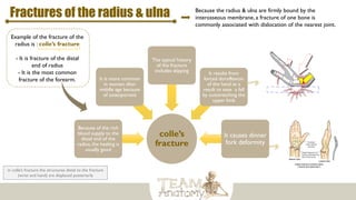
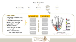
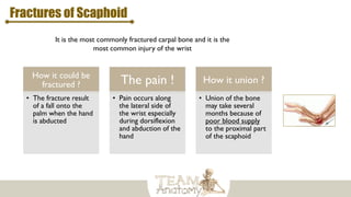
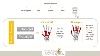
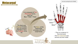
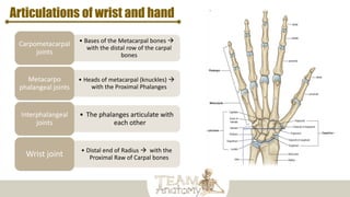
At the end of the lecture, you should be able to :
- List the different bones of the UL.
- List the characteristic features of each bone.
- Differentiate between the bones of the right and left sides.
- List the articulations between the different bones.
Bones
Bones
support and
protect the
various organs of
the body
Produce red and
white blood cells
Store
minerals
Come in a
variety of
shapes and
sizes
Provides
attachment for
muscles
Enable
movement
Example:
Thoracic cage
Protect:
Heart and lungs
From the bone
marrow
Different shapes of
bone in next slide
Classification of bones by shape
Long bones
Short bones
Flat bones
Irregular bones
Sesamoid bones
Limbs and fingers
Wrist and ankles
Skull and sternum
Spine and pelvis
Patella
Pectoral girdle
Arm
Forearm
Wrist
Hand
Bones of upper limb Clavicle
Scapula
Ulna
Radius
Metacarpals
Phalanges
Humerus
Carpal bones
Bones of upper limb
Pectoral girdle Arm Forearm Wrist Hand
- It is very light
- allows the upper limb to
have exceptionally free
movement
On the other hand, the pelvic
girdle “ in lower limb “ has
limited movement compare to
pectoral girdle
Formed
of
two
bones
Scapula
“posterior”
Clavicle
“Anterior”
General
information
• long bone lying
horizontally across the
root of the neck.
• doubly curved.
• It is subcutaneous “under
the skin” throughout its
length.
• There is NO medullary
cavity.
• It has the appearance of
an elongated letter
Capital (S) lying on one
side
Functions
• It serves as a rigid support from
which the scapula and free
upper limb are suspended
• keep them away from the trunk
so that the arm has maximum
freedom of movement
• Transmits forces from the UL to
the axial skeleton
• Provides attachment for muscles
• It forms a boundary of the
Cervicoaxillary canal for
protection of the neurovascular
bundle of the UL
Clavicle
All long bones are vertical except for the clavicle is
horizontal
- Clavicle is clear seen in thin people and disappear in
obese one.
- medullary cavity = marrow cavity
- clavicles are one of the Cervicoaxillary components
- Cervicoaxillary canal : contain the nerves that come
from the neck and supply to the upper limb
Clavicle
The clavicle has :
- 2 ends
- 2 surface
- Body (shaft) doubly curved
- 3 articulations Next slide
Two Ends
Sternal (medial)
enlarged &
triangular
Acromial
(lateral) :
flattened
Body (shaft)
Convex
forward:
23 its medial
Concave
forward:
13 its lateral
Two Surface
Superior:
smooth as it lies
just deep to the skin
Inferior:
rough because strong
ligaments bind it to
the 1st rib
- the clavicle has 2 ends MEDIAL & LATERAL, but all the
other long bones have PROXIMAL & DISTAL ends
- We said sternal end because it is near to sternum
and acromial because it is near to acromion
Articulations of Clavicle
- Any bone that form a joint is “smooth” because it is
covered by a cartilage
Sternoclavicular joint
Acromioclavicular joint
Costoclavicular Joint
Clavicle with manubrium
of sternum
Clavicle with acromion of
scapula
Clavicle with the 1st rib
COSTAE
mean rib in
Latin
Fractures of the Clavicle
- The clavicle is commonly
fractured especially in children
- as forces are impacted to the
outstretched hand during
falling
- It may be pulled medially by
the adductors of the arm
- The sagging limb is supported
by the other
The weakest part of the clavicle
is : the junction of the middle
and lateral thirds
After fracture:
- the medial fragment elevated
by the sternomastoid muscle
- lateral fragment drops because
of the weight of the UL
sternomastoid muscle
Scapula
Scapula (shoulder blade):
-it is a triangular flat bone.
-extends between the 2nd rib
till the 7th rib.
Two surface :
• convex posterior : divided by the spine of the scapula into
the :
1. supraspinous fossa (the small part above the spine of the
scapula)
2. infraspinous fossa (the largest part below the spine of the
scapula)
• concave anterior (costal) : it forms the large subscapula fossa
Function of scapula:
• Gives attachment to
muscles
• Has a considerable degree
of movement on the
thoracic wall to enable the
arm to move freely
• The glenoid cavity forms
the socket of the shoulder
joint
the scapula gives some muscles the
origin point or the insertion point
Scapula
Three borders :
• Superior
• Lateral : axillary: the thickest part of the bone, it
terminates at the lateral angle
• Medial : vertebral
Three angels :
• Inferior
• lateral : a shallow concave oval fossa that receives the
head of the humerus ( forms the glenoid cavity )
• Superior
Three processes :
•Spine : a thick projecting ridge of bone that continues
laterally
•Coracoid : a beaklike process, it has a finger like shape
pointed to the shoulder. it resembles is size shape and
direction
•Acromion : forms the subcutaneous point of the shoulder.
spine
coracoid
acromion
Winged Scapula
* Clinical appearance :
it will protrude posteriorly
* Etiology :
it's due to injury of thoracic long
nerve as in radial mastectomy
which causes paralysis of serratus
anterior muscle
* Symptoms :
the patient has difficulty in
raising the arm above the head
(difficult in rotation of the scapula).
The medial border and inferior angle of
the scapula will no longer be kept
closely applied to the chest wall
acromion ‫هو‬ ‫هنا‬ ‫للكسر‬ ‫عرضة‬ ‫األكثر‬ ‫المكان‬
‫الـ‬
because most of the scapula
is well protected by muscles
and by it’s association with
the thoracic wall, most of it's
fractures involve the
protruding subcutaneous
Acromion.
Bones of upper limb
Pectoral girdle Arm Forearm Wrist Hand
- The arm formed of one bone
called : Humerus
Humerus is :
- Typical Long bone.
- It is the largest bone in the
upper limb
Humerus has
Proximal end
Body (shaft)
Distal end
Proximal end of Humerus
Proximal end consist of :
Head, Neck, Greater and Lesser Tubercles
Head: Smooth
it forms 1/3 of a sphere, it articulates with the
glenoid cavity of the scapula.
Greater tubercle:
at the lateral margin of the Humerus
Lesser tubercle: projects anteriorly.
The two tubercles are separated by
Intertubercular Groove
Anatomical neck: formed by a groove
separating the head from the tubercles
Surgical Neck: a narrow part distal to
the tubercles.
Greater tubercle: up and lateral
Lesser tubercle: up and forward
Shaft of Humerus has two prominent features :
Deltoid tuberosity
A rough elevation
laterally for the
attachment of deltoid
muscle
Spiral (Radial) groove
Runs obliquely down
the posterior aspect
of the shaft
It lodges the
important radial
nerve and vessels
Distal end of Humerus
- Widens as the sharp medial and
lateral Supracondylar Ridges and
end in the Medial (can be felt) and
Lateral Epicondyles
- They provide muscular
attachment
Anteriorly
Trochlea:
(medial) for
articulation with
the ulna
Capitulum:
(lateral) for
articulation with
the radius.
Coronoid
fossa:
above the
trochlea
Radial fossa:
above the
capitulum.
Posteriorly
Olecranon fossa:
above the
trochlea.
Here we have 3 fossa : 2 anterior and 1 posterior
Explanation
‫كل‬fossa ‫يقابلها‬process
‫غائرة‬ ‫أجزاء‬ ‫النتوءات‬ ‫يقابل‬
- Trochlea  coronoid fossa & Olecranon Fossa
- Capitulum  Radial Fossa
Articulations of humerus
• Shoulder joint
• Elbow joint
Head of
the
humerus
glenoid
cavity of
the
scapula
Lower end
of humerus
(Trochlea &
Capitulum)
upper
ends of
the radius
and ulna
Fracture of Humerus
Most common fractures are of the
Surgical neck especially in elder
people with osteoporosis
The fracture results from falling
on the hand
(transition of force through the
bones of forearm of
the extended limb)
The body of the humerus
can be fractured by
a direct blow to the arm or
by indirect injury as falling on the
outstretched hand
In younger people,
fractures of the
greater tubercle results
from falling on the hand when
the arm is abducted
Surgical
neck
Body of the humerus .. the fracture by a DIRECT blow
‫مباشرة‬ ‫كدمة‬ ‫تلقي‬ ‫خالل‬ ‫من‬ ‫إال‬ ‫فيها‬ ‫الكسر‬ ‫حصول‬ ‫نادر‬
Nerves affected in fractures of humerus
• Ulnar
nerve
• Median
nerve
• Axillary
nerve
• Radial
nerve
spiral
groove
Surgical
neck
Medial
epicondyle
Distal
end of
humerus
(
‫بطاولة‬ ‫المرفق‬ ‫ارتطام‬ ‫عند‬
,
‫العصب‬ ‫هذا‬ ‫بسبب‬ ‫يكون‬ ‫المنطقة‬ ‫هذه‬ ‫في‬ ‫الناشئ‬ ‫األلم‬
*medial epicondyle : ulnar nerve (
Bones of upper limb
Pectoral girdle Arm Forearm Wrist Hand
Formed
of
two
bones Ulna
Radius
ULNA RADIUS
LOCATION medial lateral
SIZE longer shorter
DIAMETER
Diminishes in
diameter as it
moves distally.
Expand in diameter
as it moves distally
The stabilizing bone of forearm
Ulna Proximal end
Olecranon
process
Coronoid
process
Tuberosity
of ulna
Trochlear
notch
Radial
notch
Distal end
Small and
rounded
head: lies
distally at
the wrist
styloid process:
medial distal
projection
Body (shaft)
Superiorly :
thick and
cylindrical
Inferiorly :
diminishes in
diameter
Has three
surfaces
Has three
borders
1. Anterior
2. Medial
3. Posterior
Anterior
(rounded)
posterior
(sharp)
Lateral
interosseous
(sharp)
- Projects proximally (near to the trunk)
from the posterior aspect.
- Forms the pointed portion of the elbow
Projects anteriorly
-A smooth rounded concavity( ‫)تقعر‬
-lateral to coronoid process.
Articulates with trochlea of humerus
Inferior to coronoid process
‫؟‬ Fossa ‫بـ‬ Radial Notch ‫تسميتنا‬ ‫عدم‬ ‫سبب‬ ‫ما‬
‫لل‬
‫ـ‬
‫ألنها‬
‫عميقة‬ ‫ليست‬
See slide 22
it is a long ridge on the
anterior side of the coronoid
process where the brachialis
muscle attaches
Articulates with the head of the radius
Proximal end
Head
Neck
Radial
(biciptal)
tuberosity
Body (shaft)
Convex at it’s
lateral surface
Gradually
enlarges as it
passes distally
Distal end
Dorsal
tubercle
Radial
styloid
process
Ulnar
notch
- Small & circular.
- Has a concave upper surface to
articulate with the capitulum of
humerus.
Radius
- Directed medially.
- Separate the proximal end from
the body(shaft)
Insertion point of the biceps
brachii muscle
It is rectangular
- Concavity
- Directed medially
- Fit in with the ulna’s head
Projects from the
lateral side.
Projects dorsally (away
from the trunk)
forms the radioulnar joint
Ulna
every fossa in ulna or radius has
process in the humerus ‫صحيح‬ ‫والعكس‬
‫الـ‬ ‫هو‬ ‫االلنا‬ ‫في‬ ‫بارز‬ ‫مكان‬ ‫اكثر‬
Tuberosity of Ulna
Radius
Articulations of radius & ulna
• Head of
radius 
with radial
notch of ulna
• Head of
ulna  with
ulnar notch of
radius
• Distal end of
Humerus 
with the
proximal ends
• of radius and
• ulna
• The two bones
are connected
by the flexible
membrane
interosseous
membrane
Elbow
joint
Proximal
Radioulnar
joint
Distal
Radioulnar
joint
Fractures of the radius & ulna
colle’s
fracture
Because of the rich
blood supply to the
distal end of the
radius, the healing is
usually good
It is more common
in women after
middle age because
of osteoporosis
The typical history
of the fracture
includes slipping It results from
forced dorsiflexion
of the hand as a
result to ease a fall
by outstretching the
upper limb
It causes dinner
fork deformity
Because the radius & ulna are firmly bound by the
interosseous membrane, a fracture of one bone is
commonly associated with dislocation of the nearest joint.
Example of the fracture of the
radius is : colle’s fracture
- It is fracture of the distal
end of radius
- It is the most common
fracture of the forearm.
in colle’s fracture the structures distal to the fracture
(wrist and hand) are displaced posteriorly
Bones of upper limb
Pectoral girdle Arm Forearm Wrist Hand
Carpal bones :
• small bones make the wrist
more flexible
• 8 short bones, arranged in 2
irregular rows “each row
composed of 4 bones”
• Has two surface :
1.The concave surface :
anteriorly
2.The convex surface :
side to side posteriorly
proximal row
Scaphoid
Lunate
Triquetral
Pisiform
Distal row
Trapezium
Trapezoid
Capitate
Hamate
from
lateral
to
medial
To remember them :
Sally Left The Party To Take Cathy Home
Fractures of Scaphoid
It is the most commonly fractured carpal bone and it is the
most common injury of the wrist
How it could be
fractured ?
• The fracture result
of a fall onto the
palm when the hand
is abducted
The pain !
• Pain occurs along
the lateral side of
the wrist especially
during dorsiflexion
and abduction of the
hand
How it union ?
• Union of the bone
may take several
months because of
poor blood supply
to the proximal part
of the scaphoid
Bones of upper limb
Pectoral girdle Arm Forearm Wrist Hand
The
hand
Formed
of
metacarpal
Phalanges
Form the
skeleton of
the hand
between the
carpus and
phalanges
Each digit
has Three
Phalanges.
Except :
the Thumb
which has
only Two
Metacarpal
It is composed of Five
Metacarpal bones,
each has :
Base + Shaft + Head
The Bases of the
metacarpals
articulate with the
carpal bones
The distal ends
(Heads) articulate
with the proximal
phalanges to form
the Knuckles of the
fist - They are numbered 1-5
from the thumb
- The first metacarpal is the
shortest and most mobile
we start counting from the THUMB
Phalanges
The proximal
phalanx is the
largest
The distal ones
are the smallest,
its distal ends are:
- flattened
- expanded distally
to form the nail
beds
The middle ones
are intermediate
in size
Each phalanx has : Base Proximally + Head distally + Body in between
Articulations of wrist and hand
• Bases of the Metacarpal bones 
with the distal row of the carpal
bones
Carpometacarpal
joints
• Heads of metacarpal (knuckles) 
with the Proximal Phalanges
Metacarpo
phalangeal joints
• The phalanges articulate with
each other
Interphalangeal
joints
• Distal end of Radius  with the
Proximal Raw of Carpal bones
Wrist joint
Summary
Zoom it if it’s not clear
Video: arm and forearm
https://www.youtube.com/watch?
v=2ufqUOpm8O0
Video: bones of upper limb
https://www.youtube.com/watch
?v=715Z0XPtlDI
Application: Essential anatomy 5
you can have it for free, ask
https://twitter.com/Med_435
Quiz:
Upper Limb part.1 (15 questions)
https://www.onlineexambuilder.com/bones-of-the-upper-
limb/exam-47458
Upper Limb part.2 (15 questions)
https://www.onlineexambuilder.com/upper-limb-part-
ii/exam-52135
Upper limb and their bones with their special features

Upper limb and their bones with their special features

  • 2.
    At the endof the lecture, you should be able to : - List the different bones of the UL. - List the characteristic features of each bone. - Differentiate between the bones of the right and left sides. - List the articulations between the different bones.
  • 3.
    Bones Bones support and protect the variousorgans of the body Produce red and white blood cells Store minerals Come in a variety of shapes and sizes Provides attachment for muscles Enable movement Example: Thoracic cage Protect: Heart and lungs From the bone marrow Different shapes of bone in next slide
  • 4.
    Classification of bonesby shape Long bones Short bones Flat bones Irregular bones Sesamoid bones Limbs and fingers Wrist and ankles Skull and sternum Spine and pelvis Patella
  • 5.
    Pectoral girdle Arm Forearm Wrist Hand Bones ofupper limb Clavicle Scapula Ulna Radius Metacarpals Phalanges Humerus Carpal bones
  • 6.
    Bones of upperlimb Pectoral girdle Arm Forearm Wrist Hand - It is very light - allows the upper limb to have exceptionally free movement On the other hand, the pelvic girdle “ in lower limb “ has limited movement compare to pectoral girdle Formed of two bones Scapula “posterior” Clavicle “Anterior”
  • 7.
    General information • long bonelying horizontally across the root of the neck. • doubly curved. • It is subcutaneous “under the skin” throughout its length. • There is NO medullary cavity. • It has the appearance of an elongated letter Capital (S) lying on one side Functions • It serves as a rigid support from which the scapula and free upper limb are suspended • keep them away from the trunk so that the arm has maximum freedom of movement • Transmits forces from the UL to the axial skeleton • Provides attachment for muscles • It forms a boundary of the Cervicoaxillary canal for protection of the neurovascular bundle of the UL Clavicle All long bones are vertical except for the clavicle is horizontal - Clavicle is clear seen in thin people and disappear in obese one. - medullary cavity = marrow cavity - clavicles are one of the Cervicoaxillary components - Cervicoaxillary canal : contain the nerves that come from the neck and supply to the upper limb
  • 8.
    Clavicle The clavicle has: - 2 ends - 2 surface - Body (shaft) doubly curved - 3 articulations Next slide Two Ends Sternal (medial) enlarged & triangular Acromial (lateral) : flattened Body (shaft) Convex forward: 23 its medial Concave forward: 13 its lateral Two Surface Superior: smooth as it lies just deep to the skin Inferior: rough because strong ligaments bind it to the 1st rib - the clavicle has 2 ends MEDIAL & LATERAL, but all the other long bones have PROXIMAL & DISTAL ends - We said sternal end because it is near to sternum and acromial because it is near to acromion
  • 9.
    Articulations of Clavicle -Any bone that form a joint is “smooth” because it is covered by a cartilage Sternoclavicular joint Acromioclavicular joint Costoclavicular Joint Clavicle with manubrium of sternum Clavicle with acromion of scapula Clavicle with the 1st rib COSTAE mean rib in Latin
  • 10.
    Fractures of theClavicle - The clavicle is commonly fractured especially in children - as forces are impacted to the outstretched hand during falling - It may be pulled medially by the adductors of the arm - The sagging limb is supported by the other The weakest part of the clavicle is : the junction of the middle and lateral thirds After fracture: - the medial fragment elevated by the sternomastoid muscle - lateral fragment drops because of the weight of the UL sternomastoid muscle
  • 11.
    Scapula Scapula (shoulder blade): -itis a triangular flat bone. -extends between the 2nd rib till the 7th rib. Two surface : • convex posterior : divided by the spine of the scapula into the : 1. supraspinous fossa (the small part above the spine of the scapula) 2. infraspinous fossa (the largest part below the spine of the scapula) • concave anterior (costal) : it forms the large subscapula fossa Function of scapula: • Gives attachment to muscles • Has a considerable degree of movement on the thoracic wall to enable the arm to move freely • The glenoid cavity forms the socket of the shoulder joint the scapula gives some muscles the origin point or the insertion point
  • 12.
    Scapula Three borders : •Superior • Lateral : axillary: the thickest part of the bone, it terminates at the lateral angle • Medial : vertebral Three angels : • Inferior • lateral : a shallow concave oval fossa that receives the head of the humerus ( forms the glenoid cavity ) • Superior Three processes : •Spine : a thick projecting ridge of bone that continues laterally •Coracoid : a beaklike process, it has a finger like shape pointed to the shoulder. it resembles is size shape and direction •Acromion : forms the subcutaneous point of the shoulder. spine coracoid acromion
  • 13.
    Winged Scapula * Clinicalappearance : it will protrude posteriorly * Etiology : it's due to injury of thoracic long nerve as in radial mastectomy which causes paralysis of serratus anterior muscle * Symptoms : the patient has difficulty in raising the arm above the head (difficult in rotation of the scapula). The medial border and inferior angle of the scapula will no longer be kept closely applied to the chest wall acromion ‫هو‬ ‫هنا‬ ‫للكسر‬ ‫عرضة‬ ‫األكثر‬ ‫المكان‬ ‫الـ‬ because most of the scapula is well protected by muscles and by it’s association with the thoracic wall, most of it's fractures involve the protruding subcutaneous Acromion.
  • 14.
    Bones of upperlimb Pectoral girdle Arm Forearm Wrist Hand - The arm formed of one bone called : Humerus Humerus is : - Typical Long bone. - It is the largest bone in the upper limb Humerus has Proximal end Body (shaft) Distal end
  • 15.
    Proximal end ofHumerus Proximal end consist of : Head, Neck, Greater and Lesser Tubercles Head: Smooth it forms 1/3 of a sphere, it articulates with the glenoid cavity of the scapula. Greater tubercle: at the lateral margin of the Humerus Lesser tubercle: projects anteriorly. The two tubercles are separated by Intertubercular Groove Anatomical neck: formed by a groove separating the head from the tubercles Surgical Neck: a narrow part distal to the tubercles. Greater tubercle: up and lateral Lesser tubercle: up and forward
  • 16.
    Shaft of Humerushas two prominent features : Deltoid tuberosity A rough elevation laterally for the attachment of deltoid muscle Spiral (Radial) groove Runs obliquely down the posterior aspect of the shaft It lodges the important radial nerve and vessels
  • 17.
    Distal end ofHumerus - Widens as the sharp medial and lateral Supracondylar Ridges and end in the Medial (can be felt) and Lateral Epicondyles - They provide muscular attachment Anteriorly Trochlea: (medial) for articulation with the ulna Capitulum: (lateral) for articulation with the radius. Coronoid fossa: above the trochlea Radial fossa: above the capitulum. Posteriorly Olecranon fossa: above the trochlea. Here we have 3 fossa : 2 anterior and 1 posterior
  • 18.
    Explanation ‫كل‬fossa ‫يقابلها‬process ‫غائرة‬ ‫أجزاء‬‫النتوءات‬ ‫يقابل‬ - Trochlea  coronoid fossa & Olecranon Fossa - Capitulum  Radial Fossa
  • 19.
    Articulations of humerus •Shoulder joint • Elbow joint Head of the humerus glenoid cavity of the scapula Lower end of humerus (Trochlea & Capitulum) upper ends of the radius and ulna
  • 20.
    Fracture of Humerus Mostcommon fractures are of the Surgical neck especially in elder people with osteoporosis The fracture results from falling on the hand (transition of force through the bones of forearm of the extended limb) The body of the humerus can be fractured by a direct blow to the arm or by indirect injury as falling on the outstretched hand In younger people, fractures of the greater tubercle results from falling on the hand when the arm is abducted Surgical neck Body of the humerus .. the fracture by a DIRECT blow ‫مباشرة‬ ‫كدمة‬ ‫تلقي‬ ‫خالل‬ ‫من‬ ‫إال‬ ‫فيها‬ ‫الكسر‬ ‫حصول‬ ‫نادر‬
  • 21.
    Nerves affected infractures of humerus • Ulnar nerve • Median nerve • Axillary nerve • Radial nerve spiral groove Surgical neck Medial epicondyle Distal end of humerus ( ‫بطاولة‬ ‫المرفق‬ ‫ارتطام‬ ‫عند‬ , ‫العصب‬ ‫هذا‬ ‫بسبب‬ ‫يكون‬ ‫المنطقة‬ ‫هذه‬ ‫في‬ ‫الناشئ‬ ‫األلم‬ *medial epicondyle : ulnar nerve (
  • 22.
    Bones of upperlimb Pectoral girdle Arm Forearm Wrist Hand Formed of two bones Ulna Radius ULNA RADIUS LOCATION medial lateral SIZE longer shorter DIAMETER Diminishes in diameter as it moves distally. Expand in diameter as it moves distally The stabilizing bone of forearm
  • 23.
    Ulna Proximal end Olecranon process Coronoid process Tuberosity ofulna Trochlear notch Radial notch Distal end Small and rounded head: lies distally at the wrist styloid process: medial distal projection Body (shaft) Superiorly : thick and cylindrical Inferiorly : diminishes in diameter Has three surfaces Has three borders 1. Anterior 2. Medial 3. Posterior Anterior (rounded) posterior (sharp) Lateral interosseous (sharp) - Projects proximally (near to the trunk) from the posterior aspect. - Forms the pointed portion of the elbow Projects anteriorly -A smooth rounded concavity( ‫)تقعر‬ -lateral to coronoid process. Articulates with trochlea of humerus Inferior to coronoid process ‫؟‬ Fossa ‫بـ‬ Radial Notch ‫تسميتنا‬ ‫عدم‬ ‫سبب‬ ‫ما‬ ‫لل‬ ‫ـ‬ ‫ألنها‬ ‫عميقة‬ ‫ليست‬ See slide 22 it is a long ridge on the anterior side of the coronoid process where the brachialis muscle attaches Articulates with the head of the radius
  • 24.
    Proximal end Head Neck Radial (biciptal) tuberosity Body (shaft) Convexat it’s lateral surface Gradually enlarges as it passes distally Distal end Dorsal tubercle Radial styloid process Ulnar notch - Small & circular. - Has a concave upper surface to articulate with the capitulum of humerus. Radius - Directed medially. - Separate the proximal end from the body(shaft) Insertion point of the biceps brachii muscle It is rectangular - Concavity - Directed medially - Fit in with the ulna’s head Projects from the lateral side. Projects dorsally (away from the trunk) forms the radioulnar joint
  • 25.
    Ulna every fossa inulna or radius has process in the humerus ‫صحيح‬ ‫والعكس‬ ‫الـ‬ ‫هو‬ ‫االلنا‬ ‫في‬ ‫بارز‬ ‫مكان‬ ‫اكثر‬ Tuberosity of Ulna Radius
  • 26.
    Articulations of radius& ulna • Head of radius  with radial notch of ulna • Head of ulna  with ulnar notch of radius • Distal end of Humerus  with the proximal ends • of radius and • ulna • The two bones are connected by the flexible membrane interosseous membrane Elbow joint Proximal Radioulnar joint Distal Radioulnar joint
  • 27.
    Fractures of theradius & ulna colle’s fracture Because of the rich blood supply to the distal end of the radius, the healing is usually good It is more common in women after middle age because of osteoporosis The typical history of the fracture includes slipping It results from forced dorsiflexion of the hand as a result to ease a fall by outstretching the upper limb It causes dinner fork deformity Because the radius & ulna are firmly bound by the interosseous membrane, a fracture of one bone is commonly associated with dislocation of the nearest joint. Example of the fracture of the radius is : colle’s fracture - It is fracture of the distal end of radius - It is the most common fracture of the forearm. in colle’s fracture the structures distal to the fracture (wrist and hand) are displaced posteriorly
  • 28.
    Bones of upperlimb Pectoral girdle Arm Forearm Wrist Hand Carpal bones : • small bones make the wrist more flexible • 8 short bones, arranged in 2 irregular rows “each row composed of 4 bones” • Has two surface : 1.The concave surface : anteriorly 2.The convex surface : side to side posteriorly proximal row Scaphoid Lunate Triquetral Pisiform Distal row Trapezium Trapezoid Capitate Hamate from lateral to medial To remember them : Sally Left The Party To Take Cathy Home
  • 29.
    Fractures of Scaphoid Itis the most commonly fractured carpal bone and it is the most common injury of the wrist How it could be fractured ? • The fracture result of a fall onto the palm when the hand is abducted The pain ! • Pain occurs along the lateral side of the wrist especially during dorsiflexion and abduction of the hand How it union ? • Union of the bone may take several months because of poor blood supply to the proximal part of the scaphoid
  • 30.
    Bones of upperlimb Pectoral girdle Arm Forearm Wrist Hand The hand Formed of metacarpal Phalanges Form the skeleton of the hand between the carpus and phalanges Each digit has Three Phalanges. Except : the Thumb which has only Two
  • 31.
    Metacarpal It is composedof Five Metacarpal bones, each has : Base + Shaft + Head The Bases of the metacarpals articulate with the carpal bones The distal ends (Heads) articulate with the proximal phalanges to form the Knuckles of the fist - They are numbered 1-5 from the thumb - The first metacarpal is the shortest and most mobile we start counting from the THUMB
  • 32.
    Phalanges The proximal phalanx isthe largest The distal ones are the smallest, its distal ends are: - flattened - expanded distally to form the nail beds The middle ones are intermediate in size Each phalanx has : Base Proximally + Head distally + Body in between
  • 33.
    Articulations of wristand hand • Bases of the Metacarpal bones  with the distal row of the carpal bones Carpometacarpal joints • Heads of metacarpal (knuckles)  with the Proximal Phalanges Metacarpo phalangeal joints • The phalanges articulate with each other Interphalangeal joints • Distal end of Radius  with the Proximal Raw of Carpal bones Wrist joint
  • 34.
  • 35.
    Zoom it ifit’s not clear
  • 36.
    Video: arm andforearm https://www.youtube.com/watch? v=2ufqUOpm8O0 Video: bones of upper limb https://www.youtube.com/watch ?v=715Z0XPtlDI Application: Essential anatomy 5 you can have it for free, ask https://twitter.com/Med_435 Quiz: Upper Limb part.1 (15 questions) https://www.onlineexambuilder.com/bones-of-the-upper- limb/exam-47458 Upper Limb part.2 (15 questions) https://www.onlineexambuilder.com/upper-limb-part- ii/exam-52135