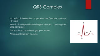
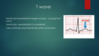
Project :PQRST Complex
Introduction
Electrocardiograph (ECG)
ECG is defined as recording of electrical activity of heart on a
graph paper ,
Or
Graphical representation of electrical activity of heart.
Significance
1. ECG gives information about rate and rhythm of the heart.
2. It is a diagnostic tool for various heart conditions like
hypertrophies, ischemia , infraction, arrhythmia ,conduction
problem and pace maker activity .
3. ECG does not provide information about mechanical activity.
P wave
This is the fiorst small , rounded upward deflection .
Atrial depolarization , initiated by the SA node , causes the p wave .
With atrial depolarization complete , the impulses delayed at the AV node .
Duration – 0.08 – 0.1 sec
QRS Complex
It consist of three sub components the Q wave , R wave
, S wave
Ventricular depolarization begins at apex , causing the
QRS complex .
This is a sharp prominent group of wave .
Atrial repolarization occurs .
T wave
Ventricular repolarization begins at apex , causing the T
wave .
Ventricular repolarization is completed .
here ventricles reset electrically after contraction.
ECG
uses
electrocardiogram Is useful in determining and diagnosing the following
• heart rate
• heart rhythm
• abnormal electrical conduction
• poor blood flow to heart muscle
• heart attack
• coronary artery disease
• hypertrophy of heart chambers
By Aradhna Meshram
B.Pharm 3rd year , SRTM University Nanded

PQRST WAVE COMPLEX - ELECTROCARDIOGRAM PPT

  • 1.
  • 2.
    Introduction Electrocardiograph (ECG) ECG isdefined as recording of electrical activity of heart on a graph paper , Or Graphical representation of electrical activity of heart. Significance 1. ECG gives information about rate and rhythm of the heart. 2. It is a diagnostic tool for various heart conditions like hypertrophies, ischemia , infraction, arrhythmia ,conduction problem and pace maker activity . 3. ECG does not provide information about mechanical activity.
  • 3.
    P wave This isthe fiorst small , rounded upward deflection . Atrial depolarization , initiated by the SA node , causes the p wave . With atrial depolarization complete , the impulses delayed at the AV node . Duration – 0.08 – 0.1 sec
  • 4.
    QRS Complex It consistof three sub components the Q wave , R wave , S wave Ventricular depolarization begins at apex , causing the QRS complex . This is a sharp prominent group of wave . Atrial repolarization occurs .
  • 5.
    T wave Ventricular repolarizationbegins at apex , causing the T wave . Ventricular repolarization is completed . here ventricles reset electrically after contraction.
  • 6.
    ECG uses electrocardiogram Is usefulin determining and diagnosing the following • heart rate • heart rhythm • abnormal electrical conduction • poor blood flow to heart muscle • heart attack • coronary artery disease • hypertrophy of heart chambers
  • 7.
    By Aradhna Meshram B.Pharm3rd year , SRTM University Nanded