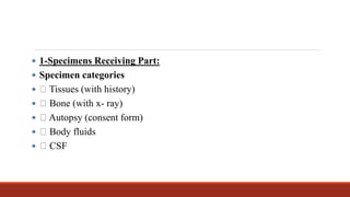
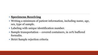
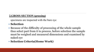
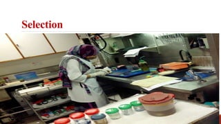
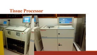
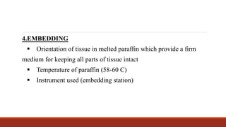
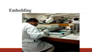
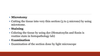
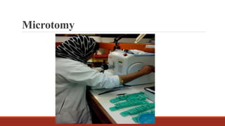
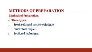
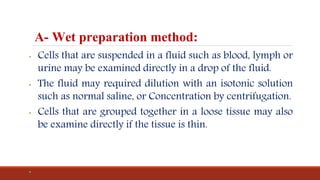
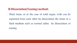
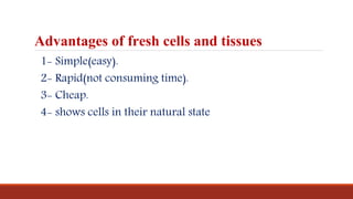
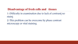
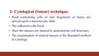
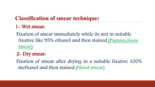
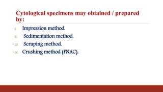
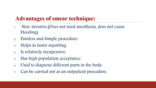
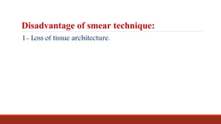
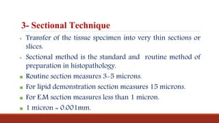
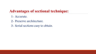
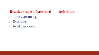
Workflow in Histopathology Laboratory
MR:MAHMOUD IBRAHIM
The objectives of the lecture
1-Recognize the steps of work in histopathology
laboratories.
2-Knowledge of methods of preparing samples in
histopathology laboratories.
 1-Specimens Receiving Part:
 Specimen categories
 Tissues (with history)
 Bone (with x- ray)
 Autopsy (consent form)
 Body fluids
 CSF
 Specimens Receiving
 Writing a minimum of patient information, including name, age,
sex, type of sample.
 Labeling with unique identification number.
 Sample transportation – covered containers, in 10% buffered
formalin.
 Strict Sample rejection criteria
Specimens Receiving Part
2.GROSS SECTION (grossing)
specimens are inspected with the bare eye
 Selection
 Because of the difficulty of processing of the whole sample
thus select part from it to process, before selection the sample
must be weighed and measured dimensions and examined by
naked eye
 Selection Criteria(Home Work)
Selection
 Tissue processing
 The aim of tissue processing is to embed the tissue in a solid medium
firm enough to support the tissue and give it sufficient rigidity to
enable thin sections to be cut and yet soft enough not to damage the
knife or tissue.
 Stages of tissue processing
 Dehydration
 Clearing
 Impregnation
 Embedding
Tissue Processor
4.EMBEDDING
 Orientation of tissue in melted paraffin which provide a firm
medium for keeping all parts of tissue intact
 Temperature of paraffin (58-60 C)
 Instrument used (embedding station)
Embedding
 Microtomy
 Cutting the tissue into very thin section (3 to 5 microns) by using
microtome.
 Staining
 Coloring the tissue by using dye (Hematoxylin and Eosin is
routine stain in histopathology lab)
 Examination
 Examination of the section done by light microscope
Microtomy
METHODS OF PREPARATION
Methods of Preparation:
 Three types:
1. Fresh cells and tissues technique.
2. Smear technique.
3. Sectional technique.
A- Wet preparation method:
• Cells that are suspended in a fluid such as blood, lymph or
urine may be examined directly in a drop of the fluid.
• The fluid may required dilution with an isotonic solution
such as normal saline, or Concentration by centrifugation.
• Cells that are grouped together in a loose tissue may also
be examine directly if the tissue is thin.
•
B-Dissociation(Teasing) method:
• Thick tissue or in the case of solid organ, Cells can be
separated from each other by dissociation the tissue in a
fluid medium such as normal saline by dissociation or
teasing.
Advantages of fresh cells and tissues
1- Simple(easy).
2- Rapid(not consuming time).
3- Cheap.
4- shows cells in their natural state
Disadvantage of fresh cells and tissues
1-Difficulty in examination due to lack of contrast( no
stain).
2-This problem can be overcome by phase contrast
microscopy or vital staining.
2- Cytological (Smear) technique:
• Fluid containing cells or tiny fragments of tissue are
spread upon a microscopic slide.
• The adherent cells fixed.
• Then the smears are stained to demonstrate cell structure.
• The examination of stained smears is the Standard method
in Cytology.
Classification of smear technique:
1- Wet smear:
Fixation of smear immediately while its wet in suitable
fixative like 95% ethanol and then stained.(Papanicolaou
smear)
2- Dry smear:
Fixation of smear after drying in a suitable fixative 100%
methanol and then stained.(blood smear)
Cytological specimens may obtained / prepared
by:
I. Impression method.
II. Sedimentation method.
III. Scraping method.
IV. Crushing method (FNAC).
Advantages of smear technique:
1. Non-invasive.(Does not need anesthesia, does not cause
bleeding)
2. Painless and Simple procedure.
3. Helps in faster reporting.
4. Is relatively inexpensive.
5. Has high population acceptance.
6. Used to diagnose different parts in the body.
7. Can be carried out as an outpatient procedure.
Disadvantage of smear technique:
1- Loss of tissue architecture.
3- Sectional Technique
 Transfer of the tissue specimen into very thin sections or
slices.
 Sectional method is the standard and routine method of
preparation in histopathology.
 Routine section measures 3-5 microns.
 For lipid demonstration section measures 15 microns.
 For E.M section measures less than 1 micron.
 1 micron = 0.001mm.
Advantages of sectional technique:
1- Accurate.
2- Preserve architecture.
3- Serial sections easy to obtain.
Disadvantages of sectional technique:
1. Time Consuming.
2. Expensive.
3. Need experience.
2 workflow

2 workflow

  • 1.
    Workflow in HistopathologyLaboratory MR:MAHMOUD IBRAHIM
  • 2.
    The objectives ofthe lecture 1-Recognize the steps of work in histopathology laboratories. 2-Knowledge of methods of preparing samples in histopathology laboratories.
  • 3.
     1-Specimens ReceivingPart:  Specimen categories  Tissues (with history)  Bone (with x- ray)  Autopsy (consent form)  Body fluids  CSF
  • 4.
     Specimens Receiving Writing a minimum of patient information, including name, age, sex, type of sample.  Labeling with unique identification number.  Sample transportation – covered containers, in 10% buffered formalin.  Strict Sample rejection criteria
  • 5.
  • 6.
    2.GROSS SECTION (grossing) specimensare inspected with the bare eye  Selection  Because of the difficulty of processing of the whole sample thus select part from it to process, before selection the sample must be weighed and measured dimensions and examined by naked eye  Selection Criteria(Home Work)
  • 7.
  • 8.
     Tissue processing The aim of tissue processing is to embed the tissue in a solid medium firm enough to support the tissue and give it sufficient rigidity to enable thin sections to be cut and yet soft enough not to damage the knife or tissue.  Stages of tissue processing  Dehydration  Clearing  Impregnation  Embedding
  • 9.
  • 10.
    4.EMBEDDING  Orientation oftissue in melted paraffin which provide a firm medium for keeping all parts of tissue intact  Temperature of paraffin (58-60 C)  Instrument used (embedding station)
  • 11.
  • 12.
     Microtomy  Cuttingthe tissue into very thin section (3 to 5 microns) by using microtome.  Staining  Coloring the tissue by using dye (Hematoxylin and Eosin is routine stain in histopathology lab)  Examination  Examination of the section done by light microscope
  • 13.
  • 14.
    METHODS OF PREPARATION Methodsof Preparation:  Three types: 1. Fresh cells and tissues technique. 2. Smear technique. 3. Sectional technique.
  • 15.
    A- Wet preparationmethod: • Cells that are suspended in a fluid such as blood, lymph or urine may be examined directly in a drop of the fluid. • The fluid may required dilution with an isotonic solution such as normal saline, or Concentration by centrifugation. • Cells that are grouped together in a loose tissue may also be examine directly if the tissue is thin. •
  • 16.
    B-Dissociation(Teasing) method: • Thicktissue or in the case of solid organ, Cells can be separated from each other by dissociation the tissue in a fluid medium such as normal saline by dissociation or teasing.
  • 17.
    Advantages of freshcells and tissues 1- Simple(easy). 2- Rapid(not consuming time). 3- Cheap. 4- shows cells in their natural state
  • 18.
    Disadvantage of freshcells and tissues 1-Difficulty in examination due to lack of contrast( no stain). 2-This problem can be overcome by phase contrast microscopy or vital staining.
  • 19.
    2- Cytological (Smear)technique: • Fluid containing cells or tiny fragments of tissue are spread upon a microscopic slide. • The adherent cells fixed. • Then the smears are stained to demonstrate cell structure. • The examination of stained smears is the Standard method in Cytology.
  • 20.
    Classification of smeartechnique: 1- Wet smear: Fixation of smear immediately while its wet in suitable fixative like 95% ethanol and then stained.(Papanicolaou smear) 2- Dry smear: Fixation of smear after drying in a suitable fixative 100% methanol and then stained.(blood smear)
  • 21.
    Cytological specimens mayobtained / prepared by: I. Impression method. II. Sedimentation method. III. Scraping method. IV. Crushing method (FNAC).
  • 22.
    Advantages of smeartechnique: 1. Non-invasive.(Does not need anesthesia, does not cause bleeding) 2. Painless and Simple procedure. 3. Helps in faster reporting. 4. Is relatively inexpensive. 5. Has high population acceptance. 6. Used to diagnose different parts in the body. 7. Can be carried out as an outpatient procedure.
  • 23.
    Disadvantage of smeartechnique: 1- Loss of tissue architecture.
  • 24.
    3- Sectional Technique Transfer of the tissue specimen into very thin sections or slices.  Sectional method is the standard and routine method of preparation in histopathology.  Routine section measures 3-5 microns.  For lipid demonstration section measures 15 microns.  For E.M section measures less than 1 micron.  1 micron = 0.001mm.
  • 25.
    Advantages of sectionaltechnique: 1- Accurate. 2- Preserve architecture. 3- Serial sections easy to obtain.
  • 26.
    Disadvantages of sectionaltechnique: 1. Time Consuming. 2. Expensive. 3. Need experience.