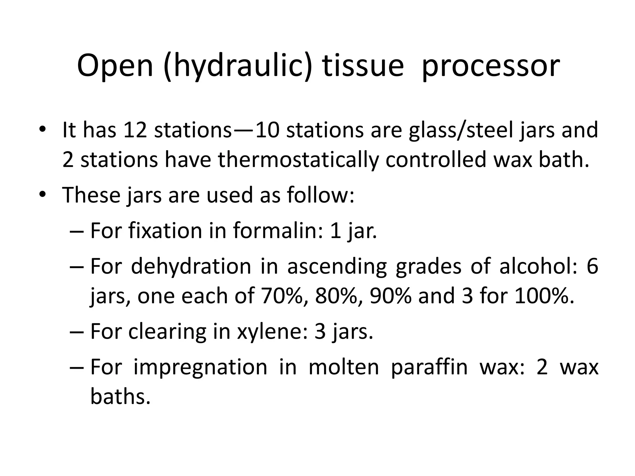
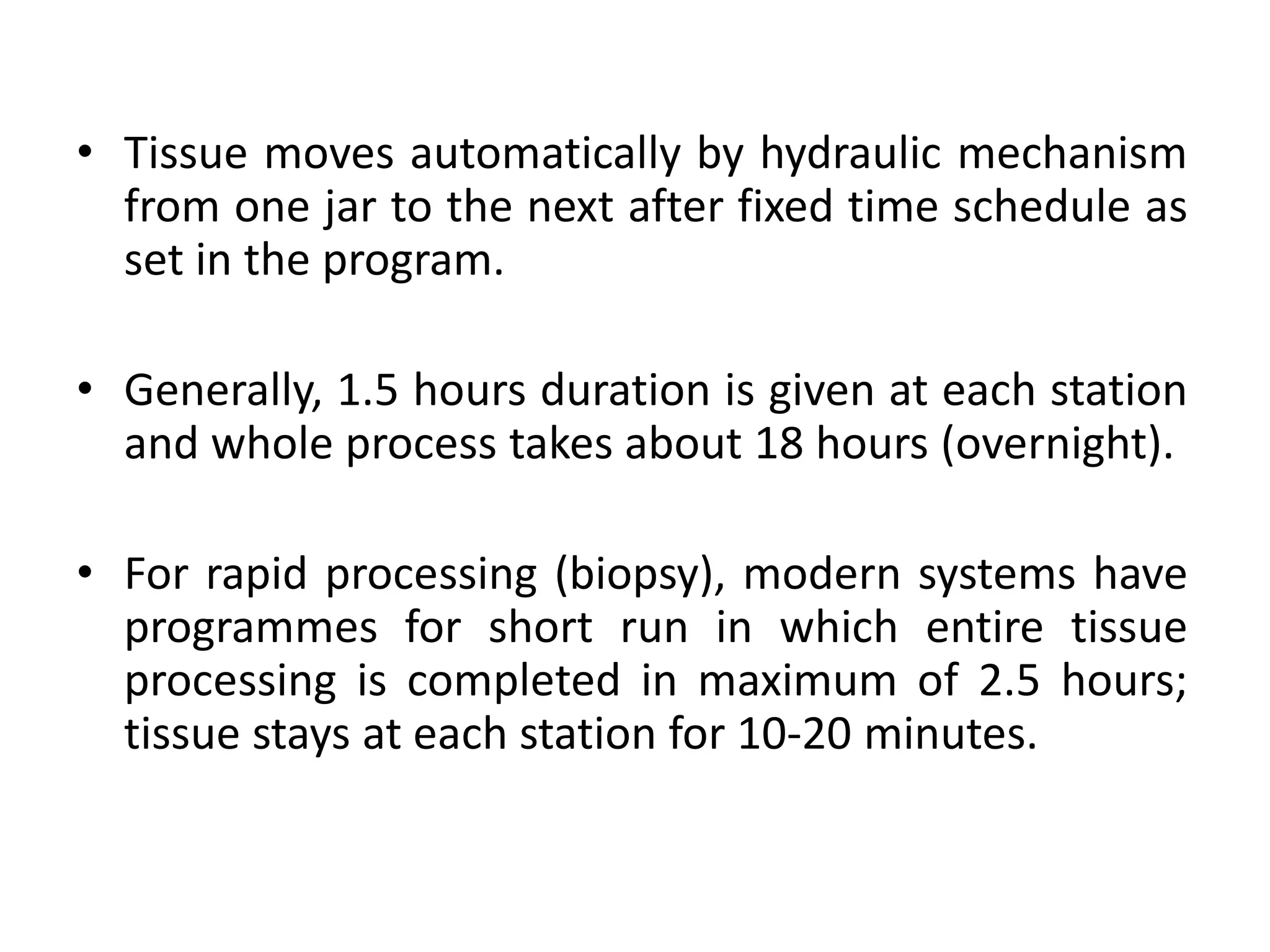
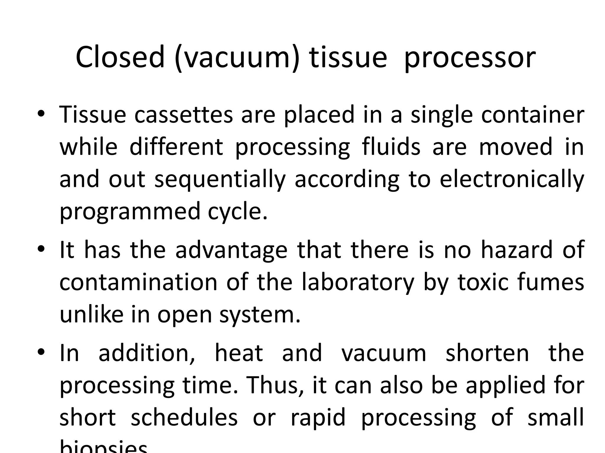
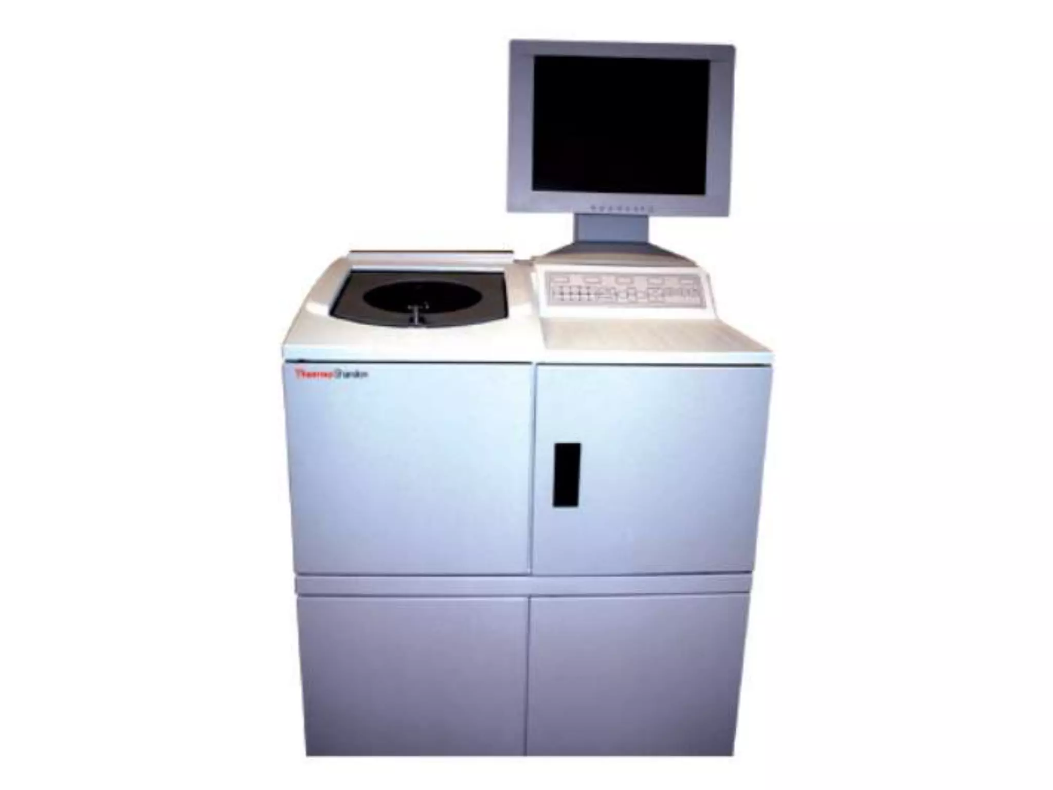
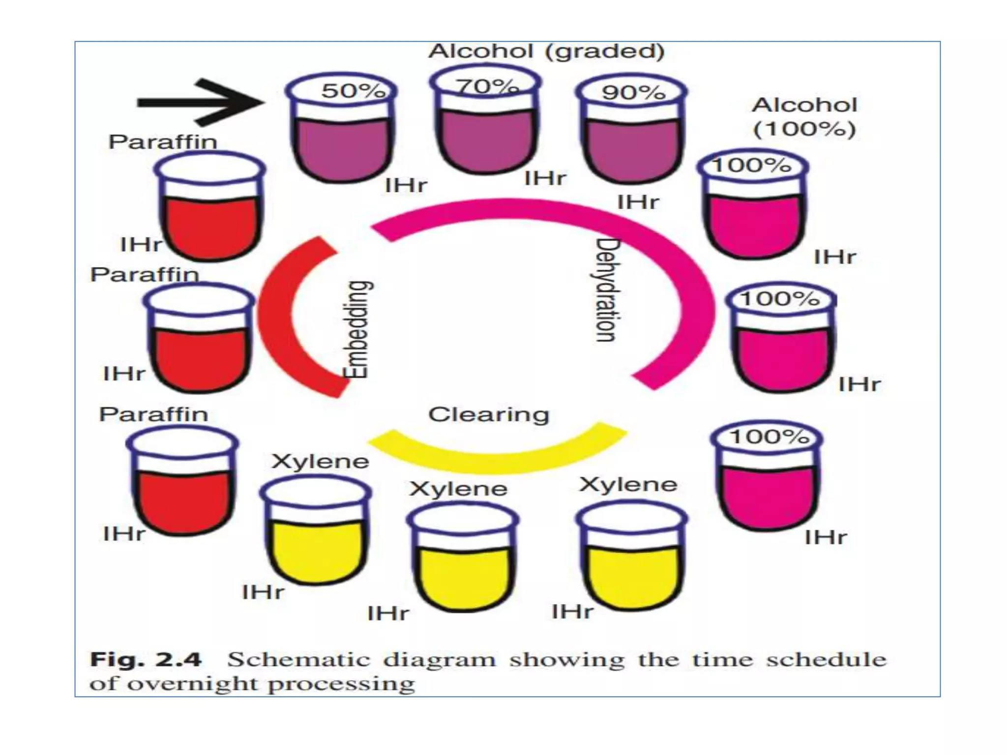
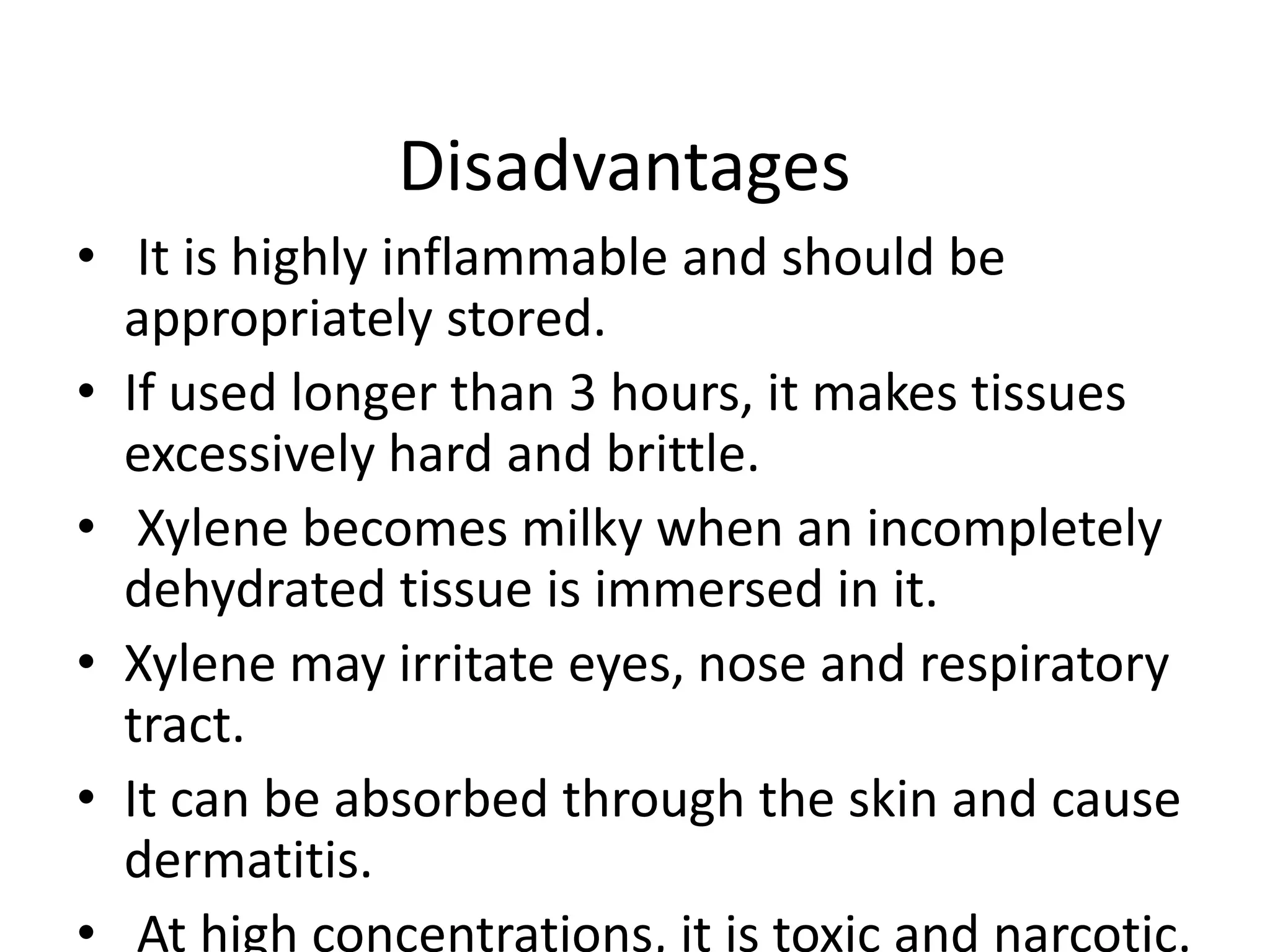
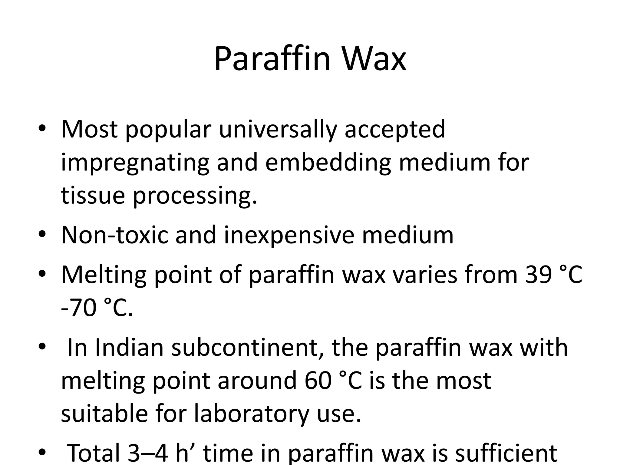
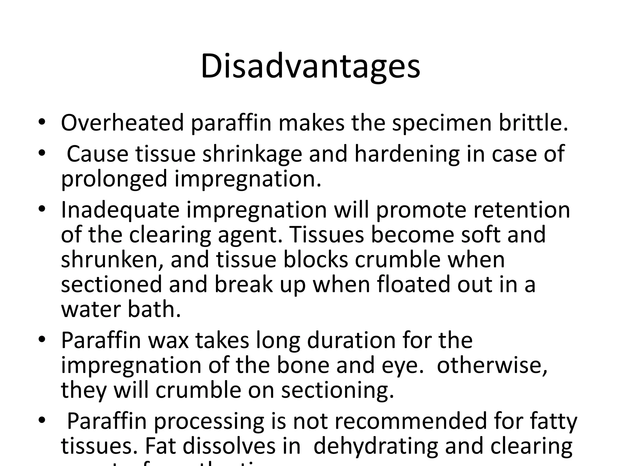
Tissue processing involves removing water from tissue and replacing it with paraffin wax to provide rigidity for microscopic examination. The main steps are fixation, dehydration using increasing concentrations of alcohol, clearing with xylene to remove alcohol, and impregnation with molten paraffin wax. Automated tissue processors complete this process overnight using different stations for each step. Factors like tissue size, agitation, heat, and vacuum pressure influence effective processing. Ethyl alcohol is most commonly used for dehydration, while xylene is used for clearing prior to paraffin wax impregnation and embedding.
This document discusses the process of decalcification, which is the removal of calcium from tissues to make them suitable for section cutting. It outlines the key steps: selection of tissue, fixation, decalcification using mineral acids, chelating agents, or electrophoresis, detection of endpoint, neutralization, and washing. Common decalcifying agents discussed include Gooding and Stewart's fluid, von Ebner's fluid, citrate-citric acid buffer, and chelating agents like EDTA. The factors that influence decalcification speed and the importance of determining the endpoint are also summarized.
This document describes the steps involved in tissue processing from fixation to embedding in wax. It discusses obtaining fresh specimens, fixation in formalin, dehydration through an alcohol series, clearing in xylene, infiltration and embedding in paraffin wax. Sections are then cut on a microtome, mounted on slides and stained, usually with hematoxylin and eosin, to visualize tissue structures microscopically. Proper processing is important to preserve tissue morphology and produce high quality stained sections for diagnostic examination.
This document discusses tissue fixation in histopathology. It describes the various stages of histopathology including fixation, processing, embedding, sectioning and staining of tissues. It explains the importance of fixation in preventing autolysis and putrefaction of tissues. The key properties and reactions of common fixatives like formaldehyde and glutaraldehyde are outlined. Factors that can affect fixation quality including temperature, pH, concentration and duration of fixation are also summarized. Finally, different classifications of fixatives are presented based on their structural and functional properties.
The major steps in tissue processing are dehydration, clearing, impregnation, and embedding. Dehydration removes water from tissues using increasing concentrations of alcohol. Clearing removes residual dehydrating agent using a substance miscible with both the dehydrating agent and paraffin wax. Impregnation completely replaces clearing agent with paraffin wax. Embedding involves orienting tissue in molten paraffin wax before it solidifies to provide structure for sectioning. Key advantages of this process are preservation of tissue structure and ability to cut thin, consistent sections for analysis.
Microtomes, Section cutting , Sharpening of Razorsvikas25187
This document discusses various types of microtomes and microtomy techniques. It describes different parts of microtomes like the block holder, knife holder, and handwheels. It explains different types of microtomes based on their cutting mechanism, including rotary, rocking, base-sledge, sliding, freezing, vibrating, saw, cryostat, and ultramicrotome. It also discusses microtome knives, sharpening techniques, section cutting for paraffin blocks, and section mounting methods.
Honing and stropping are processes used to sharpen knives. Honing removes nicks and irregularities from the cutting edge to make it straight and sharp. It is done using a hone, which must be kept clean and lubricated. The process involves pushing and pulling the knife across the hone in strokes until the edge is smooth. Stropping further polishes the edge after honing. It straightens and aligns the microscopic teeth at the edge without removing material, resulting in an even sharper, mirror-like finish.
The document discusses the principles and techniques of tissue infiltration and embedding. It describes how clearing agents are removed from tissues through diffusion and replaced with molten embedding media like wax. The wax is then cooled to solidify and provide support for sectioning thin tissue samples. Several factors influence infiltration including tissue size, type, clearing agent used, and whether vacuum embedding is performed. Common embedding media include paraffin wax which provides good support but can cause shrinkage, and DMSO-supplemented wax which speeds infiltration. Both manual and automated processors are described.
The document summarizes the process of tissue processing which involves fixing, dehydrating, clearing, infiltrating with wax, embedding, sectioning, staining, and mounting tissue samples in order to examine them microscopically. Key steps include fixation to prevent decay, dehydration using alcohol to remove water, clearing with xylene to remove alcohol, infiltration and embedding in paraffin wax, sectioning thin slices with a microtome, staining typically with hematoxylin and eosin for examination, and mounting on slides. The goal is to prepare tissue for microscopic analysis while maintaining structure.
Tissue processing involves fixing, dehydrating, clearing, and infiltrating tissue samples with paraffin wax to embed them for sectioning. The key steps are fixation to prevent degradation, dehydration using graded alcohols, clearing with solvents like xylene to remove alcohol, infiltration using paraffin wax, embedding wax blocks for sectioning, sectioning on a microtome, and staining for examination. Automated tissue processors can complete many processing steps unattended for increased efficiency and throughput in pathology laboratories. Proper handling and processing is essential to obtain an accurate histological diagnosis from tissue specimens.
The document discusses the hematoxylin and eosin stain, which is the most widely used histological stain. It stains cell nuclei blue or black using hematoxylin, and stains cell cytoplasm and connective tissue fibers pink using eosin. The purpose of staining is to identify tissue structures and the presence or absence of disease. Common stains discussed include hematoxylin and eosin, Gram's method, Ziehl-Neelson's method, and Papanicolaou stain. The document also provides details on the chemistry and procedures for hematoxylin and eosin staining.
The document discusses tissue fixation, which is the first step in preparing tissue for microscopic examination. It aims to prevent decomposition of tissues after removal from the body through chemical and physical processes. This involves using fixatives to preserve tissues in a lifelike state. Several types of fixatives are described including aldehydes, oxidizing agents, and mercurials. The effects of fixation on different cell components and factors affecting fixation are also outlined. A variety of fixatives commonly used in histopathology are then classified and their formulations and uses explained.
Frozen sections of tissue are prepared using a cryostat to quickly obtain thin sections for histological examination and diagnosis. A cryostat maintains tissue at freezing temperatures to allow sectioning without ice crystal formation. Tissue is mounted on a chuck and placed in the cryostat, then sectioned and mounted on slides for staining. Frozen sections allow rapid diagnosis but have poorer morphology and staining than fixed tissue sections.
The document discusses cryostats, which are devices used to cut thin frozen sections of tissues for examination under a microscope. Cryostats contain a microtome inside a freezer unit that can rapidly freeze tissue samples and cut sections as thin as 1 micrometer at temperatures below freezing. The cryostat process allows for quick diagnosis by freezing and sectioning tissues within minutes rather than having to dehydrate, embed in paraffin, and section as with traditional microtomes.
This document discusses tissue processing techniques. It describes how clearing agents act as intermediaries between dehydration and infiltration solutions. Xylene, toluene, and chloroform are common clearing agents that make tissues transparent. The criteria for choosing clearing agents and infiltration methods like paraffin wax embedding are also outlined. Automated tissue processors allow for standardized overnight processing through sequential alcohol, clearing, and infiltration solutions.
This document discusses tissue processing and fixation. It begins by introducing tissue fixation and its objectives, such as preventing degradation and maintaining morphology. It then describes various fixation methods and factors that affect fixation quality. Common fixatives are discussed, including formaldehyde, glutaraldehyde, Zenker's solution and Bouin's solution. Fixation protocols for specific tissues like brain, breast, lung and kidney are also reviewed. The document emphasizes the importance of proper fixation for histological examination.
The document discusses various techniques used in histopathology sample processing including decalcification, fixation, dehydration, clearing, embedding and sectioning. It covers different chemical agents used for each step along with their properties and advantages. Various methods are described such as paraffin, celloidin and vacuum embedding for optimal tissue preservation and section quality. Automatic tissue processors and freeze drying are also mentioned as techniques to reduce processing time.
This document discusses tissue fixation in pathology. It begins by describing the overall tissue processing steps and importance of fixation. The main types of fixatives are then outlined, including coagulant, cross-linking, and compound fixatives. Formalin and glutaraldehyde fixation are discussed in depth. Key factors that influence fixation quality like buffer pH, fixation duration, tissue size, and temperature are also summarized. The document provides a comprehensive overview of fixation in pathology.
Fixatives are chemicals used to preserve biological tissues from decay. They terminate biochemical reactions and may increase mechanical strength. Fixatives disable enzymes and protect samples from damage. Common fixatives include formaldehyde, glutaraldehyde, osmium tetroxide, alcohols, and picric acid. Fixation aims to inhibit autolysis, preserve tissues, harden them, and improve staining. Factors like temperature, concentration, and duration impact fixation quality. Different tissues require specific fixatives for optimal preservation. An ideal fixative kills cells quickly without damage, penetrates rapidly, prevents decay, hardens tissues, and allows long-term storage.
There are several knife profiles used for microtomy including Profile A (plano concave), Profile B (biconcave), Profile C (wedge), and Profile D (tool edge). Profile A has one flat side and one concave side and is used for soft tissues embedded in nitrocellulose. Profile B is classical with concavity on both sides and was introduced by Heiffor. Profile C is a standard wedge profile used for cutting all materials in all microtomes. Profile D is a wedge knife with a steep cutting edge used for hard objects like bone.
Decalcification is a process used to remove mineral content from calcified tissues like bone and teeth to allow for microscopic examination. It involves selecting an appropriate decalcifying agent based on factors like the tissue, required staining, and urgency. Common decalcifying agents include acids like nitric acid, formic acid, and chelating agents. The decalcification process must be monitored and the tissue properly processed, sectioned, and stained afterwards to examine it microscopically. Undecalcified sections can also be prepared to examine mineralized and non-mineralized bone ratios.
This document discusses fixatives used in histopathology. It describes the process of fixation and how fixatives preserve tissue by denaturing or precipitating proteins. The ideal properties of a fixative are described, including preventing autolysis and allowing for staining. Common fixatives are classified and their mechanisms and uses are explained. Factors that affect fixation such as temperature, size, volume ratio, time, choice of fixative, and penetration are also summarized.
This document discusses cytopreparatory techniques, including fixation of cytological samples, staining methods, and interpretation. It focuses on fixation, explaining that fixation preserves cells in a lifelike state after death by preventing autolysis and putrefaction. The key properties of a good fixative are outlined, and various fixatives are classified and examples are provided, including alcohols, formalin, and mercuric chloride, which are commonly used for cytological preparations.
Histology is the microscopic study of tissues. Key steps in processing tissues for histological examination include fixation, dehydration, clearing, embedding in paraffin wax, sectioning, and staining. Tissues are first fixed in chemicals like formaldehyde to preserve their structure. They are then dehydrated using graded alcohols to remove water. Next, tissues are cleared using solvents like xylene to make them permeable to paraffin prior to embedding. The embedded tissues can then be thinly sectioned and stained for microscopic examination. Proper tissue processing is important for high quality histological analysis.
The tissue section is colourless because the fixed protein has the same refractive index as that of glass. We use dyes that have specific affinity with the different tissue proteins and colour them differently.
Colour is seen by the eye as a result of the effect of certain electromagnetic waves on the rods and cones of the retina. These waves, which have a varying length, will determine the colour that is seen.
White light being composed of all the colours of the visible spectrum varies in wavelength from 4,000 Â to 8,000 Â.
If light of a specific wavelength is absorbed from white light the resultant light will then be coloured, the colour being dependent upon the particular wavelength that has been removed.
A tissue processor is used to prepare tissue samples for analysis by fixing, staining, dehydrating or decalcifying them.
The techniques for processing the tissue, whether biopsies, larger specimen removed at surgery
Tissue processing involves fixing, dehydrating, clearing, infiltrating, and embedding tissue samples to produce diagnostic microscope slides. It stabilizes tissues and removes water, replacing it with paraffin wax. Efficient agitation, heat up to 45°C, low viscosity reagents, and vacuum up to 50.79 kPa can reduce processing time. Stages include fixation with formalin, dehydration using graded alcohols, clearing with xylene or toluene, infiltration with paraffin wax, and embedding for microtomy. Proper orientation during embedding is important for diagnosis.
Lecture (5) processing of tissue in histopathology laboratoryHafsa Hussein
This document discusses the principles and steps of tissue processing for microscopic examination, including dehydration, clearing, and infiltration/impregnation with paraffin wax.
The key steps are:
1) Dehydration using graded alcohols to remove water from tissues, allowing infiltration with wax. Xylene is commonly used for clearing to remove alcohols.
2) Clearing with xylene or other agents to make tissues transparent before wax infiltration.
3) Infiltration and impregnation with molten paraffin wax provides rigidity to tissues for thin sectioning and microscopic examination. Proper processing is important for examining tissue structures.
Histological specimen preparation involves fixing, processing, sectioning, and staining tissue samples. Key steps include:
1. Fixation prevents tissue degradation and preserves cellular structure. Common fixatives are formaldehyde and glutaraldehyde.
2. Tissue processing involves dehydration, clearing, and embedding tissues in paraffin wax to allow thin sectioning.
3. Sections are cut on a microtome and mounted on slides for staining. Staining with hematoxylin and eosin is most common, coloring nuclei blue and cytoplasm pink.
This document discusses the process of tissue processing, which involves fixing, dehydrating, clearing, and embedding tissue samples in paraffin wax to allow for thin sectioning. The key stages are fixation using chemicals like formalin to preserve tissue structure, dehydration using increasing concentrations of alcohol, clearing using solvents like xylene to make tissues transparent, and embedding in paraffin wax for sectioning. Automated and manual methods are described. Tissue microarrays allow evaluation of multiple tissue samples on a single slide by arranging small cores in a recipient paraffin block.
1. The document discusses the various steps involved in tissue processing for microscopic examination, which includes fixation, processing, embedding, sectioning and staining of tissues.
2. Key steps include fixation of tissues using formalin to preserve structure, dehydration using increasing concentrations of alcohol, clearing with xylene, impregnation and embedding in paraffin wax.
3. Thin sections are then cut from the paraffin blocks using a microtome and stained, usually with hematoxylin and eosin, for microscopic examination.
Histopathology is examination of tissues for presence or absence of changes in their structure due to disease processes. We go through various steps in the process of converting gross sample to microscopic slides.
This document provides an overview of tissue processing techniques used in histology and histopathology. It discusses the various steps involved, including dehydration, clearing, infiltration, and embedding in paraffin wax or other mediums. It describes the purposes and methods for dehydration, clearing using agents like xylene, infiltration using paraffin wax, and embedding tissues. It also discusses alternative embedding techniques like ester wax, water soluble waxes, gelatin, and celloidin embedding as well as double embedding methods and potential processing artifacts.
This document provides an overview of the tissue processing techniques used to prepare tissue samples for microscopic examination. It describes the main steps in tissue processing as fixation, dehydration, clearing, infiltration/embedding. Dehydration involves removing water from tissues using a series of increasing concentrations of ethanol or other solvents to prevent damage. Clearing replaces the dehydrating fluid with a solvent miscible with both the dehydrating fluid and paraffin wax. The goal is to embed tissues in paraffin wax for microtomy, as it provides sufficient rigidity while being soft enough for thin sectioning without harming tissues or knives. Factors like tissue type, fixation, and desired detail influence processing parameters.
This document provides an overview of the main steps involved in tissue processing for histopathological examination, which are specimen accessioning, gross examination, tissue processing (including the paraffin technique), and potential problems. The paraffin technique is the most common method and involves fixation, dehydration via an alcohol series, clearing with xylene, infiltration and embedding in paraffin wax, then microtome sectioning of the paraffin block. Proper identification and labeling of specimens is critical to avoid errors.
4. Handling of surgical Specimens - II.pdfAhad412190
1) Tissue specimens are fixed in formalin to prevent deterioration. They then undergo dehydration, clearing, infiltration with paraffin wax, and embedding to allow sectioning.
2) Sections are cut on a microtome and stained, usually with hematoxylin and eosin, with hematoxylin staining nuclei blue and eosin staining cytoplasm pink.
3) Care must be taken to properly handle, sample, and process tissues to avoid contamination and allow accurate histological examination.
This document discusses filtration techniques used in Unani medicine. It begins by defining filtration and related terms. It then discusses factors that affect the filtration rate and different types of filter media and filter aids. Finally, it describes various filtration equipment used in Unani medicine like filter funnels, Buchner funnels, Seitz filters, filter presses, rotary filters, and vacuum filtration. The key techniques and considerations for filtration in Unani medicine are summarized in 3 sentences or less.
The document discusses various filtration techniques used in pharmaceutical processing. It defines filtration as the removal of solids from fluids or fluids from other fluids. Clarification can be achieved through filtration or centrifugation. There are two main reasons for these processes in pharmaceuticals: to remove unwanted particles and to collect solids as the final product. The document describes various types of filtration like solid/fluid, solid/gas, fluid/fluid filtration and their applications. It also discusses filtration theory, factors affecting filtration rate, various filter media types, filter aids, selection of filtration equipment and systems for different applications.
The document discusses the process of singeing textiles. Singeing involves burning off protruding fibers from fabric surfaces to improve smoothness and luster. It can be done using gas singeing machines, which pass fabric over flames, or hot plate/roller machines. Key factors that affect singeing include flame intensity, fabric speed and temperature, and fiber type. Singeing removes fuzz to create a uniform, lustrous surface that reflects light evenly.
This document discusses filtration techniques in Unani medicine. It defines filtration and describes factors that affect the filtration rate. It outlines various filter media like cloth, paper, cotton wool and membranes. It also discusses filter aids and common filtration equipment like funnels, filters, and filter presses. Vacuum filtration is described as a preferred technique in Unani to efficiently collect solids through terms like tarweeq, tasfiya and tarsheeh.
The document provides information about embedding and section cutting techniques in histopathology. It discusses the aims of embedding tissue, such as providing support and preventing distortion. Common embedding mediums include paraffin wax, resin, agar, and gelatin. Paraffin wax is the most popular due to being inexpensive, non-toxic, and allowing long term tissue storage. The document outlines the tissue embedding process and describes equipment used, including embedding molds and the Tissue-Tek system. Tissue orientation in the block is important for visualizing the desired morphology.
The document discusses the key steps in histological techniques which are used to prepare tissue samples for microscopic examination. The techniques include fixation, dehydration, clearing, embedding, sectioning, and staining of tissues. Fixation preserves the tissue structure using chemical fixatives. The tissue then undergoes dehydration, clearing and infiltration with paraffin wax before being embedded in wax blocks and sectioned for microscopic analysis. Staining helps to visualize different structures within the tissue sample under the microscope.
This document outlines the process for obtaining tissue samples for histology. It discusses collecting tissue from animal bodies or plant parts, fixing tissues in formalin to prevent decay, processing tissues by dehydrating in alcohol, clearing in xylene, infiltrating and embedding in paraffin wax, sectioning samples, staining typically with hematoxylin and eosin, and mounting slides under coverslips. The goal is to prepare tissues so they can be clearly examined microscopically while maintaining their structure.
This document discusses the process of histopathology specimen processing. There are several key steps:
1) Tissue specimens are fixed, typically in formalin, to preserve their structure. 2) The tissues then undergo dehydration using increasing concentrations of alcohol, followed by clearing with xylene to remove the alcohol. 3) The tissues are then infiltrated with paraffin wax through impregnation to harden them. 4) The tissues are embedded in paraffin blocks. 5) Sections are cut from the paraffin blocks using a microtome and placed on slides. The slides are then stained to visualize the tissue structures under a microscope.
Similar to Histotechnique - processing, embedding.pptx (20)
White blood cells, also known as leukocytes, are formed in the bone marrow and circulate in the bloodstream. They protect the body from infection by locating sites of infection and signaling other white blood cells. There are five main types of white blood cells - neutrophils, lymphocytes, eosinophils, basophils, and monocytes - each with a specialized role in the immune system such as killing bacteria, producing antibodies, or cleaning up damaged cells. A low white blood cell count indicates leukopenia while a high count signifies leukocytosis.
Hemolytic anemia is a condition where red blood cells are destroyed and removed from circulation faster than they can be replaced. The patient's medical record shows findings related to hemolytic anemia including signs and symptoms of anemia like fatigue, jaundice, and abdominal pain as well as lab results showing low hemoglobin and elevated bilirubin. Further tests are needed to determine the underlying cause of hemolysis in this patient.
This document discusses different types of transfusion reactions, including immune and non-immune reactions. Immune reactions can be immediate like hemolytic or febrile non-hemolytic reactions, or late reactions like alloimmunization or post-transfusion purpura. Non-immune reactions include circulatory overload from too large a transfusion or transmission of infectious agents. Specific reactions are defined by their pathophysiology and clinical features such as fever, chills, breathing issues, or bleeding tendencies. Transfusion reactions require recognition of symptoms to provide appropriate treatment and prevent future complications.
Dr. Sandeep Singh is a pathologist. He received his medical degree from Johns Hopkins University and completed his residency at Massachusetts General Hospital. Dr. Singh has over 15 years of experience diagnosing diseases and conditions by examining body tissues and fluids.
The document discusses the histology of the male reproductive system. It focuses on providing information about the microscopic anatomy and cellular structure of tissues that make up organs in the male reproductive tract, such as the testes, epididymis, vas deferens, seminal vesicles, prostate and penis. The author, Dr. Sandeep Singh, is a pathologist who has expertise in examining reproductive tissues.
Cartilage is a firm, flexible connective tissue found in various parts of the body like joints, trachea, and nasal septum. It provides flexibility and support. Cartilage is avascular and receives nutrients through diffusion. It is classified into hyaline, elastic, and fibrocartilage based on composition. Hyaline cartilage is present in trachea and joints. It has a homogeneous matrix and chondrocytes arranged in rows. Perichondrium covers cartilage except in some joints.
histology of epithelial and connective tissue.pptxsandeep singh
Epithelium and connective tissue are the two main types of tissues in the human body. This document discusses the histology, or microscopic anatomy, of these two tissue types. It was written by Dr. Sandeep Singh in the field of pathology to provide information on the structure and function of epithelium and connective tissues at the cellular level.
Digestive System Liver, Gall Bladder and Pancreas.pptxsandeep singh
Dr. Sandeep Singh is a pathologist. He received his medical degree from Johns Hopkins University School of Medicine. Dr. Singh has over 15 years of experience diagnosing diseases and conditions by examining body tissues and fluids.
This document discusses systemic lupus erythematosus (SLE), a chronic inflammatory disease that can affect many parts of the body including the heart, lungs, skin, joints, blood vessels, and kidneys. SLE is characterized by a rash, fever, renal issues, and abnormalities in blood counts. The disease involves the immune system inappropriately attacking the body's own tissues. A key indicator is the presence of lupus erythematosus cells, which are white blood cells that have ingested pieces of altered nuclear material from other cells. SLE most commonly affects women of childbearing age and those with African ancestry genetics.
MGG stain is used to stain blood, bone marrow, and cytological specimens. It contains basic dyes that stain nuclei and granules blue and an acidic dye that stains red blood cells and eosinophil granules red. The basic dyes stain cellular components due to their positive charge binding to negatively charged DNA and RNA, while the acidic dye stains due to its negative charge. Proper pH is critical for the staining reaction to work correctly.
The document discusses the benefits of exercise for mental health. Regular physical activity can help reduce anxiety and depression and improve mood and cognitive functioning. Exercise causes chemical changes in the brain that may help protect against mental illness and improve symptoms for those who already suffer from conditions like anxiety and depression.
The document discusses the benefits of exercise for mental health. Regular physical activity can help reduce anxiety and depression and improve mood and cognitive functioning. Exercise boosts blood flow, releases endorphins, and promotes changes in the brain which help enhance one's emotional well-being and mental clarity.
The document appears to be about Dr. Sandeep Singh and microscopes. It mentions two topics - Dr. Sandeep Singh as the first topic, and microscopes as the second topic. However, there are no other details provided about either of these topics in the very short document.
The document discusses different types of neurons, synaptic transmission, and peripheral nerves. It describes multipolar, bipolar, and pseudo-unipolar neurons. It explains how neurotransmitters are released at synapses and transmitted across the synaptic cleft. It also discusses the structure of peripheral nerves including fascicles, endoneurium, perineurium, and epineurium.
The document discusses the endocrine system. It was written by Dr. Sandeep Singh, who is likely an expert on the topic. In a very concise manner, it addresses the endocrine system without providing many details.
Investigation in mismatch blood transfusion.pptxsandeep singh
This document discusses lab investigation of transfusion reactions and mismatched blood transfusions. It begins by defining a transfusion reaction as an adverse reaction to a blood transfusion that is incompatible with the recipient's blood. It describes initial measures if a reaction occurs, such as stopping the transfusion and notifying the physician. Preliminary tests include clerical checks for errors, visual checks of blood colors, and serology checks like repeating ABO and Rh typing of pre-and post-transfusion samples. Additional tests include cross-matching samples using saline, albumin, and indirect antiglobulin techniques, as well as antibody screening. Interpretation of forward and reverse blood grouping results is also discussed.
This document discusses haemostasis, the process by which bleeding is stopped. It describes the key components of the haemostatic system, including platelets, coagulation factors, and the endothelium. The sequence of events that occurs at the site of vascular injury is outlined, from initial platelet plug formation to stabilization of the fibrin clot. The roles of platelets, coagulation factors, fibrinolysis, and the endothelium are explained. Causes of bleeding disorders include problems with platelets, coagulation factors, fibrinolysis or the endothelium. The document also covers approaches to evaluating bleeding disorders.
There are several types of glands in the human body that are classified based on their structure and secretory components. Simple glands include tubular glands with a single lumen and acinar glands that secrete into pockets. Compound glands have branched duct systems and can have tubular, acinar, or mixed secretory components like the submandibular gland. The document provides examples of each gland type including their histological appearance and secretory cell types.
This document discusses performing a differential leukocyte count from a peripheral blood smear. Key steps include making a blood smear, staining it with Leishman's stain, and counting different types of white blood cells (WBCs) under a microscope. The goal is to determine percentages of neutrophils, lymphocytes, eosinophils, monocytes, and basophils, which can indicate various health conditions if levels are abnormal. Proper technique for the blood smear and staining is described.
This document discusses red blood cell indices and methods for estimating haemoglobin. Red blood cell indices provide information about the size, shape and quality of red blood cells by measuring metrics like MCV, MCH and MCHC. Abnormal values can indicate different types of anaemia. Haemoglobin estimation is important for diagnosing anaemia. Common methods discussed are the Sahli's method (visual colorimetric), cyanmethemoglobin method (photoelectric) and specific gravity method. Modern analyzers are now commonly used to estimate blood components and parameters like haemoglobin.
Temple of Asclepius in Thrace. Excavation resultsKrassimira Luka
The temple and the sanctuary around were dedicated to Asklepios Zmidrenus. This name has been known since 1875 when an inscription dedicated to him was discovered in Rome. The inscription is dated in 227 AD and was left by soldiers originating from the city of Philippopolis (modern Plovdiv).
How to Manage Reception Report in Odoo 17Celine George
A business may deal with both sales and purchases occasionally. They buy things from vendors and then sell them to their customers. Such dealings can be confusing at times. Because multiple clients may inquire about the same product at the same time, after purchasing those products, customers must be assigned to them. Odoo has a tool called Reception Report that can be used to complete this assignment. By enabling this, a reception report comes automatically after confirming a receipt, from which we can assign products to orders.
Andreas Schleicher presents PISA 2022 Volume III - Creative Thinking - 18 Jun...EduSkills OECD
Andreas Schleicher, Director of Education and Skills at the OECD presents at the launch of PISA 2022 Volume III - Creative Minds, Creative Schools on 18 June 2024.
Level 3 NCEA - NZ: A Nation In the Making 1872 - 1900 SML.pptHenry Hollis
The History of NZ 1870-1900.
Making of a Nation.
From the NZ Wars to Liberals,
Richard Seddon, George Grey,
Social Laboratory, New Zealand,
Confiscations, Kotahitanga, Kingitanga, Parliament, Suffrage, Repudiation, Economic Change, Agriculture, Gold Mining, Timber, Flax, Sheep, Dairying,
A Visual Guide to 1 Samuel | A Tale of Two HeartsSteve Thomason
These slides walk through the story of 1 Samuel. Samuel is the last judge of Israel. The people reject God and want a king. Saul is anointed as the first king, but he is not a good king. David, the shepherd boy is anointed and Saul is envious of him. David shows honor while Saul continues to self destruct.
2. Tissue Processing
• Aims of tissue processing:
– To provide sufficient rigidity to the tissue so that
it can be cut into thin section for microscopic
examination.
• Principle of processing:
– In tissue processing the water within the tissue is
removed, and another medium (usually paraffin
wax) is impregnated in the tissue that provides the
adequate support to the tissue.
3.
4. Tissue processors
• Processes of dehydration, clearing and impregnation
are carried out in a composite equipment which is
known as automated tissue processor.
• Automated tissue processor can be:
• Open (hydraulic) system
• Closed (vacuum) type
5. Open (hydraulic) tissue processor
• It has 12 stations—10 stations are glass/steel jars and
2 stations have thermostatically controlled wax bath.
• These jars are used as follow:
– For fixation in formalin: 1 jar.
– For dehydration in ascending grades of alcohol: 6
jars, one each of 70%, 80%, 90% and 3 for 100%.
– For clearing in xylene: 3 jars.
– For impregnation in molten paraffin wax: 2 wax
baths.
6.
7.
8. • Tissue moves automatically by hydraulic mechanism
from one jar to the next after fixed time schedule as
set in the program.
• Generally, 1.5 hours duration is given at each station
and whole process takes about 18 hours (overnight).
• For rapid processing (biopsy), modern systems have
programmes for short run in which entire tissue
processing is completed in maximum of 2.5 hours;
tissue stays at each station for 10-20 minutes.
9. Closed (vacuum) tissue processor
• Tissue cassettes are placed in a single container
while different processing fluids are moved in
and out sequentially according to electronically
programmed cycle.
• It has the advantage that there is no hazard of
contamination of the laboratory by toxic fumes
unlike in open system.
• In addition, heat and vacuum shorten the
processing time. Thus, it can also be applied for
short schedules or rapid processing of small
10.
11. Factors that Influence Tissue
Processing
• Size of the tissue:
– The smaller the size, the better the processing.
– Optimum thickness of the tissue - 3–4 mm only.
• Agitation:
– The tissue gets better contact with the
surrounding medium if it is completely immersed
and gently agitated.
– Too rapid agitation may damage the soft and
delicate tissue
12. Factors that Influence Tissue
Processing
• Heat:
– Increases the better penetration of fluid.
• Viscosity:
– The higher the viscosity of the medium, lower the
penetration.
– Heat reduces the viscosity of the medium and
helps in better penetration.
• Negative pressure (Vaccume):
– Negative pressure removes trapped air in the
tissue.
13.
14. Dehydration –Removal of water
• The process of removing intercellular and
extracellular water from the tissue following
fixation and prior to wax impregnation is known
as "dehydration”, .
• Solutions utilized for this process are called
"Dehydrating Agents“.
• Sharp difference of concentration gradient of the
dehydrating fluid may damage the delicate tissue.
• Gradual dehydration is necessary from low to
high concentration of dehydration fluid. Routine
laboratory: 70, 90 and 100% alcohol for 1.5 to 2 h
15. • Common dehydrating agents:
– Ethyl alcohol
– Methylated spirit
– Methanol
– Butyl alcohol
– Isopropyl alcohol
– Dehydrating agents other than alcohol: dioxane,
ethylene glycol and acetone
16. Ethyl alcohol
• Most popular and most commonly used
dehydrating agent.
• This is a clear , colourless and flammable fluid.
• It is considered to be the best dehydrating agent
because :
– fast-acting,
– mixes with water and many organic solvents
– penetrates tissues easily.
– Little shrinkage if graded alcohols are used
– Non- poisonous
– Not very expensive .
17. • As a dehydrating agent ethyl alcohol is used in
50, 70, 90 and 100% concentration.
• For delicate tissue, the dehydration may be
started from 30% concentration of ethyl
alcohol.
• If tissue is immersed in the ethyl alcohol for
long time, then the removal of attached water
from the carbohydrate and protein molecules
causes hard and brittle tissue
18. Anhydrous Cupric Sulphate in Final
Container
• Anhydrous cupric sulphate (CuSO4) is a white
powder that draws water from the alcohol and
thereby helps in dehydration.
• About 1 cm layer of this powder is kept in the
bottom of the container.
• The cupric sulphate powder should be
covered with two to three layers of filter
paper to prevent any colouring of the tissue.
• When the CuSO4 becomes hydrated, the
colour of the powder changes to blue. This
gives warning signal to change the alcohol and
19. • Advantage:
– Increases the life span of alcohol
– Better dehydration
– Good indicator to change alcohol
20. Methanol
• Clear, colourless, volatile and inflammable
liquid.
• It can be used as a substitute of ethanol, but
it is rarely used in laboratory because of its
volatility and high cost.
21. Butanol (butyl alcohol)
• Boiling point 117.7° C
• Advantages:
– Less shrinkage and hardening than with ethyl
alcohol
– Excellent for slow processing
– Miscible with paraffin
• Disadvantages:
– Odorous
– Slow-acting
– Long periods of infiltration necessary
– Dehydrating power low
22. Isopropyl Alcohol
• Available as isopropanol (99.8%).
• This is miscible with water and liquid paraffin.
• It is a relatively rapid acting, non-toxic .
• causing minimal tissue shrinkage and
hardening.
• No government restrictions on its use.
• It is a good lipid dissolving solvent
• Less expensive than alcohol
23.
24. Clearing
• Next step of processing after dehydration.
• Removal of dehydrating agent from the
tissue because many dehydrating agents are
not miscible with the impregnating material
(paraffin wax).
• The clearing agent should be miscible with
both the dehydrating agent and the
embedding medium.
• The refractive index of the clearing agent is
similar to the tissue, and therefore it gives
25. Aims of clearing
• Removal of dehydrating agent (e.g. alcohol)
to facilitate impregnation of paraffin wax .
• To make the tissue clear and improve the
microscopic examination
26. Ideal clearing agent:
• It should be miscible with alcohol to promote rapid removal of the
dehydrating agent from the tissue.
• It should be miscible with, and easily removed by melted paraffin wax
and/or by mounting medium to facilitate impregnation and mounting of
sections.
• It should not produce excessive shrinkage, hardening or damage of tissue.
• Low viscosity and high penetration rate
• Low melting point : easily and quickly removed by the molten wax
• It should not evaporate quickly in a water bath.
• It make tissues transparent.
• Less toxic and Less inflammable
• Cheap
27. • Volume of clearing agent: 40 times the volume
of the specimen
• Total duration:
– Smaller biopsy: 1 h
– Larger tissue: Three changes in xylene or toluene
60 min each
• End point detection: Tissue becomes
transparent
• Prolonged exposure to clearing agent: The
brittle and more friable tissue
• Different clearing agents: Xylene, toluene,
chloroform, amyl nitrate, cedarwood oil and
28. Xylene
• Most commonly used clearing agent in the
laboratory.
• This is a clear and inflammable liquid.
• The small pieces of tissue are cleared rapidly
by xylene within 30–60 min.
• Prolonged exposure to xylene may make the
tissue hard and brittle
29. Advantages:
• Rapid clearing agent, suitable for urgent biopsies
which it clears within 15-30 minutes.
• It makes tissues transparent.
• miscible with absolute alcohol and paraffin.
• For mounting procedures, it does not dissolve
celloidin and can, therefore, be used for celloidin
sections.
• It evaporates quickly in paraffin oven and can,
therefore, be readily replaced by wax during
impregnation and embedding.
30. Disadvantages
• It is highly inflammable and should be
appropriately stored.
• If used longer than 3 hours, it makes tissues
excessively hard and brittle.
• Xylene becomes milky when an incompletely
dehydrated tissue is immersed in it.
• Xylene may irritate eyes, nose and respiratory
tract.
• It can be absorbed through the skin and cause
dermatitis.
• At high concentrations, it is toxic and narcotic.
31.
32. Impregnation
• Process whereby the clearing agent is
completely removed from the tissue and
replaced by a medium that will completely fill
all the tissue cavities and give a firm
consistency to the specimen.
• Allows easier handling and cutting of suitably
thin sections without any damage or
distortion to the tissue.
33. • Ideal impregnating medium:
– Miscible with clearing agent
– Liquid in higher temperature and solid in room
temperature
– Homogenous and stable
– Non-toxic, odorless and cheap
– Transparent
– Fit for sectioning the tissue
– Capable of flattening after ribboning
– Easy to handle
– Inexpensive
34. • The time duration and the number of changes
required for the impregnation in tissue
depends on:
– Size of tissue: Thicker large tissue takes more time
to impregnate with the embedding medium. It
also contains more clearing agent to remove.
– Type of tissue: Hard tissue such as bone and
cartilage takes more time for embedding than soft
tissue.
– The type of clearing agent: Certain clearing
agents are easy to remove than others. Such as
xylene and toluene are easy to remove than
36. Paraffin Wax
• Most popular universally accepted
impregnating and embedding medium for
tissue processing.
• Non-toxic and inexpensive medium
• Melting point of paraffin wax varies from 39 °C
-70 °C.
• In Indian subcontinent, the paraffin wax with
melting point around 60 °C is the most
suitable for laboratory use.
• Total 3–4 h’ time in paraffin wax is sufficient
37. Advantages:
• Easy cutting of serial sections
• very rapid processing
• Tissue blocks and unstained mounted sections
may be stored in paraffin for an indefinite period
of time after impregnation without considerable
tissue destruction.
• Because formalin-fixed, paraffin-embedded
tissues may be stored indefinitely at room
temperature, and nucleic acids (both DNA and
RNA) may be recovered from them decades after
fixation, they are an important resource for
historical studies in medicine.
38. Disadvantages
• Overheated paraffin makes the specimen brittle.
• Cause tissue shrinkage and hardening in case of
prolonged impregnation.
• Inadequate impregnation will promote retention
of the clearing agent. Tissues become soft and
shrunken, and tissue blocks crumble when
sectioned and break up when floated out in a
water bath.
• Paraffin wax takes long duration for the
impregnation of the bone and eye. otherwise,
they will crumble on sectioning.
• Paraffin processing is not recommended for fatty
tissues. Fat dissolves in dehydrating and clearing
39. • Additives and Modification of Paraffin Wax To
alter the physical characteristics of paraffin
wax, the following modifications may be done:
• To increase hardness: addition of stearic acid
• Reduction of melting point: addition of
phenanthrene
• Improving adhesiveness with tissue and wax:
addition of 0.5% of ceresin
40. Dimethyl sulphoxide DMSO
• The addition of small amount of DMSO in
paraffin wax
– reduces the infiltration time of the wax
– removes the residual clearing agent.
– It produces a homogenous matrix and better
support.
41. Embedding
• Embedding (Casting or Blocking) is the
process by which the impregnated tissue is
placed into a precisely arranged position in a
mold containing a supporting medium which
is then allowed to solidify.
• This supporting media for blocking known as
embedding media.
• Aims of embedding: Embedding medium has
three important functions:
– To give support of the tissue
42. • Done by:
– Filling mould of suitable size with molten
wax/embedding media
– Orientation of tissue in mould to ensure it will cut
in right plane
– Cooling of mass to promote solidification
44. The choice of the embedding medium
• Type of tissue:
– The density of the tissue and the embedding
medium should be close otherwise tissue may not
be sectioned properly, and tissue will be
deformed.
• Type of microtome
• Type of microscope
45. Paraplast
• Mixture of highly purified paraffin and
synthetic plastic polymers, with a melting
point of 56-57°C.
• More elastic and resilient than paraffin wax
thereby permitting large dense tissue blocks
such as bones and brain to be cut easily.
• Better ribboning of sections.
• Serial sections may be cut with ease, without
cooling the tissue block, thereby preventing
the formation of ice crystal artefact.
• Soluble in common clearing agents and
46. Carbowax
• a polyethylene glycol containing 18 or more
carbon atoms, which appears solid at room
temperature.
• It is soluble in and miscible with water; hence
does not require dehydration and clearing of
the tissue.
• The tissues are fixed, washed out and
transferred directly into the melted Carbowax.
• Processing time is reduced.
47. • For routine processing: Temperature of 56°C
are used
– I – 70% carbowax for 30 min.
– II – 90% carbowax for 45 min
– III – 100 % carbowax for 1 hr.
– IV - 100 % carbowax for 1 hr.
– Specimens are then embedded in fresh Carbowax
at 50°C and rapidly cooled in a refrigerator.
48. • Disadvantages:
– Carbowax is very easily dissolved in water. Hence
care must be taken to avoid contact of the block
with water or ice.
– Tissue sections are very difficult to float out and
mount
• Adding soap to water or using 10%
Polyethylene Glycol 900 in water will reduce
tissue distortion and promote flattening and
49. Epoxy resin
• Mainly used in electron microscopy as it
provides better resolution and greater details
of tissue.
Agar gel:
• helps in cohesion of friable and fragmented
tissue particularly in cytology sample and also
endometrial curetting and small endoscopic
biopsies.
• It does not provide good support of the tissue
50. Gelatin
• :It is also used in small friable tissues and
frozen section containing friable and necrotic
tissue.
• The melting point of gelatin is 35–40 °C, and
this low melting point makes it unsuitable for
embedding
51. Methacrylate
• Methacrylate monomer is miscible with
ethanol.
• In the presence of catalyst (benzoyl peroxide
2%),it is polymerized and provides a hard and
clear block.
• .
52. Celloidin
• Celloidin is purified form of nitrocellulose.
• Mainly used for specimens with:
– large hollow cavities which tend to collapse, f
– For hard and dense tissues such as bones and
teeth
– for large tissue sections of the whole embryo.
• Main disadvantages:
– Celloidin impregnation is very slow
– inability to cut thin sections.
– Serial sections are difficult to prepare
53. Different Types of Mould Used
for Block
• Leuckhard Embedding Moulds ( L moulds):
•
54.
55. • The two L-shaped arms are adjusted to make a
convenient size for block.
• Adequate lubricant such as glycerine is
applied to the L arms and metal plate for easy
removal of the tissue.
• The molten wax is poured in the space
between two L arms, and then the tissue is
placed within the bottom of the liquid wax.
• The wax is subsequently cooled, and the block
with tissue in one surface is removed for
57. Tissue Embedding Method
• It is essentially same for all the types of
embedding media.
• The following things are needed for tissue
embedding:
– Molten paraffin wax
– Mould with cover
– Metal plate (cold plate)
60. Steps of Tissue tek system
• Liquid paraffin is kept at a constant
temperature in the dispenser of the system.
• The tissue is put on the lower surface of the
mould by forceps. The cutting surface should
be faced down.
• Molten paraffin is poured on the metallic
mould.
• The mould is covered with peripheral plastic
ring on the upper surface.
Tissue processor: construction: all exterior parts are powder coated in RAL 9002 12 stations, 10 glass containers, 2 stainless steel containers, 1 transport basket is included, for approx. 110 cassettes, optional twin basket for approx. 220 cassettes lifting mechanism for 1 transport basket rear connection possibility for hose (100 mm diameter), with fan or active carbon filter as option electronical controls with multi power supply 85-264 V. Worldwide operation without power adjustment display and operating panel with programming keys in case of power failure the
basket is moved in a programmable