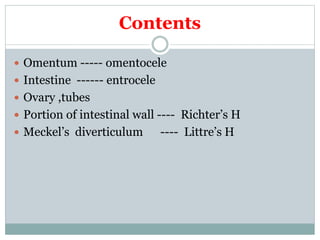
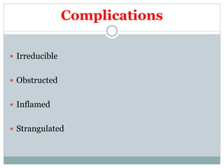
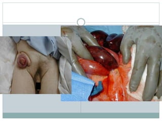
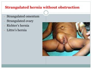
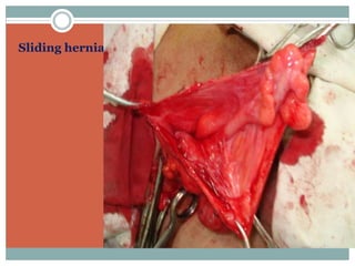
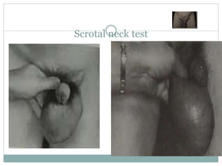
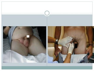
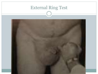
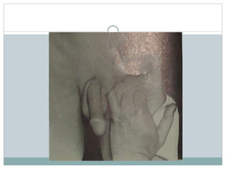
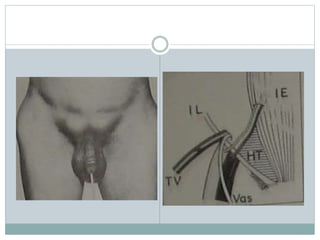
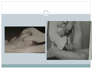
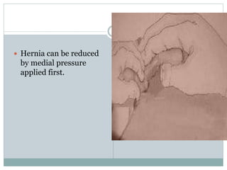
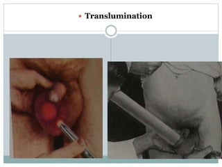
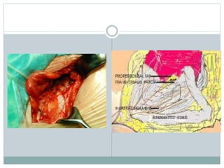
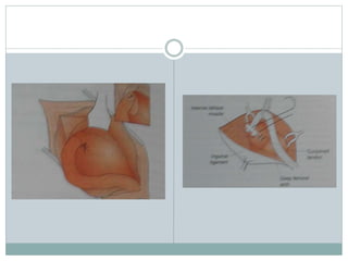
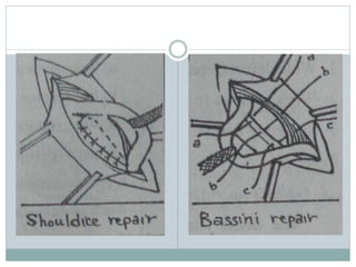
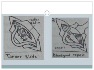
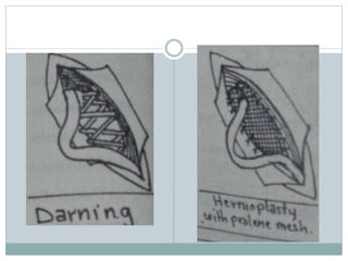
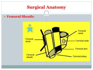
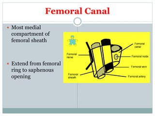
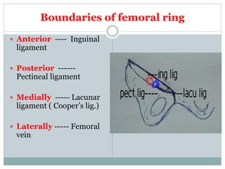
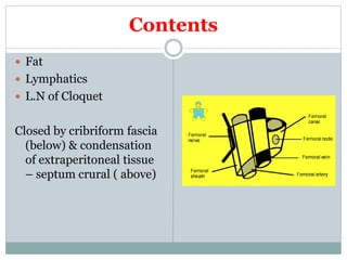
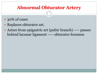
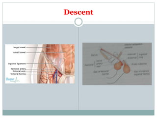
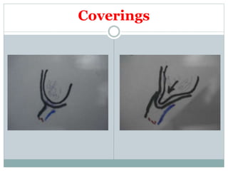
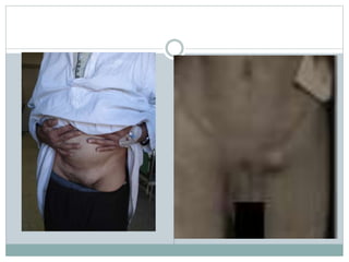
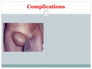
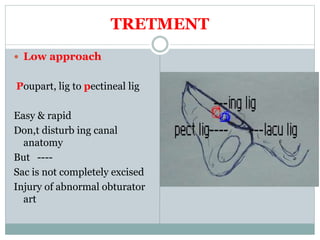
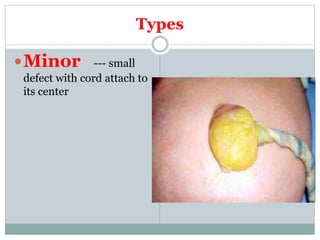
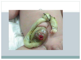
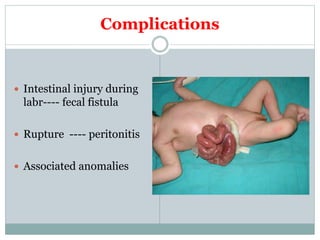
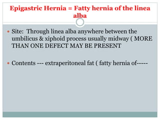
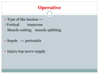
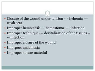
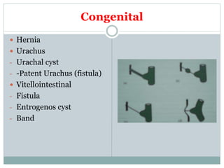
B Y
PROF. TAREK GOBRAN
PROF. OF GENERAL AND PEDIATRIC
SURGERY
HERNIA and ABDOMINAL
WALL DEFECTS
DEFINITION
 Protrusion of a viscus or part of it through a defect in
the wall of the containing cavity
 It is either internal or external
ETIOLOGY
 Predisposing factors:
- Increase of intra-abdominal pressure
- Pregnancy
- Congenital preformed sac
- Undescended testis
- Obesity
- Collagen abnormalities
COMPOSITION
 Sac
 Coverings
 Contents
Sac
 Neck
 Body
 Fundus
Coverings
 Layers of abdominal wall through which the sac
passes
Contents
 Omentum ----- omentocele
 Intestine ------ entrocele
 Ovary ,tubes
 Portion of intestinal wall ---- Richter’s H
 Meckel’s diverticulum ---- Littre’s H
Complications
 Irreducible
 Obstructed
 Inflamed
 Strangulated
 Contents can not
reduced back to
abdomen
 Causes:
- Adhesions
- Large contents and
narrow neck
IRREDUCIBLE HERNIA
 Hernia content balloons
over external ring when
reduction is attempted.
Obstructed Hernia
 Irreducible hernia with obstructed intestinal lumen
without interference with blood supply
 Clinically ----- colic, constipation, vomiting, .......
 Sometimes it is difficult to differentiate from
strangulation so it is better to be managed as
strangulated hernia
Strangulated hernia
 = Serious impairment of blood supply of the contents
with or without obstruction ----- ischemia ----- if not
treated within 5-6 hrs ------ gangrene
 In strangulation venous impairment occurs first ----
intestinal congestion & edema ------- more
congestion &edema ----- arterial impairment ------
ischemia ---- exudation of blood into the sac +
bacterial transudation through the wall ( infected
toxic fluid in the sac ) ------ gangrene -----
perforation
Clinical Features
 Sudden onset of pain +/- signs of intestinal
obstruction
 Local signs:
- Irreducible
- No impulse with cough
- Tense
- Tender
If not treated early ----- perforation ----- peritonitis ---
-- septic shock
Strangulated hernia without obstruction
- Strangulated omentum
- Strangulated ovary
- Richter’s hernia
- Littre’s hernia
Inflamed Hernia
 Source of infection:
-Inflamed contents as appendix
- From skin infection as ulcerations
 Clinical features:
- Hernia is painful, hot red and tender but not tense
TREATMENT OF HERNIA
 Truss ???????????????????????????????????????????
 Surgery
Herniotomy
Hernioplasty
Herniorrhaphy
CAUSES OF RECURRENCE
PREOPERATIVE CAUSES
 Causes of increased intra-abdominal pressure as
chronic cough ----
 Debilitating disease
 Weak musculature
OPERATIVE
 Repair undertension
 Imperfect hemostasis and devitalization of tissues ---
-- infection
 Use of absorbable suture
 Missed sac or failure to completely excise the sac
POSTOPERATIVE
 Persistence of predisposing factors as------
 Wound infection
 Lifting heavy objects early postoperatively
 Incidence:
Excluding incisional h
 75% inguinal
 15% umbilical
 8.5% femoral
 1.5% rare hernias
INGUINAL HERNIA
 Indirect Hernia (oblique inguinal hernia )
 Direct hernia
INDIRECT INGUINAL HERNIA
ANATOMY of INGUINAL CANAL
 Inguinal canal is an
oblique canal extending
from internal (deep) ring
to external (superficial)
ring
 It is about 4 cm in adult
and in infants the two
rings are opposite each
others
INTERNAL (DEEP) RING
 Opening in in fascia
transversalis ½ an inch
above the mid-inguinal
point medial to inferior
epigastric vessels
External Ring
 opening in
external oblique
apponeurosis ½ an inch
above pubic tubercle
bounded by supromedial
and infrolateral crus of
ext ob . Normally it just
admit the little finger
Contents
 Male ------ spermatic cord + ilio-inguinal n
+genital branch of genitofemoral n.
 Females: Round ligament + -------
Boundaries
 Anterior:
- External oblique
apponeurosis +
- Conjoint tendon medially
 Posterior
- Fascia tranversalis +
- Conjoint tendon laterally
Superior
- Conjoint tendon
 Inferior
-Inguinal ligament
Mechanisms that prevent hernia
 Shutter mechanism
 Valvular mechanism
 Plugging mechanism
Indirect Hernia (OIH)
 It is a hernia that pass through the internal ring and
enter inguinal canal (bubonocele) and may pass
through external ring and descend in scrotum
(complete)
INCIDENCE
 Commonest type of hernia
 Male: female 20:1
 Common in right side
 Bilateral in 30%
Etiology
 Congenital preformed sac ( patent procesus
vaginalis) ------- most accepted
- More common on the RT side
- Herniotomy only in children is curative
- PPV is found in many autopsy of individual with no
history of hernia
Incidence
 It is most common hernia
 More common on RT side ------- why?
Types of the sac
 Congenital
 Infantile
 Funicular
 Saddle hernia
 Bubonocele
 Complete hernia
Sliding hernia
Contents
 As before
Descent
 Downward, forward and medially ( reduction in
reverse direction)
Coverings
 Extrapertitonial fat
 internal spermatic fascia |(fascia tranversalis)
 cremastric muscle and fascia
(from internal oblique)
 External spermatic fascia (external oblique)
 skin and superficial fascia
Complications
Clinical features
INSPECTION
Palpation
Scrotal neck test
External Ring Test
3 fingers
Testicular exammination
 Hernia can be reduced
by medial pressure
applied first.
 Translumination
Differential diagnosis
Treatment
 Correct predisposing causes
 Surgery
DIRECT INGUINAL HERNIA
INCIDENCE
 15% of inguinal hernias
 Always in male
 More than 50% bilateral
 Hernia through weak
Hasselbach’s triangle
Lateral defect : Malgaigne
bulge
Medial defect; narrow
neck
ETIOLOGY
 Acquired
- Weak conjoint tendon
- Injury of ilioinguinal nerve
- Precipitating factors
CONTENTS
 Sliding urinary bladder is common
COVERINGS
 Extraperitonial fat
 Fascia transversalis
 Conjoint tendon
 External oblique
aponeurosis
 Skin and sc tissues
Descent
 Forward ( very rarely pass through external ring)
COMPLICATIONS
 Rare ---- why?
Treatment
 surgery
FEMORAL HERNIA
 Herniation through femoral canal
 About 20% of hernia in women & 5 % in men
 Female to male 2:1 ( elderly females and 30 to 40
years old males)
 More in multipara.
 Most liable to become strangulated and may be the
first presentation why?
More in females:
 Wider canal
 Pelvic tilt
 Repeated pregnancy
Surgical Anatomy
 Femoral Sheath:
Femoral Canal
 Most medial
compartment of
femoral sheath
 Extend from femoral
ring to saphenous
opening
Boundaries of femoral ring
 Anterior ---- Inguinal
ligament
 Posterior ------
Pectineal ligament
 Medially ----- Lacunar
ligament ( Cooper’s lig.)
 Laterally ----- Femoral
vein
Contents
 Fat
 Lymphatics
 L.N of Cloquet
Closed by cribriform fascia
(below) & condensation
of extraperitoneal tissue
– septum crural ( above)
Abnormal Obturator Artery
 30% of cases
 Replaces obturator art.
 Arises from epigastric art (pubic branch) ---- passes
behind lacunar ligament ---- obturator foramen
Descent
Coverings
Contents
Complications
TRETMENT
 Low approach
Poupart, lig to pectineal lig
Easy & rapid
Don,t disturb ing canal
anatomy
But ----
Sac is not completely excised
Injury of abnormal obturator
art
 High approach
Cooper iliopectineal), to
conjoint or
Poupert to pectinal or the
3 lig
Umbilical Hernia
Umbilical Hernia
 Congenital
 Infantile
 U.H. in adults
Congenital = Exomphlos= Omphalocele
 = Persistence of the physiologic hernia of fetal life
Coverings
 2 layers
- Inner peritonial
- Outer amniotic membrane
Types
Minor --- small
defect with cord attach to
its center
Major ----
wide defect with the cord
attach to its lower part
Contents
Complications
 Intestinal injury during
labr---- fecal fistula
 Rupture ---- peritonitis
 Associated anomalies
Treatment
 Small defect ------- primary closure
 Large defect
- Primary closure
-Skin flap closure
- Nonoperative ---- repeated painting with betdine,
gentian violot, etc ------ ventral hernia --- repair
GASTRISCHISIS
Infantile Umbilical Hernia
 Due to weak umbilical
scar
 Rarely complicates
 Spontaneous cure
 If persist for 2-4 years or
large --- repair
Umbilical Hernia in adult=
Paraumbilical
 Protrusion through linea
alba just above or may be
below the umbilicus
(supra or infra umbilical)
Sac
 The neck is often
remarkably narrow
compared to the size of
the sac ------
complication
 Longstanding -----
loculated & adhesions
Contents
 As any hernia but commonly omentum
Predisposing factors
 + Obesity , weak abdominal ms, repeated
pregnancy
Clinical features
 As any hernia
 More in women 5 times
men
 Usually obese
 35-50 years
Complications
 + dyspepsia ( dragging on colon & stomach)
 Large hernia --- intertrigo
Treatment
 Preop ----- + weight loss
 Herniorrhaphy by primary closure ( small defect)
 Mayo, repair
 Hernioplasty ---- large defects & recurrent cases
 +/- lipectomy & abdominplasty
Epigastric Hernia = Fatty hernia of the linea
alba
 Site: Through linea alba anywhere between the
umbilicus & xiphoid process usually midway ( MORE
THAN ONE DEFECT MAY BE PRESENT
 Contents --- extraperitoneal fat ( fatty hernia of-----
Clinical Features
 No symptoms
 Symptoms of peptic dyspepsis
TREATMENT
Rare Hernias
Lumbar Hernia
 Primary
- Inf lumbar triangle
( commonest)
-Between iliac crest , ext
oblique , latissmus dorsi
Sup lumbar triangle ----
12th rib ,internal oblique
, sacrospinalis
 Secondary commoner
D.D
 Lipoma
 Cold abscess
 Phntom hernia (
paralysis of muscles)
TREATMENT
Spigilian Hernia
 Hernia thr ough linea semilunaris lateral to rectus m.
midway between umbilicus and symp pubis
Divarication of the Recti
 In multiparous women, ascitis ……. Etc
 Infants
Incisional Hernia = Ventral =
Postoperative
 HERNIA at the site of abdominal scar
Aetiology
 Preoperative ----as
in rec hernia
Operative
 Type of the incsion ---
-Vertical transvrse
- Muscle cutting muscle splitting
 Sepsis --- pertonitis
 Injury top nerve supply
 Closure of the wound under tension --- ischemia ---
weak scar
 Improper hemostasis -- hemastoma --- infection
 Improper technique --- devitalization of the tissues --
-- infection
 Improper closure of the wound
 Imprpoer anaethesia
 Improper suture material
Postoperative
 As in rec hernia + wound infection
Clinical Features
Treatment
 Palliative : very poor risk patients with
uncomplicated hernia with wide neck
 Surgery
Surgery
 Preoperative:
- As U.H
Surgical Procedures
 Anatomical repair
 Cattle, 5 layers
 Keel, ( historical)
 Hernioplasty
Burst Abdoen = Abdominal Dehiscence
 Etiology as in incisional hernia
Types
 Complete
 Incomplete
Incidence
 1-2 %
Clinical Features
 6th to 8th postop day ---- serosanguinous discharge (
pathognomonic -------
Treatment
 Emergency operation
 Preoperative:
- Reassure
- - Resuscitate
- NGT
- Cover the intestine with sterile towel
Diseases of the Umbilicus
 Congenital
 Inflammatory
 Neoplastic
 Fistula
 Others
Congenital
 Hernia
 Urachus
- Urachal cyst
- -Patent Urachus (fistula)
 Vitellointestinal
- Fistula
- Entrogenos cyst
- Band
Inflammatory
 Neonatal omphalitis --- infection of umbilical stump
 Adult omphalitis
 Pilonidal sinus
Benign Neoplasms
 Adenoma (Raspbery tumor) in infants from
vitellointestinal duct mucosal reminant
 Endometriosis
Malignant Neoplastic
 Primary epithelioma (rare) ----- inguinal & axillary
LN
 Secondaries ( sister Joseph nodule) --- breast,
stomach, colon,
Fistula
 Fecal --- congenital , malignant infiltration of cancer
colon, T.B. peritonitis
 Urinary
 Biliary (subacute perforation of gall bladder)
Others
 Umbilical stone
 Umbilical polyp
D E S M O = T E N D O N L I K E
Desmoid Tumor
 Incidence Adult multiparous female (80% females)
 Site Rectus sheath usually below the umbilicus
never in the mid-line but other abdominal muscles
can be affected
Aetiology
 Female who have borne children
 Rarely arises from old abdominal scar
 May be associated with familial polposis ( Gardner
sayndrome)
Pathology
 Composed of fibrous tiossues containing
multinucleated masses resemble F.B giant cells ,
infiltrate muscles
 No distant metastasis
 Myxomatous degenration --- rapid increase in size
 Never undergoes sarcomatous changes ( unlike
fibroma)
Treatment
 Wide excision ( at least 2.5 cm safty margin)
Rupture inferior epigastric
artery
Incidence
 Old age, thin weak females
 Athletic below middle age males
 Pregnant multi female ( late in pregnancy)
Site
 Usually at the level of arcuate ligament where post
rectal sheath is defecient
Clinical features
 Severely tender rctus muscle lump following a bout
of cough or trauma to abd wall
 Sometimes, bruising
D.D.
 Twisted ov cyst
 Appendicular abscess
 Strangulated spigilian h
Treatment
 Small hematoma ---- rest
 Early operation and evacutiuon of the hematoma
and ligation of inf epigastric is safer as bleedind mar
recur and mar ruture intra peritneal

Herniaabdwalllecture 100618085852-phpapp02