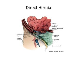
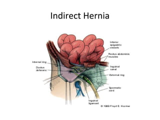
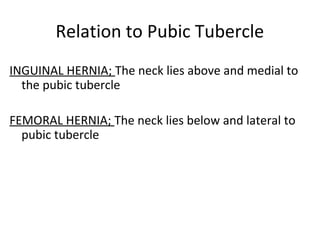
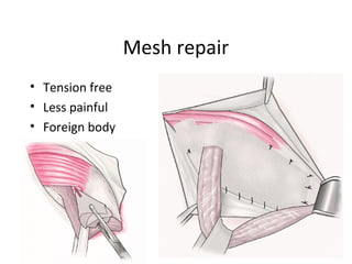
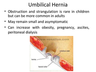
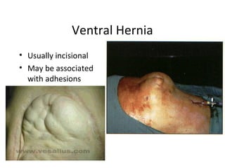
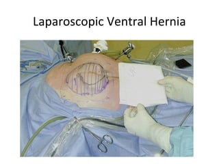
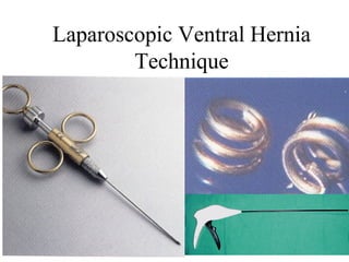
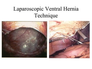
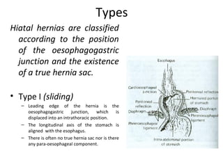
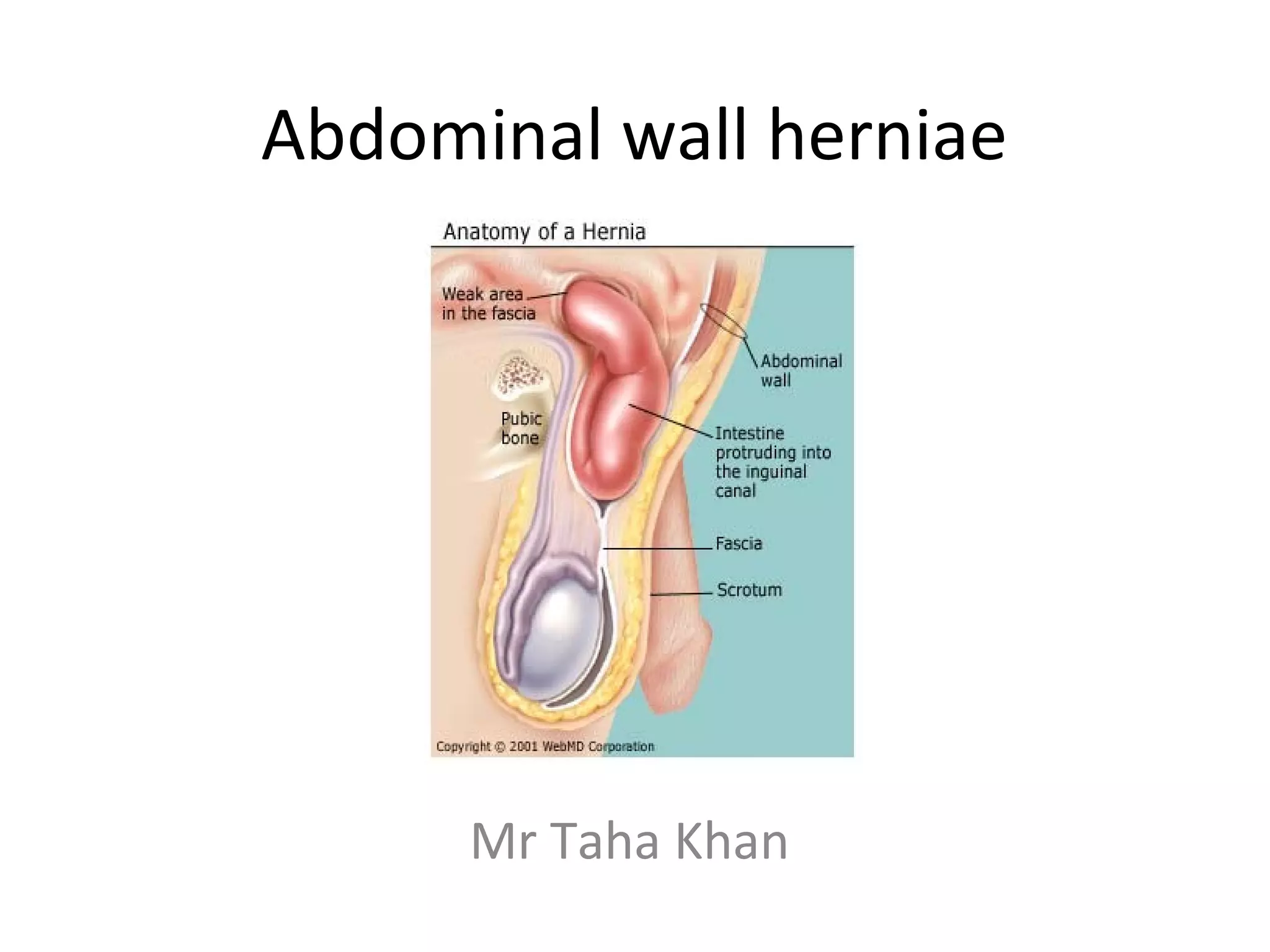
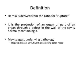
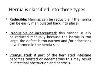
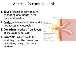
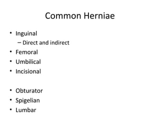
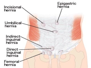
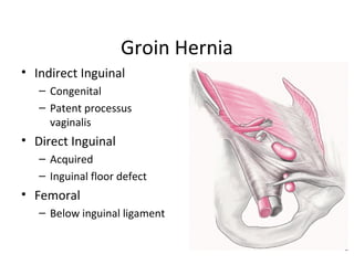
Abdominal wall herniae
Mr Taha Khan
Definition
• Hernia is derived from the Latin for "rupture“
• It is the protrusion of an organ or part of an
organ through a defect in the wall of the cavity
normally containing it.
• May suggest underlying pathology
– Hepatic disease, BPH, COPD, obstructing colon mass
Hernia is classified into three types:
• Reducible: Hernias can be reducible if the hernia
can be easily manipulated back into place.
• Irreducible or incarcerated: this cannot usually
be reduced manually because the hernia is too
large, the defect is too narrow and /or adhesions
have formed in the hernia sac.
• Strangulated: if part of the herniated intestine
becomes twisted or oedematous this may result
in intestinal obstruction and necrosis.
A hernia is composed of:
1.Sac: a folding of peritoneum
consisting of a mouth, neck,
body and fundus.
2.Body: which varies in size and is
not necessarily occupied.
3.Coverings: derived from layers
of the abdominal wall.
4.Contents: which could be
anything from the omentum,
intestines, ovary or urinary
bladder.
Common Herniae
• Inguinal
– Direct and indirect
• Femoral
• Umbilical
• Incisional
• Obturator
• Spigelian
• Lumbar
Pertinent History
• Duration/onset
• Symptoms
– Local
– Obstructive
• Nausea, emesis, pain, distension, obstipation
• Prior Incarceration
• Related comorbidity
– Cough/Urinary flow/Constipation
– Operative risk
Pertinent Exam
• Distension
– Bowel obstruction
• Scars
– Incisional hernias
– Recurrence
– Contraindications for certain approaches
• Rectal--blood/masses
Pertinent Exam
• Location
• Reducible?
• Tender?
• Skin changes?
• Palpable edges
• Genitalia
• Rectal
Groin Anatomy--Anterior
• Inguinal ligament
• Layers
• External ring
• Internal ring
• Spermatic cord
• Inferior epigastric
vessels
• Hesselbach’s triangle
• Femoral vessels
Hesselbach’s Triangle
Groin Anatomy--Posterior
Groin Anatomy--Nerves
Groin Hernia
• Indirect Inguinal
– Congenital
– Patent processus
vaginalis
• Direct Inguinal
– Acquired
– Inguinal floor defect
• Femoral
– Below inguinal ligament
Groin Hernia
• In the UK 93% are inguinal, 7% femoral
• 20% bilateral
• Most common in both sexes indirect.
• Femoral hernias more common in elderly females
• Male to female ratio in 9:1 for inguinal hernias,
1:3 for femoral hernias
Inguinal
• Superficial inguinal ring—1.25 cm
above and lateral to the pubic
tubercle
• Deep inguinal ring—1.25 cm
above and medial to the mid
point of inguinal ligament
• Length of the inguinal canal—
3.25cm
Ingiunal canal Boundaries
MALT: 2M 2A, 2L, 2T:
Superior wall [roof]: 2 Muscles:
• Internal oblique Muscle
• Transverse abdominus Muscle
Anterior wall: 2 Aponeuroses:
• Aponeurosis of external oblique
• Aponeurosis of internal oblique
Lower wall [floor]: 2 Ligaments:
• Inguinal Ligament
• Lacunar Ligament
Posterior wall: 2Ts:
• Transversalis fascia [laterally]
• Conjoint Tendon [medially]
Inguinal canal contents
Ilioinguinal nerve.
Spermatic cord, which contains:
3 arteries:
• Testicular artery.
• Ductus deferens artery.
• Cremasteric artery.
3 nerves:
• Cremasteric nerve.
• Genital branch of the genitofemoral nerve.
• Autonomics
3 other things:
• Ductus deferens
• Pampiniform plexus
• Lymphatics
Nerves
• Major nerves in the region are ilioinguinal,
iliohypogastric, genitofemoral nerves.
• Ilioinguinal provides sensory to pubic region,
upper labia, scrotum. Most commonly injured.
• Iliohypogastric supplies sensory to skin superior
to the pubis.
• Genitofemoral sensory to scrotum and thigh.
Types of inguinal herniae
Inguinal
Indirect or indirect
ď‚—Inguinal hernias can be direct which is herniation
through an area of muscle weakness, in the inguinal
canal,
ď‚—and inguinal hernias indirect herniation through the
inguinal ring. Indirect hernias, the more common form,
can develop at any age but are especially prevalent in
infants younger than age 1. This form is three times more
common in males.
ď‚—Femoral Herniation through the femoral canal
Direct Hernia
Indirect Hernia
Position
•Pt. stands, exposed area visible.
•best performed with the patient standing and in supine
•the physician seated on a stool
Prepare
• Stand at the side of the patient,
• one hand on the patients back to support him.
• hand and arm should be roughly parallel to the inguinal
ligament when palpating the lump.
Examination
• Observation of the groin area in oblique light
• Visible swelling.
• Examine as a mass; (site,skin,size,shape,…)
Mass
Most important
1. Can you get above it?
2. Reducibility test
3. Expansile cough Impulse
4. Invagination test
5. Three finger test
Zieman’s technique
6. Ring occlusion test
Also Assess
• Intra or extra abdominal
• Tension
• Composition
• Percussion and auscultation;
Bowel Sounds
• Always examine both groins
• Transillumination
1-Cough Impulse
•Pt. coughs to highlight hernia.
•May not ;if the neck is blocked by adhesions
•Visible & Palpable cough impulse.
•Reappear on straining, standing or coughing
2-Reducibility test
• Ask the patient to reduce the hernia
• Usually done in the supine position.
Relation to Pubic Tubercle
INGUINAL HERNIA; The neck lies above and medial to
the pubic tubercle
FEMORAL HERNIA; The neck lies below and lateral to
pubic tubercle
3-Get above the swelling test
•Ask the patient to stand
•At the root of the scrotum place the thumb in front
and the index behind
•Try to reach above the swelling.
• Inguinal hernia; cannot get above
• Pure scrotal swelling; will get above
4-Invagination test
•The scrotum on each side is inverted
with the examining index finger
•Entering the inguinal canal along
the course of the cord structures.
•The finger push up to the
superficial inguinal ring.
•The pulp should feel the ring.
•Patient is asked to cough,
•A palpable impulse will confirm the presence of a
hernia;
felt on the pulp then direct
felt on the tip then indirect hernia.
5-Three finger test/Zieman’s technique
Index finger; deep inguinal ring (indirect hernia)
Middle finger; superficial ing. Ring (direct hernia)
Ring finger; saphenous opening (femoral hernia)
The patient is asked to cough.
6-Ring occlusion test
•Reduce the hernia
• Occlude the deep ring with index and middle finger
• Ask the patient to stand and then cough
• If no bulging - indirect
• If bulging - direct .
Complications
• Bowel incarceration (acute, chronic): The trapping of
abdominal contents within the Hernia itself
• Strangulation: pressure on the hernial contents may
compromise blood supply (especially veins, with
their low pressure, are sensitive, and venous
congestion often results) and cause ischemia, and
later necrosis and gangrene, which may become
fatal.
• Small bowel obstruction
• Any hernia that is tender
• Nausea and vomiting;
• Caution if trying to reduce it
manually.
• An acute surgical emergency.
Strangulation
Nyhus Classification
• I indirect, internal ring normal (kids)
• II indirect, dilated internal ring
• III posterior wall defects, direct inguinal hernia,
dilated internal ring, massive scrotal, sliding,
femoral hernia
• IV recurrent hernia
Indications for Operative Repair
• Early repair is justified when potential for
strangulation is weighed against minimal risks for
surgery.
• Not warranted in terminally ill without
incarceration
• Patients with ascites should have it controlled
before surgery
• Incarceration, strangulation
Surgical Techniques
• Open anterior repair (Bassini, McVay, Shouldice).
• Open posterior repair (Nyhus, preperitoneal)
• Tension-free repair with mesh (Liechtenstein,
Rutkow)
• Laparoscopic
Open Anterior Repair
• Transversalis opened, hernia sac ligated, canal
reconstructed using permanent sutures.
• Tension of the repair can lead to recurrence.
Bassini Repair
• Conjoined tendon
(internal oblique,
transverse abdominal,
transversalis fascia)
• Shelving edge Inguinal
ligament
McVay (Cooper’s Ligament)
• Like Bassini but medial
approximation to the
pectineal ligament
• Inguinal and Femoral
hernia repair
• Femoral Vessels
Shouldice Repair
• Imbricated, running
repair
Open Posterior Repair
• Divide the layers of the abdominal wall superior
to the internal ring, enter preperitoneal space.
Dissection continues behind and deep to the
entire inguinal region.
• Suture tension problems.
Tension-Free Repair
• Same initial approach as anterior repair
• Instead of sewing fascial layers together to repair
defect, a prosthetic mesh onlay used
• Simple to learn, easy to perform, suited for local
anesthesia, excellent results with recurrence less
than 4%.
Techniques
• Coined by Liechtenstein in 1989
• Central feature is polypropylene mesh over
unrepaired floor.
• Gilbert repair uses a cone shaped plug placed
thru deep ring.
• Slit placed in mesh for cord structures
Mesh repair
• Tension free
• Less painful
• Foreign body
Kugel Patch
Bard Perfix Plug and Patch
Prolene Hernia System
Techniques
• The genital branch of the femoral nerve, and the
ilioinguinal nerve are allowed to pass through the
newly constructed deep ring.
• Suturing the plug is not necessary.
• Preformed plugs have no advantage over a hand
fashioned one.
Techniques
• Small indirect sacs are dissected and inverted,
large one are transected and ligated.
• Direct sacs are inverted.
• If plugs are placed to repair direct defects, a
mesh only must be placed over the plug to
prevent expulsion.
Laparoscopic Procedures
• Increasingly popular, controversial
• Early in the development, hernias were repaired
by placing very large mesh over entire inguinal
region on top of the peritoneum. Was
abandoned because of contact with bowel.
• Today, most performed TEP or TAPP
Laparoscopic Procedures
• In the TEP procedure, an inflatable balloon is
placed in the preperitoneal space, and the repair
is done preperitoneal. More skill required.
• In both TAPP and TEP, the hernia sac is reduced,
and a large piece of mesh is placed to cover
defects.
Balloon Dissector
Laparoscopic Inguinal Hernia
Technique
Unilateral Preperitoneal repair
Transperitoneal Anatomy
Laparoscopic Procedures
• The argued advantage of these procedures
was less pain and disability, faster return to
work.
• Great for bilateral hernia, with no increase in
morbidity.
• For recurrent hernia
• Disadvantages are cost, time.
Recurrence
Type of repair Recurrence
McVay 9%
Shouldice 7-11%
Liechtenstein 0-4%
Laparoscopic 0-1%
Complications of surgical repair -
Immediate
• Bleeding
• Neurovascular damage
• Damage to the testicular artery leading to
testicular atrophy and possibly orchidectomy
• Injury to the Vas Deferens
• Bowel perforation
• Visceral injury
Complications of surgical repair -
Early
• Urinary retention
• Bleeding
• Discomfort
Complications of surgical repair -
Intermediate
• Infection
• Bleeding
• Discomfort
• DVT/PE
• Chest infection
Complications of surgical repair -
Late
• Chronic discomfort
• Recurrence
Femoral Hernia
ď‚—I. Epidemiology
A. Accounts for 4% of Groin Hernias (96% are
inguinal)
B. More common in elderly women
C. Gender predisposition: Female by 3 to 1 ratio
Femoral herniae are seen less than inguinal
hernia - even in women
ď‚—II. Pathophysiology
A. Associated with increased intra-abdominal
pressure
B. Hernia sac bulges into femoral canal
Femoral Hernia
• Femoral hernias are more common in women,
• Present as a groin lump.
• The cause of unexplained small bowel obstruction.
• An absent Cough impulse
• Globular lump than the pear shaped lump of the inguinal
hernia.
• Differential Diagnoses:
• Inguinal Hernia.
• Femoral Artery Aneurism.
• Femoral Lymphadenopathy.
• Psoas Abscess.
Femoral Hernia Repair
Umbilical Hernia
• Obstruction and strangulation is rare in children
but can be more common in adults
• May remain small and asymptomatic
• Can increase with obesity, pregnancy, ascites,
peritoneal dialysis
Umbilical Hernia
• In infants & children.
• Boys more than girls.
• Tend to resolve without any treatment by around the
age of 5 years.
Umbilical Hernia Repair
Paraumbilical Hernia
• Affects adults.
• Either supra or infraumbilical through
the linea alba.
• The female to male ratio is 20:1.
• Clolicky pain and/or irreducibilty due to
omental adhesions.
Inicisional hernia
I. Pathophysiology
A. Type of Ventral Hernia
B. Develops in scar of prior laparotomy or drain site
C. Risks for postoperative hernia development
1. Vertical scar more commonly affected than horizontal
2. Wound infection
3. Wound dehiscence
4. Malnutrition
5. Obesity
6. Tobacco abuse
Incisional Hernia
• More along a straight line from the sternum down to
the pubis.
• Swelling at the incisional site +/- pain.
Epigastric Hernia
• As result of a defect in the linea alba between the
xiphoid process and umbilicus
• Starts as a protrusion of the extraperitoneal fat
• Swelling +/- pain
Ventral Hernia
• Usually incisional
• May be associated
with adhesions
Laparoscopic Ventral Hernia
Technique
Laparoscopic Ventral Hernia
Technique
Rare external Hernias
Spigelian Hernia:
• Occurs in the spaces of the semilunar line and
the lateral edge of the rectus muscle (inferior to
the arcuate line).
• The posterior rectus sheath is weak
• Preoperative diagnosis is difficult
• USS & CT may be helpful
Lumbar Hernia
• Broad bulging hernia
• Not vulnerable to incarceration.
A. Petit’s hernia: inferior lumbar triangle.
B. Grynfeltt’s Hernia: superior lumbar triangle and is
less common than Petit’s.
Hiatus herniaHiatus hernia
Greater curve
of stomach
Pylorus
Types
Hiatal hernias are classified
according to the position
of the oesophagogastric
junction and the existence
of a true hernia sac.
• Type I (sliding)
– Leading edge of the hernia is the
oesophagogastric junction, which is
displaced into an intrathoracic position.
– The longitudinal axis of the stomach is
aligned with the esophagus.
– There is often no true hernia sac nor is there
any para-oesophageal component.
Types
Type II & Type III are referred to as “para-oesophageal
herniae”.
• Type II (rolling)
– The oesophagogastric junction is in its normal intra-
abdominal location
– The hernia sac (containing portions of the gastric fundus and
body) develops alongside the oesophagus
• Type III
– The oesophagogastric junction is displaced into the thorax
and like a Type II, the hernia sac contains portions of the
gastric fundus or body.
Type II & Type III = Type IV
Management
• Evaluation
– Endoscopy
– Esophageal Motility Studies
– Manometry & pH Monitoring
• 1/3 of pts will have atypical peristalsis of the esophageal
body
• ½ of symptomatic patients will have abnormal pH results
Management
• Indications for Operation
– Type I
– Type II & III
• Associated with a high-risk of complications
• “catastrophic” in 20 – 30% of pts
• Symptoms do not predict risk…
Management
• Findings that may prompt surgery
(even in those patients that are “not
optimal”)
– Symptoms of obstruction
– Reflux
– Anemia
• Trying to avoid:
– Further aspiration
– Hemorrhage
– Transfusion requirements
Surgical Techniques
• Principles similar to other hernia operations
• Need to anchor the stomach
• The need for fundoplication varies
• Transthoracic vs. Transabdominal…

Abdominal wall herniae

  • 1.
  • 2.
    Definition • Hernia isderived from the Latin for "rupture“ • It is the protrusion of an organ or part of an organ through a defect in the wall of the cavity normally containing it. • May suggest underlying pathology – Hepatic disease, BPH, COPD, obstructing colon mass
  • 3.
    Hernia is classifiedinto three types: • Reducible: Hernias can be reducible if the hernia can be easily manipulated back into place. • Irreducible or incarcerated: this cannot usually be reduced manually because the hernia is too large, the defect is too narrow and /or adhesions have formed in the hernia sac. • Strangulated: if part of the herniated intestine becomes twisted or oedematous this may result in intestinal obstruction and necrosis.
  • 4.
    A hernia iscomposed of: 1.Sac: a folding of peritoneum consisting of a mouth, neck, body and fundus. 2.Body: which varies in size and is not necessarily occupied. 3.Coverings: derived from layers of the abdominal wall. 4.Contents: which could be anything from the omentum, intestines, ovary or urinary bladder.
  • 5.
    Common Herniae • Inguinal –Direct and indirect • Femoral • Umbilical • Incisional • Obturator • Spigelian • Lumbar
  • 7.
    Pertinent History • Duration/onset •Symptoms – Local – Obstructive • Nausea, emesis, pain, distension, obstipation • Prior Incarceration • Related comorbidity – Cough/Urinary flow/Constipation – Operative risk
  • 8.
    Pertinent Exam • Distension –Bowel obstruction • Scars – Incisional hernias – Recurrence – Contraindications for certain approaches • Rectal--blood/masses
  • 9.
    Pertinent Exam • Location •Reducible? • Tender? • Skin changes? • Palpable edges • Genitalia • Rectal
  • 10.
    Groin Anatomy--Anterior • Inguinalligament • Layers • External ring • Internal ring • Spermatic cord • Inferior epigastric vessels • Hesselbach’s triangle • Femoral vessels
  • 11.
  • 12.
  • 13.
  • 14.
    Groin Hernia • IndirectInguinal – Congenital – Patent processus vaginalis • Direct Inguinal – Acquired – Inguinal floor defect • Femoral – Below inguinal ligament
  • 15.
    Groin Hernia • Inthe UK 93% are inguinal, 7% femoral • 20% bilateral • Most common in both sexes indirect. • Femoral hernias more common in elderly females • Male to female ratio in 9:1 for inguinal hernias, 1:3 for femoral hernias
  • 16.
    Inguinal • Superficial inguinalring—1.25 cm above and lateral to the pubic tubercle • Deep inguinal ring—1.25 cm above and medial to the mid point of inguinal ligament • Length of the inguinal canal— 3.25cm
  • 17.
    Ingiunal canal Boundaries MALT:2M 2A, 2L, 2T: Superior wall [roof]: 2 Muscles: • Internal oblique Muscle • Transverse abdominus Muscle Anterior wall: 2 Aponeuroses: • Aponeurosis of external oblique • Aponeurosis of internal oblique Lower wall [floor]: 2 Ligaments: • Inguinal Ligament • Lacunar Ligament Posterior wall: 2Ts: • Transversalis fascia [laterally] • Conjoint Tendon [medially]
  • 18.
    Inguinal canal contents Ilioinguinalnerve. Spermatic cord, which contains: 3 arteries: • Testicular artery. • Ductus deferens artery. • Cremasteric artery. 3 nerves: • Cremasteric nerve. • Genital branch of the genitofemoral nerve. • Autonomics 3 other things: • Ductus deferens • Pampiniform plexus • Lymphatics
  • 19.
    Nerves • Major nervesin the region are ilioinguinal, iliohypogastric, genitofemoral nerves. • Ilioinguinal provides sensory to pubic region, upper labia, scrotum. Most commonly injured. • Iliohypogastric supplies sensory to skin superior to the pubis. • Genitofemoral sensory to scrotum and thigh.
  • 20.
    Types of inguinalherniae Inguinal Indirect or indirect ď‚—Inguinal hernias can be direct which is herniation through an area of muscle weakness, in the inguinal canal, ď‚—and inguinal hernias indirect herniation through the inguinal ring. Indirect hernias, the more common form, can develop at any age but are especially prevalent in infants younger than age 1. This form is three times more common in males. ď‚—Femoral Herniation through the femoral canal
  • 22.
  • 23.
  • 24.
    Position •Pt. stands, exposedarea visible. •best performed with the patient standing and in supine •the physician seated on a stool Prepare • Stand at the side of the patient, • one hand on the patients back to support him. • hand and arm should be roughly parallel to the inguinal ligament when palpating the lump. Examination
  • 25.
    • Observation ofthe groin area in oblique light • Visible swelling. • Examine as a mass; (site,skin,size,shape,…) Mass
  • 26.
    Most important 1. Canyou get above it? 2. Reducibility test 3. Expansile cough Impulse 4. Invagination test 5. Three finger test Zieman’s technique 6. Ring occlusion test
  • 27.
    Also Assess • Intraor extra abdominal • Tension • Composition • Percussion and auscultation; Bowel Sounds • Always examine both groins • Transillumination
  • 28.
    1-Cough Impulse •Pt. coughsto highlight hernia. •May not ;if the neck is blocked by adhesions •Visible & Palpable cough impulse. •Reappear on straining, standing or coughing
  • 29.
    2-Reducibility test • Askthe patient to reduce the hernia • Usually done in the supine position.
  • 30.
    Relation to PubicTubercle INGUINAL HERNIA; The neck lies above and medial to the pubic tubercle FEMORAL HERNIA; The neck lies below and lateral to pubic tubercle
  • 31.
    3-Get above theswelling test •Ask the patient to stand •At the root of the scrotum place the thumb in front and the index behind •Try to reach above the swelling. • Inguinal hernia; cannot get above • Pure scrotal swelling; will get above
  • 32.
    4-Invagination test •The scrotumon each side is inverted with the examining index finger •Entering the inguinal canal along the course of the cord structures. •The finger push up to the superficial inguinal ring. •The pulp should feel the ring. •Patient is asked to cough, •A palpable impulse will confirm the presence of a hernia; felt on the pulp then direct felt on the tip then indirect hernia.
  • 34.
    5-Three finger test/Zieman’stechnique Index finger; deep inguinal ring (indirect hernia) Middle finger; superficial ing. Ring (direct hernia) Ring finger; saphenous opening (femoral hernia) The patient is asked to cough.
  • 35.
    6-Ring occlusion test •Reducethe hernia • Occlude the deep ring with index and middle finger • Ask the patient to stand and then cough • If no bulging - indirect • If bulging - direct .
  • 36.
    Complications • Bowel incarceration(acute, chronic): The trapping of abdominal contents within the Hernia itself • Strangulation: pressure on the hernial contents may compromise blood supply (especially veins, with their low pressure, are sensitive, and venous congestion often results) and cause ischemia, and later necrosis and gangrene, which may become fatal. • Small bowel obstruction
  • 37.
    • Any herniathat is tender • Nausea and vomiting; • Caution if trying to reduce it manually. • An acute surgical emergency. Strangulation
  • 38.
    Nyhus Classification • Iindirect, internal ring normal (kids) • II indirect, dilated internal ring • III posterior wall defects, direct inguinal hernia, dilated internal ring, massive scrotal, sliding, femoral hernia • IV recurrent hernia
  • 39.
    Indications for OperativeRepair • Early repair is justified when potential for strangulation is weighed against minimal risks for surgery. • Not warranted in terminally ill without incarceration • Patients with ascites should have it controlled before surgery • Incarceration, strangulation
  • 40.
    Surgical Techniques • Openanterior repair (Bassini, McVay, Shouldice). • Open posterior repair (Nyhus, preperitoneal) • Tension-free repair with mesh (Liechtenstein, Rutkow) • Laparoscopic
  • 41.
    Open Anterior Repair •Transversalis opened, hernia sac ligated, canal reconstructed using permanent sutures. • Tension of the repair can lead to recurrence.
  • 42.
    Bassini Repair • Conjoinedtendon (internal oblique, transverse abdominal, transversalis fascia) • Shelving edge Inguinal ligament
  • 43.
    McVay (Cooper’s Ligament) •Like Bassini but medial approximation to the pectineal ligament • Inguinal and Femoral hernia repair • Femoral Vessels
  • 44.
  • 45.
    Open Posterior Repair •Divide the layers of the abdominal wall superior to the internal ring, enter preperitoneal space. Dissection continues behind and deep to the entire inguinal region. • Suture tension problems.
  • 46.
    Tension-Free Repair • Sameinitial approach as anterior repair • Instead of sewing fascial layers together to repair defect, a prosthetic mesh onlay used • Simple to learn, easy to perform, suited for local anesthesia, excellent results with recurrence less than 4%.
  • 49.
    Techniques • Coined byLiechtenstein in 1989 • Central feature is polypropylene mesh over unrepaired floor. • Gilbert repair uses a cone shaped plug placed thru deep ring. • Slit placed in mesh for cord structures
  • 50.
    Mesh repair • Tensionfree • Less painful • Foreign body
  • 51.
  • 52.
  • 53.
  • 54.
    Techniques • The genitalbranch of the femoral nerve, and the ilioinguinal nerve are allowed to pass through the newly constructed deep ring. • Suturing the plug is not necessary. • Preformed plugs have no advantage over a hand fashioned one.
  • 55.
    Techniques • Small indirectsacs are dissected and inverted, large one are transected and ligated. • Direct sacs are inverted. • If plugs are placed to repair direct defects, a mesh only must be placed over the plug to prevent expulsion.
  • 56.
    Laparoscopic Procedures • Increasinglypopular, controversial • Early in the development, hernias were repaired by placing very large mesh over entire inguinal region on top of the peritoneum. Was abandoned because of contact with bowel. • Today, most performed TEP or TAPP
  • 57.
    Laparoscopic Procedures • Inthe TEP procedure, an inflatable balloon is placed in the preperitoneal space, and the repair is done preperitoneal. More skill required. • In both TAPP and TEP, the hernia sac is reduced, and a large piece of mesh is placed to cover defects.
  • 58.
  • 59.
  • 60.
  • 61.
  • 62.
    Laparoscopic Procedures • Theargued advantage of these procedures was less pain and disability, faster return to work. • Great for bilateral hernia, with no increase in morbidity. • For recurrent hernia • Disadvantages are cost, time.
  • 63.
    Recurrence Type of repairRecurrence McVay 9% Shouldice 7-11% Liechtenstein 0-4% Laparoscopic 0-1%
  • 64.
    Complications of surgicalrepair - Immediate • Bleeding • Neurovascular damage • Damage to the testicular artery leading to testicular atrophy and possibly orchidectomy • Injury to the Vas Deferens • Bowel perforation • Visceral injury
  • 65.
    Complications of surgicalrepair - Early • Urinary retention • Bleeding • Discomfort
  • 66.
    Complications of surgicalrepair - Intermediate • Infection • Bleeding • Discomfort • DVT/PE • Chest infection
  • 67.
    Complications of surgicalrepair - Late • Chronic discomfort • Recurrence
  • 68.
    Femoral Hernia ď‚—I. Epidemiology A.Accounts for 4% of Groin Hernias (96% are inguinal) B. More common in elderly women C. Gender predisposition: Female by 3 to 1 ratio Femoral herniae are seen less than inguinal hernia - even in women ď‚—II. Pathophysiology A. Associated with increased intra-abdominal pressure B. Hernia sac bulges into femoral canal
  • 69.
    Femoral Hernia • Femoralhernias are more common in women, • Present as a groin lump. • The cause of unexplained small bowel obstruction. • An absent Cough impulse • Globular lump than the pear shaped lump of the inguinal hernia. • Differential Diagnoses: • Inguinal Hernia. • Femoral Artery Aneurism. • Femoral Lymphadenopathy. • Psoas Abscess.
  • 70.
  • 71.
    Umbilical Hernia • Obstructionand strangulation is rare in children but can be more common in adults • May remain small and asymptomatic • Can increase with obesity, pregnancy, ascites, peritoneal dialysis
  • 72.
    Umbilical Hernia • Ininfants & children. • Boys more than girls. • Tend to resolve without any treatment by around the age of 5 years.
  • 73.
  • 74.
    Paraumbilical Hernia • Affectsadults. • Either supra or infraumbilical through the linea alba. • The female to male ratio is 20:1. • Clolicky pain and/or irreducibilty due to omental adhesions.
  • 75.
    Inicisional hernia I. Pathophysiology A.Type of Ventral Hernia B. Develops in scar of prior laparotomy or drain site C. Risks for postoperative hernia development 1. Vertical scar more commonly affected than horizontal 2. Wound infection 3. Wound dehiscence 4. Malnutrition 5. Obesity 6. Tobacco abuse
  • 76.
    Incisional Hernia • Morealong a straight line from the sternum down to the pubis. • Swelling at the incisional site +/- pain.
  • 77.
    Epigastric Hernia • Asresult of a defect in the linea alba between the xiphoid process and umbilicus • Starts as a protrusion of the extraperitoneal fat • Swelling +/- pain
  • 78.
    Ventral Hernia • Usuallyincisional • May be associated with adhesions
  • 79.
  • 80.
  • 81.
  • 83.
    Rare external Hernias SpigelianHernia: • Occurs in the spaces of the semilunar line and the lateral edge of the rectus muscle (inferior to the arcuate line). • The posterior rectus sheath is weak • Preoperative diagnosis is difficult • USS & CT may be helpful
  • 85.
    Lumbar Hernia • Broadbulging hernia • Not vulnerable to incarceration. A. Petit’s hernia: inferior lumbar triangle. B. Grynfeltt’s Hernia: superior lumbar triangle and is less common than Petit’s.
  • 86.
    Hiatus herniaHiatus hernia Greatercurve of stomach Pylorus
  • 87.
    Types Hiatal hernias areclassified according to the position of the oesophagogastric junction and the existence of a true hernia sac. • Type I (sliding) – Leading edge of the hernia is the oesophagogastric junction, which is displaced into an intrathoracic position. – The longitudinal axis of the stomach is aligned with the esophagus. – There is often no true hernia sac nor is there any para-oesophageal component.
  • 88.
    Types Type II &Type III are referred to as “para-oesophageal herniae”. • Type II (rolling) – The oesophagogastric junction is in its normal intra- abdominal location – The hernia sac (containing portions of the gastric fundus and body) develops alongside the oesophagus • Type III – The oesophagogastric junction is displaced into the thorax and like a Type II, the hernia sac contains portions of the gastric fundus or body.
  • 90.
    Type II &Type III = Type IV
  • 91.
    Management • Evaluation – Endoscopy –Esophageal Motility Studies – Manometry & pH Monitoring • 1/3 of pts will have atypical peristalsis of the esophageal body • ½ of symptomatic patients will have abnormal pH results
  • 92.
    Management • Indications forOperation – Type I – Type II & III • Associated with a high-risk of complications • “catastrophic” in 20 – 30% of pts • Symptoms do not predict risk…
  • 93.
    Management • Findings thatmay prompt surgery (even in those patients that are “not optimal”) – Symptoms of obstruction – Reflux – Anemia • Trying to avoid: – Further aspiration – Hemorrhage – Transfusion requirements
  • 94.
    Surgical Techniques • Principlessimilar to other hernia operations • Need to anchor the stomach • The need for fundoplication varies • Transthoracic vs. Transabdominal…