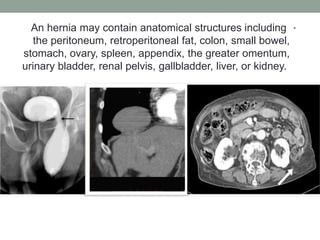
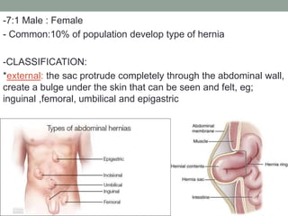
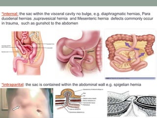
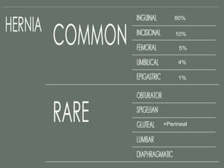
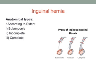
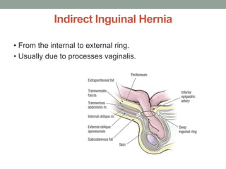
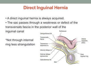
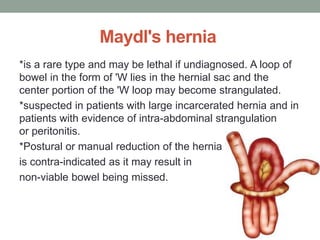
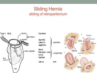
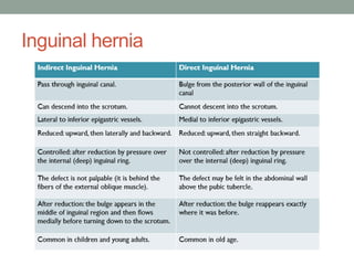
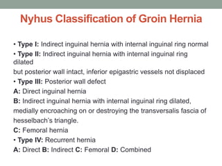
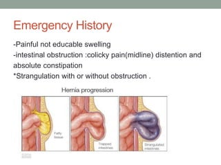
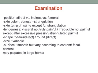
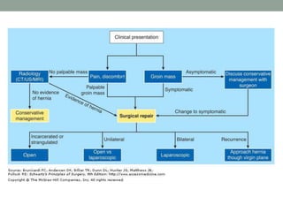
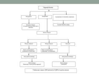
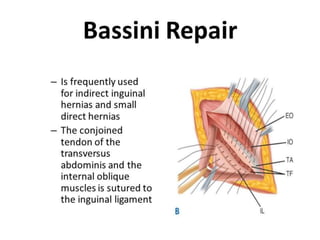
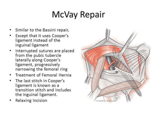
Call Girls Thane Just Call 9910780858 Get High Class Call Girls Service
Â
Hernias
1. PROTRUSION OF A VISCUS OR A PART OF VISCUS
THROUGH A NORMAL OR ABNORMAL OPENING
IN THE WALLS OF
ITS CONTAINING CAVITY
The term “hernia” is derived from the Greek
word hernios, which means “budding.”
By Marwan Nassar
2. Etiologies
• Increased abdominal pressure
– Cough, urinary trouble, constipation, straining, ascites,
intra abdominal malignancies, pregnancy
• Weakness of abdominal wall
– Congenital
• Patent processes vaginalis, patent canal of nuck in
females
– Acquired
• Excess fat (obesity)
• Post Pregnancy
• Surgical incisions
• Connective tissue disorders like Marfan’s syndrome
3. Composition of a hernia
• A hernia consists of three parts –
– the sac
– the coverings of the sac
– the contents of the sac
• The sac is a diverticulum of peritoneum, consisting of
– Mouth
– Neck
– Body
– Fundus
• The coverings are derived from the layers of the
abdominal wall through which the sac passes.
4. Contents
• omentum = omentocele;
• intestine = enterocele; more commonly small
bowel but may be large intestine or appendix;
• a portion of the circumference of the intestine =
Richter’s hernia;
• a portion of the bladder (or a diverticulum);
• ovary with or without the corresponding fallopian
tube;
• a Meckel’s diverticulum = a Littre’s hernia;
• fluid, as part of ascites or peritoneal fluid.
5. •
An hernia may contain anatomical structures including
the peritoneum, retroperitoneal fat, colon, small bowel,
stomach, ovary, spleen, appendix, the greater omentum,
urinary bladder, renal pelvis, gallbladder, liver, or kidney.
6. -7:1 Male : Female
- Common:10% of population develop type of hernia
-CLASSIFICATION:
*external: the sac protrude completely through the abdominal wall,
create a bulge under the skin that can be seen and felt, eg;
inguinal ,femoral, umbilical and epigastric
7. *internal :the sac within the visceral cavity no bulge, e.g. diaphragmatic hernias, Para
duodenal hernias ,supravesical hernia and Mesenteric hernia defects commonly occur
in trauma, such as gunshot to the abdomen
the sac is contained within the abdominal wall e.g. spigelian hernia
:
*intraparital
10. inguinal triangle (Hesselbach’s triangle)
Borders
The inguinal triangle is located within the inferimedial aspect of the
abdominal wall. It has the following boundaries:
Medial – lateral border of the rectus abdominals muscle.
Lateral – inferior epigastric vessels.
Inferior – inguinal ligament.
Contents
transversals fascia,
which forms the floor of this triangle,
A weakness or defect transversals fascia
is a potential weakness in the abdominal wall
11. triangle of Grynfeltt
The superior lumbar triangle, is one of the locations for a
lumbar hernia.
Boundaries
medially: the quadratus lumborum muscle
superiorly: twelfth rib
laterally: internal oblique muscle
floor: transversals fascia and the aponeurosis of
the transverses abdominal muscle
roof: external oblique and
latissimus dorsi muscle
12. Petit triangle
•
composed of the iliac crest inferiorly and the margins of
external oblique m. (anteriorly). The floor of the inferior
lumbar triangle is the internal oblique m .
13. The occurrence of an inguinal hernia
in boys is related to the development
and descent of the testes. The testes
develop within the abdomen and at
around the seventh month of
pregnancy, they descend into the
scrotum. On their way through the
abdominal wall, they pass through the
inguinal canal. After they reach the
scrotum, the opening behind should
close.
-Failure to close adequately results in
a hernia with an opening remaining in
the abdominal wall at this
point.
-Short canal
-Not oblique
Indirect hernia
14. Inguinal canal
Anterior wall: external oblique aponeurosis throughout the
length of the canal; its lateral part is reinforced by muscle
fibers of the internal oblique.
Posterior wall: transversals fascia; its medial part is
reinforced by pubic attachments of the internal oblique and
transverses abdominals aponeuroses that frequently merge to
variable extents into a common tendon—the inguinal falx
(conjoint tendon)—and the reflected inguinal ligament.
Roof: laterally by the transversals fascia, centrally by
musculoaponeurotic arches of the internal oblique and
transverses abdominals, and medially by the medial crus of the
external oblique aponeurosis.
Floor: laterally by the iliopubic tract, centrally by gutter formed
by the infolded inguinal ligament, and medially by the lacunar
ligament.
16. Inguinal hernia
-The most common site
-Male: female ratio 25:1 /testicular descend wide canal(indirect)
-Male: indirect> direct
-Female: direct is rare
-Cause 15-20% of intestinal obstruction
-female predisposition to femoral hernia wide true pelvic and
femoral canal
-connective tissue destruction(aponeurosis and fascia)
*pressure=physical stress
*Smoking *aging *connective tissue disease
*collagen and elastin alteration *distention *ascites
*peritoneal *dialysis *Heavy lifting
*Physical exertion *upright position
18. Inguinal hernia
According to its site of Exit :
i) Indirect hernia.
ii) Direct hernia.
Indirect(oblique) Hernia :
• 80 % of cases
• Almost all pediatric and women cases comprise this
group
• Often a complete variety
• Two forms Congenital and Acquired
Congenital
1) Congenital vaginal(complete)
2) congenital funicular
Acquired
Differentiated from above by as it wont form complete hernia
19. Inguinal hernia
According to its contents:
1) Enterocele
2) Epiplocele or Omentocele
3) Cystocele
Clinical types:
i) Reducible
ii) Irreducible
iii) Obstructed or Incarcerated (irreducibility + obstruction)
iv) Strangulated
v) Inflammed
21. Direct Inguinal Hernia
• A direct inguinal hernia is always acquired.
• The sac passes through a weakness or defect of the
transversals fascia in the posterior wall of the
inguinal canal
*Not through internal
ring less strangulation
22. Maydl's hernia
*is a rare type and may be lethal if undiagnosed. A loop of
bowel in the form of 'W lies in the hernial sac and the
center portion of the 'W loop may become strangulated.
*suspected in patients with large incarcerated hernia and in
patients with evidence of intra-abdominal strangulation
or peritonitis.
*Postural or manual reduction of the hernia
is contra-indicated as it may result in
non-viable bowel being missed.
25. Nyhus Classification of Groin Hernia
• Type I: Indirect inguinal hernia with internal inguinal ring normal
• Type II: Indirect inguinal hernia with internal inguinal ring
dilated
but posterior wall intact, inferior epigastric vessels not displaced
• Type III: Posterior wall defect
A: Direct inguinal hernia
B: Indirect inguinal hernia with internal inguinal ring dilated,
medially encroaching on or destroying the transversalis fascia of
hesselbach’s triangle.
C: Femoral hernia
• Type IV: Recurrent hernia
A: Direct B: Indirect C: Femoral D: Combined
26. Diagnosis
History
-Age :at any age
-Occupation: high physical effort
-Local symptoms notice swelling(asymptomativ) or with
/dragging or aching /pain.
-Abdominal symptom :change bowel habit
-Other disease increase straining : left colon carcinoma
,diverticulosis , COPD and UC
27. Emergency History
-Painful not educable swelling
-intestinal obstruction :colicky pain(midline) distention and
absolute constipation
*Strangulation with or without obstruction .
28. Examination
-position :direct vs. indirect vs. femoral
-skin color :redness =strangulation
-skin temp :in same except for strangulation
-tenderness: visceral not truly painful / irreducible not painful
except after excessive pressing/strangulated painful
-shape :pear(indirect) / round (direct)
-size : variable
-surface : smooth but vary according to content/ fecal
content
may palpated in large hernia
31. Inguinal hernia in female
-Hydrocele of canal of nuck
smooth ,fluctuant, transilluminate
-hematoma of round ligament
Resolve after pregnancy
Reducible in size
Cough impulse +ve
37. Surgical Treatment
• Herniotomy (excision of hernia sac)
– Usually done in children
• Herniorrhaphy (herniotomy with strengthening of the posterior
wall)
– Bassini repair
– Shouldice repair
– McVay repair
• Hernioplasty (herniorrhaphy with application of prosthesis)
– Lichtenstein repair
– Plug and patch repair
• Laparoscopic repair
– TEP (Total Extra Peritoneal)
– TAPP (Trans Abdominal PrePeritoneal)
38.
39.
40.
41.
42.
43. Shouldice Repair
• Multilayer repair of the posterior wall of the inguinal canal
• Double breasting of transversals fascia
• Transverse abdominals aponeurotic arch to the iliopubic
tract
and Conjoined tendon to the inguinal ligament
44. Lichtenstein Repair
• A piece of prosthetic nonabsorbable mesh is placed to fit
the canal
• The mesh is sutured to the aponeurotic tissue overlying
the pubic bone medially, continuine superiorly along the
transversus abdominis or conjoined tendon.
• The inferolateral edge of the mesh is sutured to the
inguinal ligament
45. Surgery complications
Immediate Complications
• Primary hemorrhage
• Basal atelectasis
• Shock
• Complications of Anasthesia
Early Complications
• Secondary hemorrhage
• Wound dehiscence
• Urinary tract infection
• Postoperative wound infection
• Bowel obstruction due to fibrinous
adhesions
Delayed
• Bowel obstruction due to fibrous
adhesions
• Persistent sinus
• Recurrence
• Keloid formation
46. Femoral Hernia
• Femoral hernia enters the femoral ring,
traverses the femoral canal and comes out
through the saphenous opening.
• The female to
male ratio is
about 2:1.
• The right side is
affected twice
as often as the
left.
47. Femoral Ring
Boundaries
• Anteriorly inguinal Ligament
• Posteriorly: Iliopectineal ligament, pubic bone
and fascia over pectinious muscle
• Medially lacunar Ligament
• Laterally septum separating from
femoral vein.
• Oval opening ½” to 1” in diameter
48. Femoral Canal
• Most medial compartment of femoral sheath.
• Conical in shape, 1.25 cm long and 1.25 cm wide at base
(Femoral ring).
• Extends from Femoral ring (above) up to saphenous
opening
(below).
• Contents- fats, lymphatic and lymph nodes of Cloquet.
49. Femoral hernia
-uncommon 2.5%
-history :
age >50 rare in children
female >male 2:1 wide canal
-examination :
*Position below inguinal ligament lateral to pubic tubercle
*normal skin and temp. even if strangulated thick walled superficial
fascia and sac
*composition :firm/ thick wall
*irreducible
*narrow neck :high incarceration 45% after 21 months
*content : extraperitoneal fat , or peritoneal sac or bowel
*right side 2:1/ left side sigmoidal colon
*no cough impulse
50. Surgical Treatment
• The low (Lockwood) operation- the inguinal
ligament to the iliopectineal ligament.
• The high (McEvedy) operation- the conjoint
tendon to the iliopectineal ligament.
• Lotheissen’s operation- the conjoint tendon
or inguinal ligament to the iliopectineal
ligament through inguinal canal
51. Congenital Umbilical hernia
Congenital-Infantile umbilical hernias result from failure of
the umbilical ring to close. The umbilical cord structures fail
to fuse with the umbilical foramen, therefore leaving a
patent umbilical ring. In contrast, anterior abdominal wall
defects such as gastroschisis and omphalocele result
from disruption in the development of the abdominal wall
structures without skin coverage .
52. Congenital Umbilical hernia
History
-age: at birth Start small
-ethnicity : more common in african-caribbian
-symptoms :very rare /parental anxiety
Examination
-hemispherical -palpable defect
-Soft -reducible -compressible resonant
+ve cough impulse – content Omentum, small bowel, and colon
Management
with observation; these defects typically close by age 4. Any
defects that persist beyond this age should undergo surgical
repair.
53. Acquired Umbilical hernias
paraumbilical
History
Age :middle and old age
Female>male: obesity /parity
symptom :is pain at the umbilicus (44% of cases) increase with standing and
exercise. Other complaints include pressure (20%) and nausea and vomiting
(9%).
examination
*Content; peritoneal fat and omentum may contain bowel resonant
*Surface :smooth easily defined edges
*Crescent shaped/ difficult to clean ompholith + foul smell
*+ve cough impulse *
*Reducible but may adhere
* Complications such as irreducibility, obstruction, strangulation, skin
ulceration, and rupture /small with a narrow neck, a configuration that
increases the risk of strangulation and incarceration.
*Look for cause of distension
55. epigastric hernia
-Site between the xiphoid process and umbilicus
- content extraperitonial fat and sometime peritoneal sac
-Symptoms epigastric pain associate with eating -
-Palpate superficially not deep
-Firm consistency
-Usually Not reducible usually –ve cough impulse
-Not lipoma its position in midline
56. incisional hernia
caused by an incompletely-healed surgical wound.
Since median incisions in the abdomen are frequent for
abdominal exploratory surgery , ventral incisional hernias
are often also classified as ventral hernias due to their
location.
57. incisional hernia
History
-surgery or wound
-wound hematoma and infection
-obesity and steroid
-chronic cough
-age mainly elderly
-symptom :lump and pain can cause strangulation and
Obstruction
Examinaion
+ve cough impulse
Beneath old scar
Adherent usually
Not reducible
Appear during first year of surgery but may during 15 years
Note :hematoma ,chronic
abscess ,and cancer met
appear after surgery
58. Lumber hernia
Lumbar hernias are rare They are classified as congenital, generally associated with other
malformations, or acquired, manifesting in adults spontaneously or secondary to trauma or
surgical incision.
Lumbar hernia may occur in two weak areas of the poster lateral abdominal wall: the superior
lumbar triangle of Grynfeltt, which is the more common site, and the inferior lumbar triangle of
Petit. In large hernias the defect wall can affect all of the lumbar region.
Symptomatology frequently consists of only lower back pain. Small hernias may be
asymptomatic except for a palpable mass. In less than 10% of cases, the onset is acute with
bowel obstruction
Anamnesis is helpful for diagnosis in post-traumatic or postsurgical lumbar hernias while in
spontaneous adult hernias, misdiagnosis may occur.
Clinical suspicion is fundamental to guide imaging diagnosis because extraperitoneal fat
herniated through a wall defect may mimic a lipoma. Computed tomography (CT) or magnetic
resonance imaging (MRI) in patients with a suspected hernia can confirm the diagnosis adding
information on parietal defect size, hernia content and muscular trophism.
The hernia may contain retroperitoneal fat, kidney, colon or less commonly small bowel,
omentum, ovary, spleen or appendix. On auscultation, bowel sounds may be audible over the
swelling if the hernia contains bowel loops
Conclusion
Although a rare pathology, knowledge of lumbar hernia is important to avoid misdiagnosis as
lipoma . Ultrasound and CT may confirm the diagnosis. Appropriate surgical treatment should
be planned on the basis of etiology and hernia size.