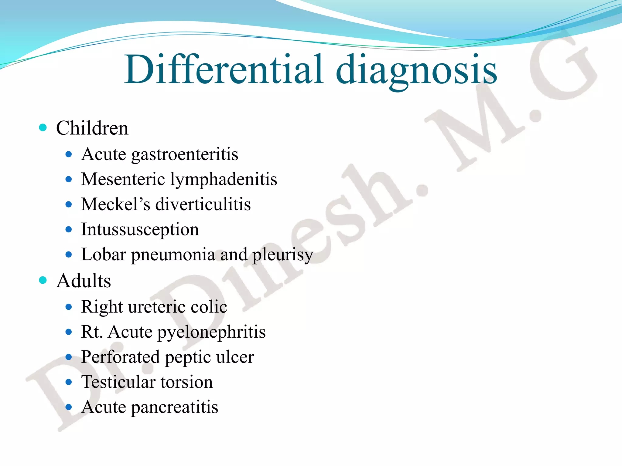
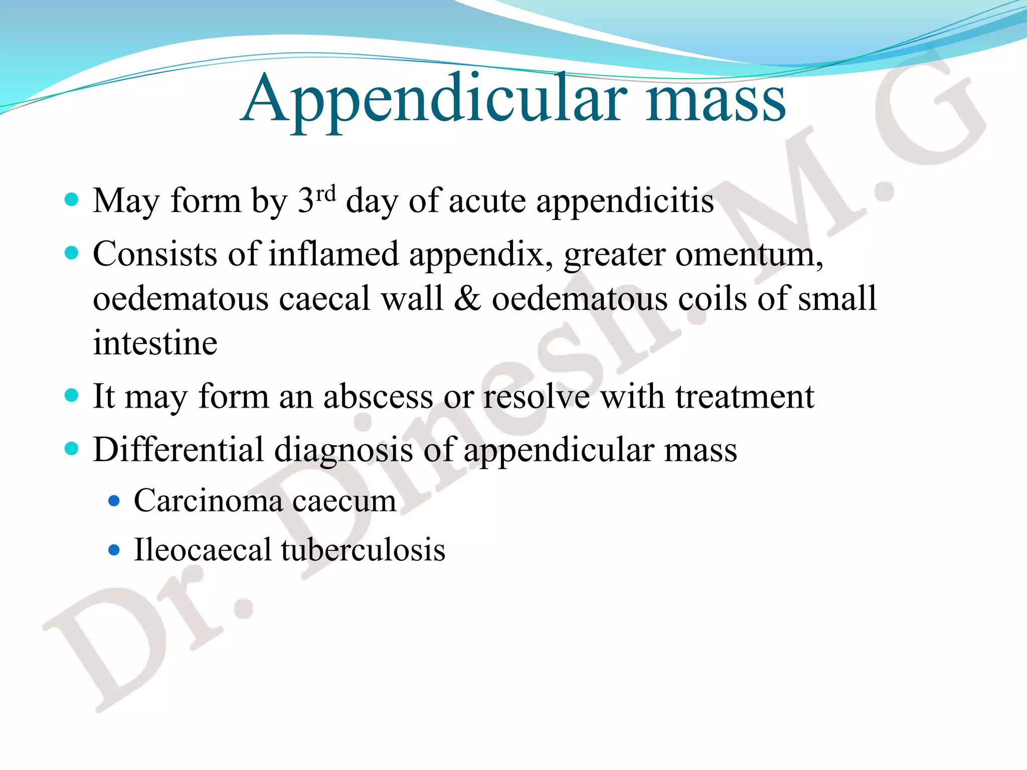
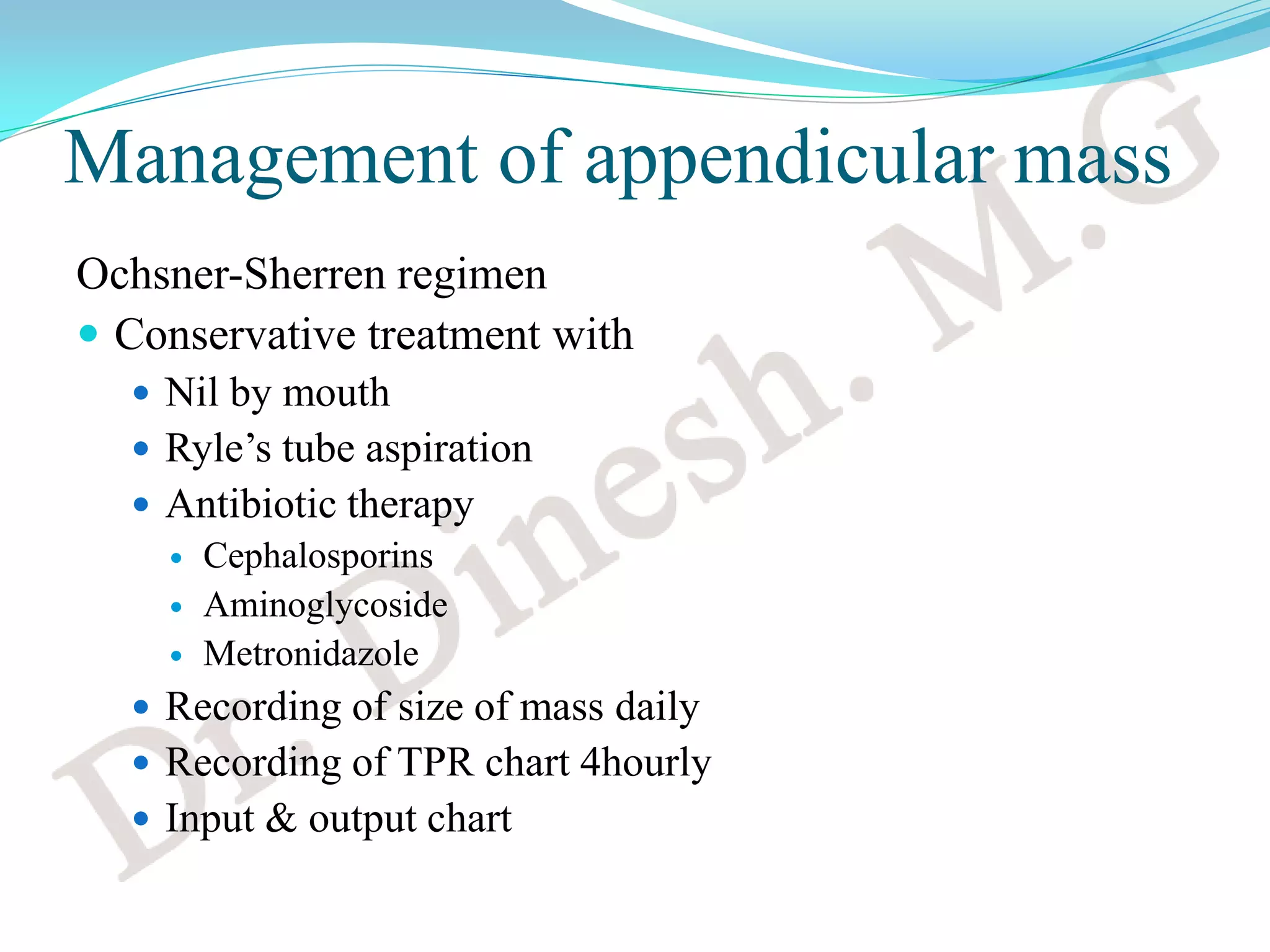
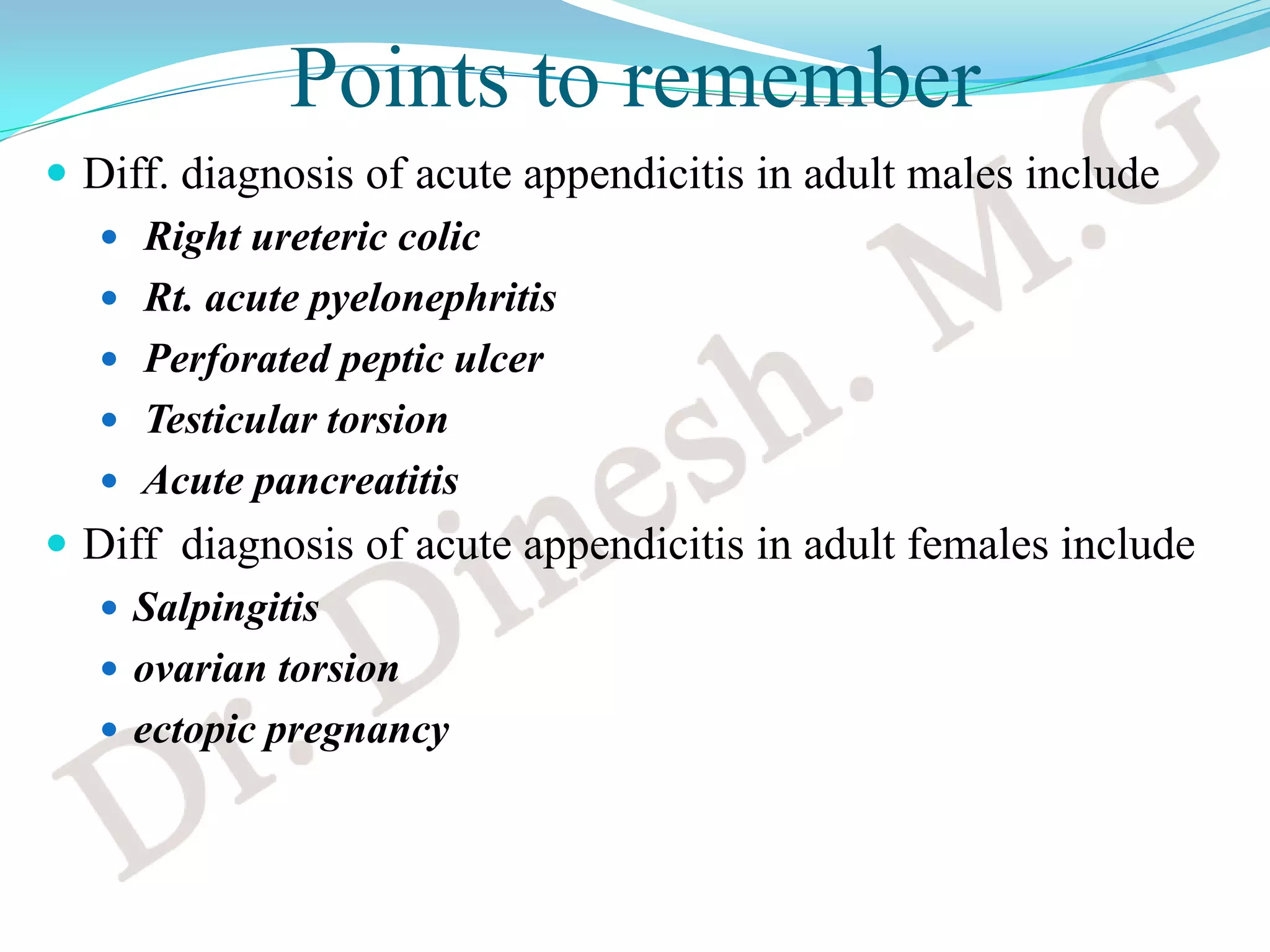
Dr. Dinesh. M.G
Professor of Surgery
J.J.M.M.C.
Davangere
Introduction
 Considered by most to be a vestigial organ
 Its importance in surgery
 Acute appendicitis
 Carcinoid tumour
Anatomy
 Blind muscular tube
 Mucosa, submucosa, muscularis propria and serosa
 Positions
 Retrocaecal 74%
 Pelvic 21%
 Paracaecal 2%
 Pre ileal 1%
 Post ileal 0.5%
 Subcaecal 1.5%
Anatomy
 Appendicular artery runs in mesoappendix
 It is an ‘end-artery’ & a branch of ileocolic artery
 Microscopic anatomy
 Lined by columnar cell of colonic type 
 Crypts are present in the mucosa in which lie the
argentaffin (Kultschitzsky) cells
Acute appendicitis
 Reginald Fitz first published paper on appendicular
perforation in 1886
 Charles McBurney described clinical features including
the point of maximum tenderness in Rt. iliac fossa
 McBurney’s point
Aetiology
 No definite single aetiology
 Obstruction of lumen by
 Faecolith
 Pin worms
 Carcinoma caecum etc.
Clinical features
Murphy’s triad
 Periumbilical pain shifting to Rt.iliac fossa
 Anorexia, nausea and vomiting
 Fever
Signs
 Tachycardia, pyrexia
 Tenderness or rebound tenderness in Rt. iliac fossa
 Guarding or rigidity
 Rovsing’s sign
 Positive ‘Psoas sign’
 Positive ‘Obturator test’
Psoas test
Obturator test
Differential diagnosis
 Children
 Acute gastroenteritis
 Mesenteric lymphadenitis
 Meckel’s diverticulitis
 Intussusception
 Lobar pneumonia and pleurisy
 Adults
 Right ureteric colic
 Rt. Acute pyelonephritis
 Perforated peptic ulcer
 Testicular torsion
 Acute pancreatitis
Differential diagnosis
Adult females
 Salpingitis
 Mittelschmerz
 Torsion /heamorrhage of an ovarian cyst
 Ectopic pregnancy
Elderly
 Carcinoma of caecum
Investigations
 CBC
 leucocytosis
 Urine analysis
 Serum creatinine and electrolytes
 X ray erect abdomen in diffuse peritonitis
 Usg abdomen
 CT abdomen
 Urine pregnancy test
Investigations
 Usg abdomen
CT scan
Treatment
 Appendicectomy
 Open
 Laparoscopic
Appendicular mass
 May form by 3rd day of acute appendicitis
 Consists of inflamed appendix, greater omentum,
oedematous caecal wall & oedematous coils of small
intestine
 It may form an abscess or resolve with treatment
 Differential diagnosis of appendicular mass
 Carcinoma caecum
 Ileocaecal tuberculosis
Management of appendicular mass
Ochsner-Sherren regimen
 Conservative treatment with
 Nil by mouth
 Ryle’s tube aspiration
 Antibiotic therapy
 Cephalosporins
 Aminoglycoside
 Metronidazole
 Recording of size of mass daily
 Recording of TPR chart 4hourly
 Input & output chart
Management of appendicular mass
 Interval appendicectomy if mass resolves after 6-8 weeks
 Early laparotomy if appendicular abscess develops
Management of appendicular abscess
 Controversial
 Early laparotomy and drainage of abscess with
appendicectomy in one sitting
 Percutaneous US or CT guided catheter drainage followed
by elective appendicectomy 8-12 weeks later
Complications of appendicitis
 Perforation
 Postoperative wound infection
 Intra abdominal and pelvic abscess
 Pyelephlebitis
 Enterocutaneous fistula
 Small bowel obstruction
Points to remember
 Diff. diagnosis of acute appendicitis in adult males include
 Right ureteric colic
 Rt. acute pyelonephritis
 Perforated peptic ulcer
 Testicular torsion
 Acute pancreatitis
 Diff diagnosis of acute appendicitis in adult females include
 Salpingitis
 ovarian torsion
 ectopic pregnancy
Points to remember
 Complications of acute appendicitis include
 Perforation
 Postoperative wound infection
 Intra abdominal and pelvic abscesses
 Pyelephlebitis
 Enterocutaneous fistula
 Small bowel obstruction
 Treatment of choice in acute appendicitis is
 Open or laparoscopic appendicectomy
 Regimen for conservative treatment of appendicular mass is
 Ochsner- Sherren regimen
Thank you

Acute appendicitis

  • 1.
    Dr. Dinesh. M.G Professorof Surgery J.J.M.M.C. Davangere
  • 2.
    Introduction  Considered bymost to be a vestigial organ  Its importance in surgery  Acute appendicitis  Carcinoid tumour
  • 3.
    Anatomy  Blind musculartube  Mucosa, submucosa, muscularis propria and serosa  Positions  Retrocaecal 74%  Pelvic 21%  Paracaecal 2%  Pre ileal 1%  Post ileal 0.5%  Subcaecal 1.5%
  • 4.
    Anatomy  Appendicular arteryruns in mesoappendix  It is an ‘end-artery’ & a branch of ileocolic artery  Microscopic anatomy  Lined by columnar cell of colonic type  Crypts are present in the mucosa in which lie the argentaffin (Kultschitzsky) cells
  • 5.
    Acute appendicitis  ReginaldFitz first published paper on appendicular perforation in 1886  Charles McBurney described clinical features including the point of maximum tenderness in Rt. iliac fossa  McBurney’s point
  • 6.
    Aetiology  No definitesingle aetiology  Obstruction of lumen by  Faecolith  Pin worms  Carcinoma caecum etc.
  • 7.
    Clinical features Murphy’s triad Periumbilical pain shifting to Rt.iliac fossa  Anorexia, nausea and vomiting  Fever Signs  Tachycardia, pyrexia  Tenderness or rebound tenderness in Rt. iliac fossa  Guarding or rigidity  Rovsing’s sign  Positive ‘Psoas sign’  Positive ‘Obturator test’
  • 8.
  • 9.
  • 10.
    Differential diagnosis  Children Acute gastroenteritis  Mesenteric lymphadenitis  Meckel’s diverticulitis  Intussusception  Lobar pneumonia and pleurisy  Adults  Right ureteric colic  Rt. Acute pyelonephritis  Perforated peptic ulcer  Testicular torsion  Acute pancreatitis
  • 11.
    Differential diagnosis Adult females Salpingitis  Mittelschmerz  Torsion /heamorrhage of an ovarian cyst  Ectopic pregnancy Elderly  Carcinoma of caecum
  • 12.
    Investigations  CBC  leucocytosis Urine analysis  Serum creatinine and electrolytes  X ray erect abdomen in diffuse peritonitis  Usg abdomen  CT abdomen  Urine pregnancy test
  • 13.
  • 14.
  • 15.
  • 16.
    Appendicular mass  Mayform by 3rd day of acute appendicitis  Consists of inflamed appendix, greater omentum, oedematous caecal wall & oedematous coils of small intestine  It may form an abscess or resolve with treatment  Differential diagnosis of appendicular mass  Carcinoma caecum  Ileocaecal tuberculosis
  • 17.
    Management of appendicularmass Ochsner-Sherren regimen  Conservative treatment with  Nil by mouth  Ryle’s tube aspiration  Antibiotic therapy  Cephalosporins  Aminoglycoside  Metronidazole  Recording of size of mass daily  Recording of TPR chart 4hourly  Input & output chart
  • 18.
    Management of appendicularmass  Interval appendicectomy if mass resolves after 6-8 weeks  Early laparotomy if appendicular abscess develops Management of appendicular abscess  Controversial  Early laparotomy and drainage of abscess with appendicectomy in one sitting  Percutaneous US or CT guided catheter drainage followed by elective appendicectomy 8-12 weeks later
  • 19.
    Complications of appendicitis Perforation  Postoperative wound infection  Intra abdominal and pelvic abscess  Pyelephlebitis  Enterocutaneous fistula  Small bowel obstruction
  • 20.
    Points to remember Diff. diagnosis of acute appendicitis in adult males include  Right ureteric colic  Rt. acute pyelonephritis  Perforated peptic ulcer  Testicular torsion  Acute pancreatitis  Diff diagnosis of acute appendicitis in adult females include  Salpingitis  ovarian torsion  ectopic pregnancy
  • 21.
    Points to remember Complications of acute appendicitis include  Perforation  Postoperative wound infection  Intra abdominal and pelvic abscesses  Pyelephlebitis  Enterocutaneous fistula  Small bowel obstruction  Treatment of choice in acute appendicitis is  Open or laparoscopic appendicectomy  Regimen for conservative treatment of appendicular mass is  Ochsner- Sherren regimen
  • 22.