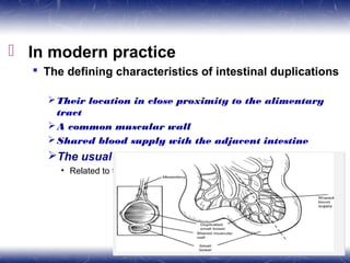
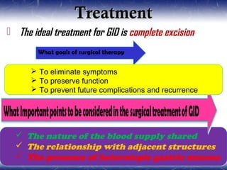
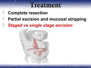
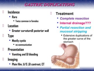
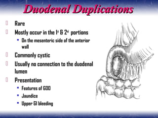
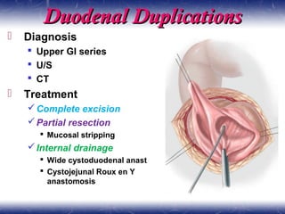
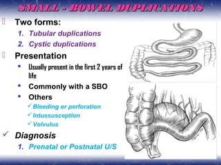
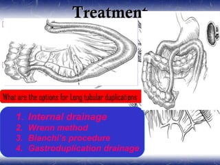
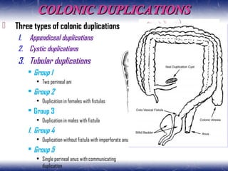
Saleamlak T.(PSR-IV)
April, 2019
MODERATOR
Prof. Milliard D. ( consultant pediatrics surgeon)
OUTLINESOUTLINES
 IntroductIon
 EpIdEmIology
 Embryology
 AnAtomy of duplIcAtIons
 clAssIfIcAtIon
 AssocIAtEd AnomAlIEs
 clInIcAl prEsEntAtIon And dIAgnosIs
 prIncIplEs of mAnAgEmEnt
 prognosIs And outcomEs
 summAry
 rEfErEncEs
IntroductionIntroduction
 Alimentary tract duplications are rare congenital developmental
abnormalities that can occur anywhere from the mouth to the anus
 Ladd's inclusive characteristics:
The presence of a well-developed smooth muscle component
The epithelial lining represents some portion of the alimentary
tract
Most duplications are intimately attached to some portion of
the gastrointestinal (GI) tract
 In modern practice
 The defining characteristics of intestinal duplications
Their location in close proximity to the alimentary
tract
A common muscular wall
Shared blood supply with the adjacent intestine
The usual location is dorsal to the normal intestine
• Related to the mesenteric aspect
EpidemiologyEpidemiology
 Incidence
 Rare lesions
 In 1 : 4,500 autopsies
 More common in whites
 Sex
 More common in males
 The exception being complex hindgut duplications
• which are more common in females
 Types
 Two types
 Cystic and tubular
• cystic being the most common
 Sites
 Small intestine and mediastinum (posterior)
 Synchronous duplications -15%
EMBRYOLOGYEMBRYOLOGY
1)Split Notochord Theory1)Split Notochord Theory
ANATOMY OF DUPLICATIONSANATOMY OF DUPLICATIONS
 Distribution
 Location
 Within the abdominal cavity- 75%
 Intrathoracic -20%
 Thoracoabdominal -5%
 Site
Jejunal and ileal (53%)
Mediastinal (18%)
Colonic (13%)
Gastric (7%)
Duodenal (6%)
Rectal (4%)
Thoracoabdominal (2%)
Cervical (1%)
ClassificationClassification
I. Based on physical appearance
a. Cystic :75% of duplications
b. Tubular :25%
II. Pathological classification
a. Parenteral cystic type
b. Parenteral canal type
c. Parietal cyst type
d. Enteral septum type
e. Solitary type
III. Vascular classification
a. Type 1: parallel type
b. Type 2: intramesentric type
ASSOCIATED ANOMALIESASSOCIATED ANOMALIES
 Thoracic and thoracoabdominal duplications
 Vertebral anomalies
 Missing , bifid, or fused vertebrae
 Other esophageal malformations
 EA and TEF
 Small intestine cystic duplications
 Coexisting intestinal atresia and malrotation
 Tubular hindgut duplications
 Extremely complex
 Genitourinary and other severe malformations
CLINICAL PRESENTATIONCLINICAL PRESENTATION
 Vary greatly depending on
Size
Location
Respiratory symptoms or dysphagia
A palpable abdominal mass and features of GOO
Abdominal Pain
Features of intestinal obstruction
Presence of gastric mucosa
GI bleeding
Perforation
Communication with the normal bowel
Type of duplication
Cystic vs tubular
ImagingImaging
1. Plain radiographs
 Mediastinal mass
1. Contrast studies
 Mass effect or communication with the alimentary tract
1. U/S
 Intraabdominal duplications
 Typical appearance
 Inner hyperechoic rim of mucosa-submucosa
 Outer hypoechoic muscular layer
¥ Scintigraphy
¥ CT
¥ MRI
TreatmentTreatment
 The ideal treatment for GID is complete excision
 The nature of the blood supply shared
 The relationship with adjacent structures
 The presence of heterotopic gastric mucosa
 The nature of the blood supply shared
 The relationship with adjacent structures
 The presence of heterotopic gastric mucosa
What goals of surgical therapyWhat goals of surgical therapy
 To eliminate symptoms
 To preserve function
 To prevent future complications and recurrence
 To eliminate symptoms
 To preserve function
 To prevent future complications and recurrence
Oral and lingual duplicationsOral and lingual duplications
 Presentation
 Asymptomatic mass in the oral region
 Diagnosis
 Usually made by the pathologist
 Treatment
 Excision with closure of the remaining
mucosa
Esophageal DuplicationsEsophageal Duplications
 Presentation
 Cervical
 Asymptomatic Neck mass or
 Symptom of UAO
 The middle portion
 Respiratory distress
 Dysphagia
 Distal esophageal
 Commonest
 Dx
 CXR
 Esophagogram ,
 CT
 MRI
TreatmentTreatment
 Complete excision
 Preferred treatment
 Partial excision & mucosal
stripping
 To avoid compromise of the esophageal
lumen
 Resection& esophagotomy
closure
 Buttressing the closure with
a pleural or intercostal
muscle flap
THORACOABDOMINAL DUPLICATIONSTHORACOABDOMINAL DUPLICATIONS
 Usually a tubular lesion that traverses the
diaphragm from the chest to the abdomen
 In the chest
 The lesions are retropleural
 May extend to the neck
 The abdominal components
 May connect to the stomach, pylorus,
duodenum, pancreas, jejunum, or ileum
 Commonly present with respiratory distress
or vomiting
TreatmentTreatment
 Complete resection
 Partial excision and mucosal stripping
 Staged vs single stage excision
GASTRIC DUPLICATIONSGASTRIC DUPLICATIONS
 Incidence
 Rare
 Twice common in females
 Location
 Greater curvature& posterior wall
 Type
 Mostly cystic
 no communication
 Presentation
 Vomiting and GI bleeding
 Imaging
 Plain film, U/S ,GI contrast, CT
 Treatment
 Complete resection
 Internal drainage???
 Partial resection and
mucosal stripping
 Extensive duplications of
the greater curve of the
stomach
DUODENALDUODENAL
DUPLICATIONSDUPLICATIONS
Duodenal DuplicationsDuodenal Duplications
 Rare
 Mostly occur in the 1st
& 2nd
portions
 On the mesenteric side of the anterior
wall
 Commonly cystic
 Usually no connection to the duodenal
lumen
 Presentation
 Features of GOO
 Jaundice
 Upper GI bleeding
Duodenal DuplicationsDuodenal Duplications
 Diagnosis
 Upper GI series
 U/S
 CT
 Treatment
Complete excision
Partial resection
 Mucosal stripping
Internal drainage
 Wide cystoduodenal anast
 Cystojejunal Roux en Y
anastomosis
SMALL - BOWELSMALL - BOWEL
DUPLICATIONSDUPLICATIONS
SMALL - BOWEL DUPLICATIONSSMALL - BOWEL DUPLICATIONS
 Two forms:
1. Tubular duplications
2. Cystic duplications
 Presentation
 Usually present in the first 2 years of
life
 Commonly with a SBO
 Others
Bleeding or perforation
Intussusception
Volvulus
 Diagnosis
1. Prenatal or Postnatal U/S
TreatmentTreatment
 Enucleation of the duplication
 Simplest
 Shared blood supply
 Common wall???
 Resection and primary anastomosis
Cystic duplication
Short tubular duplication
What are the options for Long tubular duplicationsWhat are the options for Long tubular duplications
1. Internal drainage
2. Wrenn method
3. Bianchi’s procedure
4. Gastroduplication drainage
1. Internal drainage
2. Wrenn method
3. Bianchi’s procedure
4. Gastroduplication drainage
COLONIC DUPLICATIONSCOLONIC DUPLICATIONS
 Three types of colonic duplications
1. Appendiceal duplications
2. Cystic duplications
3. Tubular duplications
 Group 1
• Two perineal ani
 Group 2
• Duplication in females with fistulas
 Group 3
• Duplication in males with fistula
1. Group 4
• Duplication without fistula with imperforate anus
 Group 5
• Single perineal anus with communicating
duplication
Associated ConditionsAssociated Conditions
 Urologic anomalies
Double bladder and urethra
Bladder exstrophy
septated bladder
 Genital anomalies
Double vagina,
Didelphic uterus
Double vulva
Double penis
Bifid scrotum
 Types
 Type I
 Type II
 Diagnosis
 Cystic colonic duplication
 Often at laparotomy
 Tubular
 Perineal inspection
 Imagings
• u/s, contrast, CT
 Treatment
 Cystic duplications
 Resection with an
anastomosis.
 Tubular colonic duplications
 Connection of distal part of duplication with
normal colon
 Resection of the distal part
RECTAL DUPLICATIONSRECTAL DUPLICATIONS
 More common in females
 Mostly occur posterior to the rectum
 Rarely they may occur anteriorly
 commonly present with constipation
 Asymptomatic mass, rectal bleeding, rectal prolapse
 Fistula to the rectum or skin(20-40%)
1. Size and mass effect
2. The presence of fistula
3. Presence of infection
4. Presence of ectopic gastric mucosa
5. Malignant degeneration
1. Size and mass effect
2. The presence of fistula
3. Presence of infection
4. Presence of ectopic gastric mucosa
5. Malignant degeneration
TreatmentTreatment
 Aim
 Resection of the entire
lesion or its mucosa
 Approaches
 Transanal
 Posterior sagittal
 Both posterior sagittal
and abdominal
Prognosis and OutcomePrognosis and Outcome
 The outcome of surgical treatment of duplications is
good
 Poor outcomes are observed
 when there are associated severe malformations
 carry a high morbidity and mortality
SummarySummary
 Gastrointestinal duplications are rare
 GIDs can arise from the mouth to the anus
 The etiology of gastrointestinal duplications is
heterogenous
 About half of all duplications are located in the
small bowel
 Most patients with gastrointestinal duplications
will present before 2 years of age
 Ultrasonography should be the first line of
investigation
 The ideal treatment is total excision
Since GID is a benign disease, therapy should
ReferencesReferences
 Coran Pediatric Surgery, 7th
ed
 Ashcraft’s pediatric surgery, 6th
edition
 Operative pediatric Surgery , Moritz, 2nd
ed.
 Principles and practice of pediatrics
surgery, 4th
ed
 Pediatric Surgery (SpringerSurgery Atlas
Series)
 Operative pediatric surgery, Lewis Spitz,
Arnold G Coran. 7th
edition
Thank YouThank You

Alimentary Tract Duplications

  • 1.
    Saleamlak T.(PSR-IV) April, 2019 MODERATOR Prof.Milliard D. ( consultant pediatrics surgeon)
  • 2.
    OUTLINESOUTLINES  IntroductIon  EpIdEmIology Embryology  AnAtomy of duplIcAtIons  clAssIfIcAtIon  AssocIAtEd AnomAlIEs  clInIcAl prEsEntAtIon And dIAgnosIs  prIncIplEs of mAnAgEmEnt  prognosIs And outcomEs  summAry  rEfErEncEs
  • 3.
    IntroductionIntroduction  Alimentary tractduplications are rare congenital developmental abnormalities that can occur anywhere from the mouth to the anus  Ladd's inclusive characteristics: The presence of a well-developed smooth muscle component The epithelial lining represents some portion of the alimentary tract Most duplications are intimately attached to some portion of the gastrointestinal (GI) tract
  • 4.
     In modernpractice  The defining characteristics of intestinal duplications Their location in close proximity to the alimentary tract A common muscular wall Shared blood supply with the adjacent intestine The usual location is dorsal to the normal intestine • Related to the mesenteric aspect
  • 5.
    EpidemiologyEpidemiology  Incidence  Rarelesions  In 1 : 4,500 autopsies  More common in whites  Sex  More common in males  The exception being complex hindgut duplications • which are more common in females  Types  Two types  Cystic and tubular • cystic being the most common  Sites  Small intestine and mediastinum (posterior)  Synchronous duplications -15%
  • 6.
  • 7.
    ANATOMY OF DUPLICATIONSANATOMYOF DUPLICATIONS  Distribution  Location  Within the abdominal cavity- 75%  Intrathoracic -20%  Thoracoabdominal -5%  Site Jejunal and ileal (53%) Mediastinal (18%) Colonic (13%) Gastric (7%) Duodenal (6%) Rectal (4%) Thoracoabdominal (2%) Cervical (1%)
  • 8.
    ClassificationClassification I. Based onphysical appearance a. Cystic :75% of duplications b. Tubular :25% II. Pathological classification a. Parenteral cystic type b. Parenteral canal type c. Parietal cyst type d. Enteral septum type e. Solitary type III. Vascular classification a. Type 1: parallel type b. Type 2: intramesentric type
  • 9.
    ASSOCIATED ANOMALIESASSOCIATED ANOMALIES Thoracic and thoracoabdominal duplications  Vertebral anomalies  Missing , bifid, or fused vertebrae  Other esophageal malformations  EA and TEF  Small intestine cystic duplications  Coexisting intestinal atresia and malrotation  Tubular hindgut duplications  Extremely complex  Genitourinary and other severe malformations
  • 11.
    CLINICAL PRESENTATIONCLINICAL PRESENTATION Vary greatly depending on Size Location Respiratory symptoms or dysphagia A palpable abdominal mass and features of GOO Abdominal Pain Features of intestinal obstruction Presence of gastric mucosa GI bleeding Perforation Communication with the normal bowel Type of duplication Cystic vs tubular
  • 13.
    ImagingImaging 1. Plain radiographs Mediastinal mass 1. Contrast studies  Mass effect or communication with the alimentary tract 1. U/S  Intraabdominal duplications  Typical appearance  Inner hyperechoic rim of mucosa-submucosa  Outer hypoechoic muscular layer ¥ Scintigraphy ¥ CT ¥ MRI
  • 14.
    TreatmentTreatment  The idealtreatment for GID is complete excision  The nature of the blood supply shared  The relationship with adjacent structures  The presence of heterotopic gastric mucosa  The nature of the blood supply shared  The relationship with adjacent structures  The presence of heterotopic gastric mucosa What goals of surgical therapyWhat goals of surgical therapy  To eliminate symptoms  To preserve function  To prevent future complications and recurrence  To eliminate symptoms  To preserve function  To prevent future complications and recurrence
  • 16.
    Oral and lingualduplicationsOral and lingual duplications  Presentation  Asymptomatic mass in the oral region  Diagnosis  Usually made by the pathologist  Treatment  Excision with closure of the remaining mucosa
  • 17.
    Esophageal DuplicationsEsophageal Duplications Presentation  Cervical  Asymptomatic Neck mass or  Symptom of UAO  The middle portion  Respiratory distress  Dysphagia  Distal esophageal  Commonest  Dx  CXR  Esophagogram ,  CT  MRI
  • 18.
    TreatmentTreatment  Complete excision Preferred treatment  Partial excision & mucosal stripping  To avoid compromise of the esophageal lumen  Resection& esophagotomy closure  Buttressing the closure with a pleural or intercostal muscle flap
  • 19.
  • 20.
    THORACOABDOMINAL DUPLICATIONSTHORACOABDOMINAL DUPLICATIONS Usually a tubular lesion that traverses the diaphragm from the chest to the abdomen  In the chest  The lesions are retropleural  May extend to the neck  The abdominal components  May connect to the stomach, pylorus, duodenum, pancreas, jejunum, or ileum  Commonly present with respiratory distress or vomiting
  • 21.
    TreatmentTreatment  Complete resection Partial excision and mucosal stripping  Staged vs single stage excision
  • 22.
  • 23.
    GASTRIC DUPLICATIONSGASTRIC DUPLICATIONS Incidence  Rare  Twice common in females  Location  Greater curvature& posterior wall  Type  Mostly cystic  no communication  Presentation  Vomiting and GI bleeding  Imaging  Plain film, U/S ,GI contrast, CT  Treatment  Complete resection  Internal drainage???  Partial resection and mucosal stripping  Extensive duplications of the greater curve of the stomach
  • 24.
  • 25.
    Duodenal DuplicationsDuodenal Duplications Rare  Mostly occur in the 1st & 2nd portions  On the mesenteric side of the anterior wall  Commonly cystic  Usually no connection to the duodenal lumen  Presentation  Features of GOO  Jaundice  Upper GI bleeding
  • 26.
    Duodenal DuplicationsDuodenal Duplications Diagnosis  Upper GI series  U/S  CT  Treatment Complete excision Partial resection  Mucosal stripping Internal drainage  Wide cystoduodenal anast  Cystojejunal Roux en Y anastomosis
  • 27.
    SMALL - BOWELSMALL- BOWEL DUPLICATIONSDUPLICATIONS
  • 28.
    SMALL - BOWELDUPLICATIONSSMALL - BOWEL DUPLICATIONS  Two forms: 1. Tubular duplications 2. Cystic duplications  Presentation  Usually present in the first 2 years of life  Commonly with a SBO  Others Bleeding or perforation Intussusception Volvulus  Diagnosis 1. Prenatal or Postnatal U/S
  • 29.
    TreatmentTreatment  Enucleation ofthe duplication  Simplest  Shared blood supply  Common wall???  Resection and primary anastomosis Cystic duplication Short tubular duplication What are the options for Long tubular duplicationsWhat are the options for Long tubular duplications 1. Internal drainage 2. Wrenn method 3. Bianchi’s procedure 4. Gastroduplication drainage 1. Internal drainage 2. Wrenn method 3. Bianchi’s procedure 4. Gastroduplication drainage
  • 30.
  • 31.
    COLONIC DUPLICATIONSCOLONIC DUPLICATIONS Three types of colonic duplications 1. Appendiceal duplications 2. Cystic duplications 3. Tubular duplications  Group 1 • Two perineal ani  Group 2 • Duplication in females with fistulas  Group 3 • Duplication in males with fistula 1. Group 4 • Duplication without fistula with imperforate anus  Group 5 • Single perineal anus with communicating duplication
  • 32.
    Associated ConditionsAssociated Conditions Urologic anomalies Double bladder and urethra Bladder exstrophy septated bladder  Genital anomalies Double vagina, Didelphic uterus Double vulva Double penis Bifid scrotum  Types  Type I  Type II
  • 33.
     Diagnosis  Cysticcolonic duplication  Often at laparotomy  Tubular  Perineal inspection  Imagings • u/s, contrast, CT  Treatment  Cystic duplications  Resection with an anastomosis.  Tubular colonic duplications  Connection of distal part of duplication with normal colon  Resection of the distal part
  • 34.
  • 35.
    RECTAL DUPLICATIONSRECTAL DUPLICATIONS More common in females  Mostly occur posterior to the rectum  Rarely they may occur anteriorly  commonly present with constipation  Asymptomatic mass, rectal bleeding, rectal prolapse  Fistula to the rectum or skin(20-40%) 1. Size and mass effect 2. The presence of fistula 3. Presence of infection 4. Presence of ectopic gastric mucosa 5. Malignant degeneration 1. Size and mass effect 2. The presence of fistula 3. Presence of infection 4. Presence of ectopic gastric mucosa 5. Malignant degeneration
  • 36.
    TreatmentTreatment  Aim  Resectionof the entire lesion or its mucosa  Approaches  Transanal  Posterior sagittal  Both posterior sagittal and abdominal
  • 37.
    Prognosis and OutcomePrognosisand Outcome  The outcome of surgical treatment of duplications is good  Poor outcomes are observed  when there are associated severe malformations  carry a high morbidity and mortality
  • 38.
    SummarySummary  Gastrointestinal duplicationsare rare  GIDs can arise from the mouth to the anus  The etiology of gastrointestinal duplications is heterogenous  About half of all duplications are located in the small bowel  Most patients with gastrointestinal duplications will present before 2 years of age  Ultrasonography should be the first line of investigation  The ideal treatment is total excision Since GID is a benign disease, therapy should
  • 39.
    ReferencesReferences  Coran PediatricSurgery, 7th ed  Ashcraft’s pediatric surgery, 6th edition  Operative pediatric Surgery , Moritz, 2nd ed.  Principles and practice of pediatrics surgery, 4th ed  Pediatric Surgery (SpringerSurgery Atlas Series)  Operative pediatric surgery, Lewis Spitz, Arnold G Coran. 7th edition
  • 40.

Editor's Notes

  • #4 Ladd suggested the term duplication of the alimentary tract be used to encompass this constellation of abnormalities Ladd's inclusive terminology emphasized that these lesions were congenital developmental anomalies that shared several characteristics: Alimentary tract duplications are relatively rare congenital anomalies found anywhere from the mouth to the anus, and can present with obstruction or be discovered incidentally. While most duplications are benign, ectopic gastric mucosa and the potential for malignant degeneration remain concerns. Most duplications are discovered by 2 years of age. However, with the increased use of prenatal ultrasound (US), more are being diagnosed in utero. The goal of operative management is to remove the duplication and prevent its recurrence. Since most share a common blood supply to the native alimentary tract, simple resection is usually adequate. Long tubular or thoracoabdominal duplications may present a more difficult scenario as radical resection can carry significant morbidity or even mortality. Overall prognosis is generally favorable but associated malformations or the presenting illness can factor into the final outcome. Alimentary tract duplications have been described for hundreds of years and multiple terms have been used in the literature. The current term duplication of the alimentary tract and a common description of the congenital malformation was applied by William Ladd in 1937.1 Three common findings were described: a well-developed smooth muscle coat, an epithelial lining, and attachment to the alimentary tract. The first large series to appear in the literature by Gross et al. in 1952 supported these finding as well
  • #5 Their location in close proximity to the alimentary tract and In contrast to vitelline duct remnants Meckel's diverticula which lie on the antimesenteric aspect of the bowel A small bowel duplication in cross section, demonstrating the common wall, shared blood supply, and intramesenteric location
  • #6 Incidence Rare lesions Their exact incidence is difficult to determine They are reported In 1 of every 4,500 autopsies They appear to be most often seen in white males The exception being complex hindgut duplications which are more common in females There does not appear to be a familial incidence Alimentary tract duplications are most commonly located in Small intestine and mediastinum (posterior), Whereas rectal, duodenal, gastric, and thoracoabdominal locations are extremely rare Synchronous duplications are reported in as many as 15% of patients
  • #7 The embryogenesis of intestinal duplications is not known There are many theories But none adequately explains the origin of all lesions in this diverse constellation of anomalies, Suggesting that more than one pathogenic possibility may be necessary to explain the anatomic variability Persistent embryonic diverticulum -Failure of the normal regression of embryonic diverticula Suggests that failure of the normal regression of embryonic diverticula may occur Diverticula are common in the developing human GI tract Their finding at numerous sites around the circumference of the gut wall Provides a potential explanation For small cystic duplications noted in the intestinal wall, and For enteric cysts located in the presacral space However, this theory does not explain The propensity for the location of enteric cysts within the leaves of the bowel mesentery, or The finding of multiple types of mucosa lining the wall of some enteric duplications The wall of adjacent fetal bowel may be flattened by Extrinsic compression with subsequent adherence and fusion Resulting in doubling of the lumen Explains the occurrence of side by side or adjacent tubular duplications But no embryonic evidence for this theory
  • #8 Approximately 75% of duplications have been reported as located within the abdominal cavity, while the remaining are intrathoracic (20%) or thoraco-abdominal (5%). Jejunal and ileal lesions are the most commonly encountered abdominal lesions (53%), followed by mediastinal (18%), colonic (13%), gastric (7%), duodenal (6%), rectal (4%), thoracoabdominal (2%) and cervical (1%).[14]
  • #9 Based on physical appearance Cystic :75% of duplications No communication with adjacent intestine Tubular :25% May or may not have communication Communication usually located at the distal end Seventy-five percent of duplications are cystic with no communication with the adjacent intestine, while the remaining duplications are tubular structures that may or may not have one or more direct communications through the common septum. Usually, such communications are located at the distal end. Parenteral cystic type (in the mesentery, cystic, most common), • Parenteral canal type (in the mesentery, tubular, second most common) • Parietal cyst type (intramural, third commonest) • Enteral septum type (intraluminal due to septae, rare) • Solitary type(seems pedunculated, not intimately attached to the gut, rare).[15] Type 1 (Parallel type): Duplication is on the mesenteric border of the intestine and has independent blood supply through the vasa recta, which is separated from the straight artery of the bowel. It is not covered by mesentery. Therefore, such duplications can be resected easily without compromising the adjoining bowel. • Type 2: In the Intramesenteric type, the duplication is in-between the leaves of the mesentery and the straight arteries arch over the duplication to reach the bowel. It is sometimes technically demanding to resect these duplications without compromising/ sacrifi cing the adjoining bowel as the vasculature is sometimes common to both or is diffi cult to separate the vessels from the cyst. Classification of small intestinal duplications based on their blood supply. In type 1, the duplication is located on one side of the mesentery, and the main artery of the duplication is parallel to the main artery of the bowel. Type 1a, the duplication has a separate mesentery; type 1b, the duplication shares common mesentery with the gut; and type 1c, the duplication shares common muscular coat with the gut. In type 2, the lesion is located between the two layers of the mesentery and supplied by vessels from both sides of the mesentery that pass over from both surfaces of the duplication to reach the native bowel. Type 2a, the duplication is separate from the bowel, and type 2b, the duplication shares common muscular coat with the bowel
  • #10 common with alimentary tract duplications Thoracic and thoracoabdominal duplications With vertebral anomalies such as missing, bifid, or fused vertebrae Esophageal duplications are seen in association with other esophageal malformations such as esophageal atresia and TEF and pulmonary agenesis Small intestine cystic duplications have been reported with coexisting intestinal atresias and malrotation Tubular hindgut duplications Are an extremely complex and diverse group of anomalies frequently associated with genitourinary and other severe malformations
  • #12 The majority of alimentary tract duplications that cause clinical symptoms are diagnosed in infancy Greater than 80% before 2 years of age Older infants and children may experience Indolent and vague abdominal complaints over prolonged periods or Intermittent GI bleeding and anemia The symptoms and clinical presentation of intestinal duplications vary greatly depending on size, location, presence of gastric mucosa, and communication with the normal bowel. A mass discovered on physical examination or radiographic examination of the chest or abdomen is a common mode of discovery. Specific symptoms are often related to the location of the duplication. Specific symptoms are often related to the location of the duplication Cervical and thoracic duplications May cause respiratory symptoms or dysphagia, or May be an incidental finding on chest radiograph Gastric and duodenal duplications May present As a palpable abdominal mass or Due to symptoms of gastric outlet obstruction Cervical and thoracic duplications may cause respiratory symptoms or dysphagia, or may be an incidental finding on chest radiograph (15,16). Gastric and duodenal duplications may present as a palpable abdominal mass or due to symptoms of gastric outlet Pain The most common symptom May be caused by Distension of the duplication or Result from a complication such as Intestinal obstruction, Peptic ulceration, or Perforation Intestinal obstruction May result from compression of the adjacent bowel lumen or Due to the mass effect of the duplication leading to Volvulus or Intussusception obstruction. Pain is the most common symptom and may be caused by distension of the duplication or result from a complication such as intestinal obstruction, peptic ulceration, or perforation. Intestinal obstruction may result from compression of the adjacent bowel lumen or due to the mass effect of the duplication leading to volvulus or intussusception. GI bleeding Also a common complication of enteric duplications The bleeding can be acute and severe, presenting as Hematemesis , Melena , or Hematochezia Depending on location and magnitude of bleeding Chronic occult bleeding may present as anemia The cystic lesions Most often do not communicate with the normal intestine These may achieve a large size Create a mass effect on plain radiographs or Be noted as a palpable mass on physical exam The tubular duplications of the small intestine More frequently communicate with the native bowel Have a high incidence of heterotopic gastric mucosa Peptic ulceration can lead to Perforation and free intraperitoneal hemorrhage or Fistulization into adjacent structures GI bleeding is also a common complication of enteric duplications. The bleeding can be acute and severe, presenting as hematemesis, melena, or hematochezia, depending on location and magnitude of bleeding. Chronic occult bleeding may present as anemia. The cystic lesions most often do not communicate with the normal intestine. These may achieve a large size and create a mass effect on plain radiographs or be noted as a palpable mass on physical exam. The tubular duplications of the small intestine more frequently communicate with the native bowel and have a high incidence of heterotopic gastric mucosa (17,18). Peptic ulceration can lead to perforation and free intraperitoneal hemorrhage or fistulization into adjacent structures. The majority of alimentary tract duplications that cause clinical symptoms are diagnosed in infancy (greater than 80% before 2 years of age) (19). Older infants and children with intestinal duplications may experience indolent and vague abdominal complaints over prolonged periods or intermittent GI bleeding and anemia. Colonic or presacral duplications typically present due to obstruction, constipation, or prolapse through the anus. Enteric duplications are frequently associated with vertebral abnormalities such as spina bifida or missing, fused, or hemivertebrae. Rarely, there may be an intraspinal component causing neurologic symptoms from spinal cord compression. Myelomeningocele is associated with some enteric duplications, especially in the thoracic cavity. Some duplications remain silent and persist into adulthood (20). Neoplastic changes have been reported in duplications diagnosed in adulthood—most in duplications of the colon and rectum (21,22). Hindgut duplications may be associated with splitting of the lower vertebrae and sacrum and severe urogenital abnormalities, such as doubling of the external genitalia or other perineal abnormality
  • #14 Multiple imaging modalities are utilized to make the diagnosis. Plain radiographs may reveal a mediastinal mass, suggesting an esophageal duplication. Contrast studies may show a mass effect or communication with the alimentary tract. ultrasound is radiation free and noninvasive, making it a useful test, particularly for intra-abdominal duplications.16 A typical sonographic appearance of duplications demonstrates an inner hyperechoic rim of muscosa–submucosa and an outer hypoechoic muscular layer (Fig. 39-3).17 A history of anemia or bleeding with a suspected duplication suggests ectopic gastric mucosa, and technetium-99m (99mTc) scintigraphy is a useful imaging modality.18,19 In cases where a combined thoracoabdominal duplication is suspected, computed tomography (CT) may aid in diagnosis. The presence of vertebral abnormalities and esophageal duplications is best investigated with magnetic resonance imaging (MRI).20 Duplication of the cervical esophagus in a 3-year-old boy with a neck mass. (a) Esophagogram shows the mass in the thoracic inlet, displacing the esophagus to the left. Portions of the cyst wall show the characteristic doublelayered appearance with echogenic mucosa internally (arrows) and hypoechoic muscle externally Gastric duplication cyst.An ultrasound image of the upper mid abdomen along the long axis of the body and antrum of the stomach demonstrates a complex cystic mass contiguous with the greater curvature of the stomach, with a hypoechoic wall suggesting the muscular layer of gut signature
  • #15 Duplications in the pediatric population are benign lesions therefore, the surgical therapy should aim primarily To eliminate symptoms To preserve function, and To prevent future complications and recurrence The goal is to make a prompt diagnosis and provide treatment before the onset of symptoms or the development of complications. The ideal treatment for GID is complete excision. However, GID in children is a benign disease, and any treatment should not be more radical than to eliminate the patient’s complaints and prevent further recurrence. Important points to be considered in the surgical treatment of GID include: 1. the nature of the blood supply shared between the duplication and native bowel; 2. the presence of heterotopic gastric mucosa, which will negate internal drainage due to the risk of peptic ulceration; and 3. the relationship with adjacent structures, such as the biliary tract in duodenal duplications. The treatment of GID is best considered by location of the duplication. However, in selected cases, an intraoperative frozen section may give further information on the absence or presence of heterotopic components. Important points to be considered in the surgical treatment of GID include: 1. the nature of the blood supply shared between the duplication and native bowel; 2. the presence of heterotopic gastric mucosa, which will negate internal drainage due to the risk of peptic ulceration; and 3. the relationship with adjacent structures, such as the biliary tract in duodenal duplications. The treatment of GID is best considered by location of the duplication. However, in selected cases, an intraoperative frozen section may give further information on the absence or presence of heterotopic components. The nature of the blood supply shared between the duplication and native bowel The relationship with adjacent structures such as the biliary tract in duodenal duplications The presence of heterotopic gastric mucosa Which will negate internal drainage due to the risk of peptic ulceration
  • #17 Oral and lingual duplications present as an asymptomatic mass in the oral region The diagnosis is usually made by the pathologist after the excision is completed These lesions may contain ectopic gastric mucosa The presence of an oral cyst or mass lesion in the mouth or pharynx is an indication for excision Treatment involves excision with closure of the remaining mucosa Image: Foregut duplication cyst presenting as lingual swelling
  • #18 Presenting Signs and Symptoms Account 23% of esophageal duplication Asymptomatic Neck mass or symptom of upper airway obstruction Ddx Cystic hygroma, thyroglossal duct cyst, branchial cleft cyst, or solid tumors such as teratomas. The middle portion comprise 17% of esophageal duplications Present with compression of the trachea at a young age The main differential diagnosis is bronchogenic cysts Distal esophageal duplications The most common Are often asymptomatic CXR usually suspected based on a CXR Revealing a posterior mediastinal mass Esophagogram Reveals an indentation on the esophagus by the lesion rarely shows a connection between the esophagus and duplication CT scan Provide much better anatomic details MRI Esophageal duplications are relatively uncommon (Table 5 1 - 1). In a review of 495 alimentary tract duplications, t he esophagus was the site of 19% of the lesions. An additional 4% of the lesions were thoracoabdominal. Most esophageal duplications are cystic in shape and do not have a shared muscular wall with the esophagus although, rarely, they do connect to the esophageal lumen (Fig. 5 1 - 1 ). The mucosal lining of esophageal duplications contains gastric mucosa in 43% of cases, but may contain pseudostratified columnar, small intestinal, or large intestinal mucosa. This gastric mucosa may lead to ulceration, pain, and hemorrhage. The esophagus can be divided into 3 parts: the cervical third, the middle third, and the lower third. Duplications occur in all segments of the esophagus; however, the presentation may vary. Cervical duplications account for 23% of esophageal duplications. They may present as an asymptomatic neck mass or with symptoms of upper-airway obstruction. These can be confused with cystic hygroma, thyroglossal duct cyst, branchial cleft cyst, or solid tumors such as teratomas. Duplications of the middle portion comprise 17% of esophageal duplications. These usually present with compression of the trachea at a young age. The main differential diagnosis is bronchogenic cysts. Distal esophageal duplications are the most common and are often asymptomatic
  • #19 Complete excision Preferred treatment Straight forward for non communicating type Technically challenging and formidable task in the more complex lesion Partial excision and mucosal stripping To avoid compromise of the esophageal lumen Resection and esophagotomy closure Buttressing the closure with a pleural or intercostal muscle flap The presence of an esophageal duplication or cystic mediastinal mass is an indication for operation. Even if the lesion is an incidental finding and is asymptomatic, it should be removed for diagnostic purposes and to prevent complications such as bleeding, perforation, and airway compromise. Lesions in patients exhibiting respiratory distress or dysphagia should be removed to correct these symptoms. Resection is the treatment of choice. The majority of esophageal duplications noted in large series were treated with simple resection via thoracotomy and no other techniques were needed. Although neuroenteric connections are unusual, careful observation for these is wise. Thoracoscopic resection of these lesions is an excellent technique that is much more common over the past 10 years. Thoracoscopic resection is technically demanding but these technical skills are much more common than in the past. Other cystic mediastinal lesions, such as bronchogenic cysts, have been removed using thoracoscopic technique. Other options described include resection of the lesion with a segment of esophagus. This should only be needed in unusual circumstances since the surgeon is usually able to separate the duplication from the esophagus. Rarely, when the duplication connects to the esophageal lumen, resection of the duplication with closure of the esophagotomy and buttressing the closure with a pleural or intercostal muscle flap is indicated. Other treatments, such as marsupialization of the cyst to the chest wall and destruction of the mucosa with packing and curettage, are of historical interest only. The long-term outcome for these patients is excellent if the lesion is resected without complications. Complications of resection include anastomotic strictures if a segment of the esophagus is resected. Phrenic nerve and thoracic duct injuries have also been reported. Deaths from resection are unusual but are reported
  • #21 Communicate with the intestinal tract below the diaphragm Most often through the esophageal hiatus Generally connect to the duodenum and the jejunum Often passing behind the stomach and pancreas Diagram of a thoracoabdominal duplication with an abdominal connection to the gastric antrum. Commonly present with respiratory distress or vomiting Usually present with a tubular lesion that traverses the diaphragm from the chest to the abdomen May be associated with vertebral abnormalities Have a 29% incidence of ectopic gastric mucosa Thoracoabdominal duplications are rare Presenting symptoms include Respiratory distress Vomiting Gastrointestinal bleeding and Meningitis The thoracic portion of these lesions occur most commonly on the right side
  • #22 Resection of thoracoabdominal duplications is more complex than resection of duplications confined to one body cavity. A variety of approaches have been described, including resection of the chest and abdominal portions at separate procedures several weeks apart. Some children require laminectomy in association with the resection. The current trend appears to be a combination of thoracotomy and laparotomy with complete resection of the lesion in 1 stage. Minimally invasive techniques can be used to resect these lesions. The spinal component, if present, must always be considered when planning the operation
  • #24 Extensive duplications of the greater curve of the stomach 5-7% Typically cystic Located on the greater curvature or posterior wall No communication with the stomach Variable in sizes More common in boys Typically present in the early life 1st year
  • #27 The treatment choice is complete excision with the preservation of the duodenum But total excision is not always possible if the cyst is in close proximity with biliary system and pancreatic dudct The walls of the ducts may be included with common wall of duplication. There may be shared bllod vessels
  • #29 75% of patients present in the first 2yrs( half during neonatal period, half during 1m to 2yrs) In small-bowel duplications an abdominal mass is present most commonly in the ileocecal area 75% Present in the first 2 years of life With half presenting in neonates and half presenting between 1 month and 2 years of age 80% Present with signs and symptoms of a bowel obstruction Other presenting signs include melena, abdominal mass, and peritonitis from perforation. Rectal bleeding and intussusception are common presentations in children older than 2 years of age Patients who present with bleeding usually Have a tubular duplication that contains ectopic gastric mucosa Tubular duplications Follow the intestine for all or part of its length may be separate from the bowel, adherent with a separate but attached wall, or May share a common wall usually occur on the mesenteric side and share a blood supply with the intestine Although they occasionally have a separate blood supply Cystic duplications Most commonly occur at the ileocecal junction share a common blood supply and wall with the ileum The most common ectopic mucosa is gastric and occurs in 33% of ileal and 50% of jejunal duplications Ectopic gastric mucosa is more likely to occur in Tubular duplications (80%) than in cystic duplications (20%) Untreated small-bowel duplications are associated with malignant degeneration. Image< Cystic ileal duplication along the mesenteric border of the bowel. Image< Tubular duplication of the small bowel with a shared common wall and a distal communication Prenatal diagnosis The double-wall configuration seen in duplications and in the Meckel diverticula The "Y'' configuration of the vasculature entering the duplication is characteristic for a small bowel duplications Ultrasound or CT scan can be helpful in cystic duplications Occasionally, ectopic mucosa in a duplication is seen on a Meckel scan performed for bleeding
  • #30 Operative resection of these lesions can take many forms. The simplest treatment is enucleation of the duplication. Enucleation of some duplications is possible because the blood supply to the small bowel divides and half goes to one side and half to the other side. Because of this, a few of the vessels on one side may be ligated and the bowel remains viable. This can allow resection of the duplication while leaving the normal bowel in situ. Despite this fact, enucleation is rarely possible because the duplications often have a shared blood supply with the intestine and they often share a common wall. For this reason, cystic duplications and short tubular duplications are most easily treated with resection of the duplication and juxtaposed small intestine. Internal drainage Stripping with multiple enterotomies Wrenn method Mucosal dissection as soave procedure Bianchi’s procedure To separate from the native bowel Gastroduplication drainage The intestine is then reconstructed with an end-to-end anastomosis ( Fig. 5 1 - 1 1 ) . In 1 study, 46 of 47 patients with jejunal and ileal duplications were able to be treated with this type of resection. FIGURE S 1 - 1 1 Cystic i l ea l d u p l ication. A. Marg i n s of resection outli ned taki ng care to preserve the proximal and d i sta l bowel mesentery. B. End-to-end bowel a n astomosis comp leted FIGURE S 1 -1 3 The gastroduplication method of treatment of an obstructed extensive ileal tubular duplication. A. The distal end of the duplication is resected and an end-to-end anastomosis is done between the proximal and distal normal ileum . B. The distal end of the duplication is then anastomosed in a side-to-side fashion t o the stomach to allow duplication content to drain into the stomach. No adverse effect of gastric acid on the small-bowel mucosa was noted after 1 0 years of patient follow-up.
  • #32 Three types of colonic duplications Appendiceal duplications Which are treated with appendectomy Cystic duplications Tubular duplications Group 1- Two perineal ani Group 2- Duplication in females with fistulas Group 3- Duplication in males with fistula Group 4- Duplication without fistula with imperforate anus Group 5- Single perineal anus with communicating duplication Colonic duplications are divided into 3 types: duplications of the appendix, which are treated with appendectomy; cystic duplications of the colon; and tubular duplications. Rectal duplications will be considered separately. Cystic duplications may present with vomiting, volvulus, abdominal mass, urinary retention, abdominal pain, or perforation presenting as an acute abdomen. Tubular colonic duplications present in various ways. Many are noted at birth in association with duplications of the genitals or bladder. Others present with acute or chronic lower intestinal obstruction, rectovaginal fistula, rectal prolapse, and urinary tract infections. The duplicated colon may be on the mesenteric or antimesenteric side of the colon. Those on the antirnesenteric side may terminate as a fistula or end blindly in the pelvis (Fig. 5 1 - 1 4A) , those on the mesenteric side usually terminate as a fistula (Fig. S l - 14B) . The mucosal lining of these lesions is usually normal colonic mucosa. Tubular colonic duplications have been divided into 5 groups: 1 . Group 1: Two perineal ani. This anomaly is diagnostic for long-segment colon duplication. Most of these patients have normal anal sphincters and are asymptomatic. They have the highest rate of genitourinary duplications, malrotation, omphalocele, and the Meckel diverticulum. 2. Group 2: Duplication in females with fistula( e). This anomaly is seen if the patient has a rectovaginal or rectoperineal fistula in addition to a perineal anus. Other forms include the patient with 2 fistulae or if a fistula is noted in a patient with duplication of the genital organs or bladder. These patients may have constipation because of complete or partial obstruction of the duplicated colon filling with feces and compressing the normal colon. 3 . Group 3: Duplication in males with fistula( e). Presence of 1 functioning perineal anus and a rectourinary fistula is the hallmark of this group of patients. They may have bifid scrotum or a double penis Group 4: Duplication without fistula with imperforate anus. These patients have the highest incidence of skeletal and urinary anomalies. Almost all have intestinal obstruction. These patients have either an imperforate a nus with 2 anal dimples or 1 functioning anus and a second anal dimple. In the latter group, the patent rectum may be obstructed and pushed laterally by a large mass of feces in the obstructed rectum. 5. Group 5: Single perineal anus with communicating duplication. Patients with this form may be asymptomatic. The diagnosis may be suspected if the patient has urethral or bladder duplication. Line diagram of triplicated colon and duplicated bladder. All of the coli communicated with the ileum above. The first of the triplications ended at a normally situated anus. The second ended blindly in an atresia in the region of the sigmoid colon and the third entered the left hemi-vescicae through a colovesical fistula.
  • #33 Classifi cation by Kottra and Dodds[20] of colonic duplications provides practical guidance for the management. Type I Duplications limited to the alimentary tract and situated above the peritoneal reflection. These could be cystic (type Ia), tubular (type Ib) or double barrel type (Ic) with one or multiple communication/s with the gut. Type II These are tubular colonic duplications associated with duplication of genitourinary tracts. Type IIa Tubular duplications with double genitalia, double urethra and bladder. There are separate perianal ani on either side of midline. There is no communication with genitourinary system. Type IIb There is an internal fistula between the duplication and the genitourinary system. Type IIc Tubular duplications with imperforate anus. There is no internal fistula between genitourinary tract and the duplication. However, the lower urinary tract may be duplicated.
  • #34 Examination under anesthesia showing two urethral openings (small arrows at the top), two vaginal openings (large arrows), ectopic anal opening (star), and normal anal opening at the bottom (arrow below) complete duplication of the colon with malrotation Exploratory laparotomy showing duplicated cecum (large arrows) and appendix (small arrows) Duplicated colon at the end of the operation. The vaginal opening is separated and a wide opening is created on the common wall. We used a simple technique on our patient. The first laparotomy gave us a chance to do a detailed exploration of the gastrointestinal tract. At the second surgery, the vaginal termination of the duplication was closed as it is done in rectovaginal fistula repair, and also a wide oval opening was created on the common wall to permit the passage between the two colons. We believe this technique is simple and applicable to this kind of malformation. Surgical treatment of cystic duplications o f the colon is much like that of cystic small-bowel duplications. Resection of t he adjacent colon is the simplest treatment. The colon is reconstructed with an anastomosis. Treatment of tubular colonic duplications is operative. Each case must be individualized. The main cause of symptoms is the distal obstructed pouch of the duplication that usually connects to the normal colon proximally and fills with feces, causing compression of the normal colon in the pelvis. This can be treated in either of 2 ways. The distal part of the duplicated colon may be connected to the normal colon with a linear stapler. If this option is chosen, the connection must be distal enough to ensure that the duplication will no longer collect feces and produce an obstructing mass. Alternatively, the distal part of the duplication may be resected. This is an especially good option if the normal rectum and the duplication are separated. If the duplicated colon connects to the urinary tract or vagina, the duplication can be divided and an endorectal mucosectomy of the distal duplication done. The muscular cuff is then closed and an end-to-side anastomosis connects the remaining distal end of the duplication to the normal colon. Because colonic duplications rarely have ectopic gastric mucosa, resection of the duplicated segment is rarely necessary; however, malignancy has been reported in untreated cases of colonic duplication, but no such reports are found in cases treated with distal diversion
  • #36 . Rectal duplications may present as a posterior rectal mass that must be differentiated from both an anterior meningomyelocele and sacrococcygeal teratoma. 4. Rectal duplications often have a separate anal opening The presentation of these lesions is related to five factors:
  • #37 If the lesions are large They may require a combination of abdominal and posterior sagittal approaches. Treatment includes resection of the entire lesion or its mucosa. The approach to this resection may be transanal or posterior sagittal ( Figs. 5 1 - 1 6 and 5 1 - 1 7) . Both approaches have good results. If the lesions are large, they may require a combination of abdominal and posterior sagittal approaches. Marsupialization of the duplication into the rectum with a stapler has been described. Because adenocarcinoma in rectal duplications has been reported, and its incidence is unknown, mucosal resection should be done if possible. Lesions with ectopic gastric mucosa may cause perineal skin excoriation if the mucosa is not resected. Laparoscopic resection has been reported. Rectal Duplications. Rectal duplications often present in the neonatal period as a perineal mucosal swelling and/or a fistula extending to the perianal area or anorectum. Some are cystic lesions with no external or internal communication. They extend into the retro rectal space and do not communicate with the urinary tract. Rarely, they first manifest as rectal prolapse in an infant. They may be excised by one of several approaches Small submucosal rectal lesions can be excised endorectally. As the anus is dilated with an anal retractor, the cyst bulges forward. The rectal mucosa over the cyst is incised and, working within the submucosal plane of the rectum, the cyst is gradually dissected free. Local injection of 1:200,000 adrenaline and bipolar diathermy are helpful in the dissection. The cyst should not be decompressed until near the end of the dissection as the wall tension can facilitate the dissection. The incision in the rectal mucosa is repaired with interrupted absorbable sutures. A limited perineal excision is all that is required for a localised small mucosal duplication. In the case of larger or more complex duplications, some of which have a lateral or cranial diverticulum, the posterior sagittal approach provides excellent exposure of the retrorectal space. With attention to bowel preparation and antibiotic prophylaxis, a covering colostomy is rarely necessary. Infected rectal duplication cysts are best treated by preliminary drainage and then resected once the acute inflammation has settled. Rectal duplications should be completely excised if possible because numerous examples of late malignant degeneration of these cysts have been described