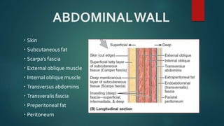
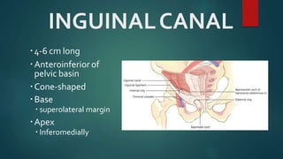
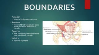
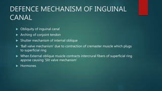
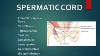
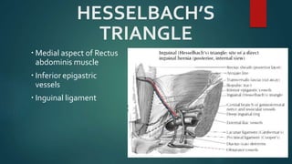
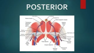
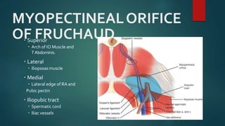
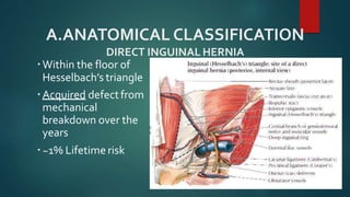
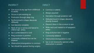
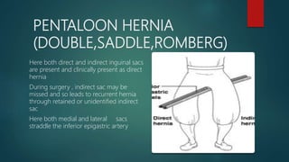
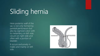
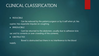
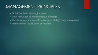
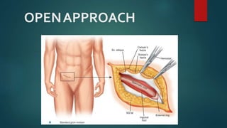
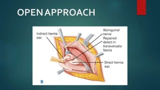
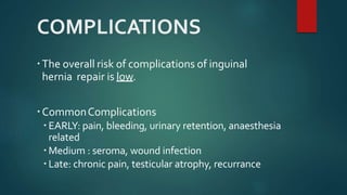
INGUINAL HERNIA
Dr Faisal Ali
PGR SU2
WHAT IS AN
INGUINAL HERNIA?
Protrusion of a peritoneal sac through a
musculoaponeurotic barrier in inguinal
area
ANATOMY
Inguinal Hernia
ABDOMINALWALL
 Skin
 Subcutaneous fat
 Scarpa’s fascia
 External oblique muscle
 Internal oblique muscle
 Transversus abdominis
 Transveralis fascia
 Preperitoneal fat
 Peritoneum
INGUINALCANAL
4-6 cm long
Anteroinferior of
pelvic basin
Cone-shaped
Base
 superolateral margin
Apex
 Inferomedially
BOUNDARIES
 Anterior
 external oblique aponeurosis
 Posterior
 fusion of the transversalis fascia
and transversus abdominus
muscle,
 Superior
 arch formed by the fibers of the
internal oblique muscle.
 Inferior
 inguinal ligament
DEFENCE MECHANISM OF INGUINAL
CANAL
 Obliquity of inguinal canal
 Arching of conjoint tendon
 Shutter mechanism of internal oblique
 ‘Ball valve mechanism’ due to contraction of cremaster muscle which plugs
to superficial ring
 When External oblique muscle contracts intercrural fibers of superficial ring
appose causing ‘Slit valve mechanism’
 Hormones
SPERMATICCORD
 Cremasteric muscle
fibers
 Vas deferens
 Testicular artery
 Testicular
pampiniform
venous plexus
 Genital branch of
the genitofemoral
HESSELBACH’S
TRIANGLE
 Medial aspect of Rectus
abdominis muscle
 Inferior epigastric
vessels
 Inguinal ligament
POSTERIOR
MYOPECTINEALORIFICE
OF FRUCHAUD Superior
 Arch of IO Muscle and
TAbdominis.
 Lateral
 Iliopsoas muscle
 Medial
 Lateral edge of RA and
Pubic pectin
 Iliopubic tract
 Spermatic cord
 Iliac vessels
TRIANGLE OF DOOM
 External iliac vessels
 Deep circumflex iliac vein
 Femoral nerve
 Genital branch ofGFnerve
TRIANGLE OF PAIN
Nerves
 Lateral femoral cutaneous
 Femoral branch of GF nerve
 Femoral nerve
CLASSIFICATION
Inguinal Hernia
A.ANATOMICAL CLASSIFICATION
DIRECT INGUINAL HERNIA
Within the floor of
Hesselbach’s triangle
Acquired defectfrom
mechanical
breakdown over the
years
~1% Lifetime risk
INDIRECT INGUINAL HERNIA
Through the internal ring
of inguinal canal
Congenital
Patent processus
vaginalis
~5% Lifetime risk
Higher risk of
strangulation than direct
INDIRECT INGUINAL HERNIA
ACCORDING TO EXTENT
INDIRECT INGUINAL HERNIA
 INCOMPLETE
BUBONOCELE: Here sac is confined to the inguinal canal
FUNICULAR: Here sac crosses the superficial ring but does not
reach the bottom of the scrotum.
 Complete : Here sac descends to the bottom of scrotum
INDIRECT
 Can occur at any age from childhood
to adults
 Occurs in pre-existing sac
 Protrusion through deep ring
 Pyriform/oval in shape, descends
obliquely
 Can become complete
 Neck of sac is narrow and lateral to
inferior epi- artery
 Sac is anterolateral to cord
 Ring occlusion is positive
 Commonly unilateral but can be
bilateral
 Obstruction/strangulation is common
 Sac should be opened during surgery
DIRECT
 Common in elderly
 Always acquired
 Herniation through posterior wall
 Globular/round in shape, descends
directly forward
 Descent down in the scrotum is rare
 Wide neck and medial to inf epigastric
artery
 Ring occlusion test is negative
 Commonly bilateral
 Obstruction/strangulation is rare
 Sac is not necessarily opened unless
obstruction is present
PENTALOON HERNIA
(DOUBLE,SADDLE,ROMBERG)
Here both direct and indirect inguinal sacs
are present and clinically present as direct
hernia
During surgery , indirect sac may be
missed and so leads to recurrent hernia
through retained or unidentified indirect
sac
Here both medial and lateral sacs
straddle the inferior epigastric artery
Sliding hernia
Here posterior wall of the
sac is not only formed by
parietal peritoneum but
also by sigmoid colon with
its mesentery on left side;
cecum on right side and
often with a portion of
bladder.
It occurs exclusively in
males and mainly on left
side.
CLINICAL CLASSIFICATION
 REDUCIBLE
Can be reduced by the patient,surgeon or by it self when pt. lies
supine. Has expansile impulse on coughing.
 IRREDUCIBLE
Cant be returned to the abdomen, usually due to adhesion b/w
sac and its contents or over crowding of the contents.
 OBSTRUCTED
Bowel is obstructed but there in no interference to the blood
supply.
STRANGULATION
 When blood supply of contents of hernia is seriously impaired leading to
formation of gangrene.
Obstruction impaired venous return Congestion Further
dilatation of bowel which becomes purple colored Fluid collects in the
sac Eventually arterial supply is compromised Bowel becomes dark
brownish with friable wall Bacteria migrate to fluid of sac
Perforation occurs at site of constriction ring Peritonitis
CAUSES OF STRANGULATION
 Narrow neck
 Adhesions
 Irreducibility
 Long time large hernia with adhesions
RICHTERS HERNIA
 This is the partial enterocele when only the anti-mesenteric margin of the
gut is strangulated in the sac.
MAYDLS HERNIA
 When a W-shaped loop of gut lies in the hernia sac and the intervening
loop is strangulated within the main abdominal cavity
 Hernial sac containing a strangulated MECKELS diverticulum. It can
progress to gangrene .
LITTRES HERNIA
NEWER CLASSIFICATION
 Gilbert classification
 NYHUS classification
 BENDAVID classification
 Halverson and McVay classification
 Ponka classification
NYHUS CLASSIFICATIONSYSTEM
Type I INDIRECT HERNIA; internal abdominal ring normal; typically in infants, children, smalladults
Type II
INDIRECT HERNIA; internal ring enlarged without impingement on the floor of theinguinal
canal; does not extend to the scrotum
Type IIIA DIRECT HERNIA; size is not taken intoaccount
Type IIIB
INDIRECT HERNIA that has enlarged enough to encroach upon the posterior inguinalwall;
INDIRECT SLIDINGOR SCROTAL HERNIAS are usually placed in this category because theyare
commonly associated with EXTENSIONTOTHE DIRECT SPACE; also includes PANTALOON
HERNIAS
Type IIIC FEMORAL HERNIA
Type IV
RECURRENT HERNIA; modifiers A–D are sometimes added, which correspondTOINDIRECT,
DIRECT, FEMORAL,AND MIXED,RESPECTIVELY
EPIDEMIOLOGY
One of the most common surgical procedures
Incidence:
~5-10% lifetime
75% of abdominal wall hernias
Male > Female
Indirect > Direct
Right > Left
1/3 may develop a contralateral inguinalhernia
ETIOLOGY
 Multifactorial
 Weakness in abdominal wall musculature
PRESUMEDCAUSESOF GROIN HERNIATION
Coughing Valsalva's maneuvers
Chronic obstructive pulmonary disease Ascites
Obesity Upright position
Straining Congenital connective tissue disorders
Constipation Defective collagen synthesis
Prostatism Previous right lower quadrant incision
Pregnancy Arterial aneurysms
Birthweight <1500 g Cigarette smoking
Family history of a hernia Heavy lifting
Physical exertion (?)
DIAGNOSIS
HISTORY
 More common in males(20:1)
 Dragging pain and swelling in
the groin which is better seen
while coughing and standing.
 History of
Constipation
Cough and Smoking
Urological symptoms
 Duration
 Progressiveness
 Any h/o surgery
(appendectomy)
 Reccurent hernia or
contralateral
PHYSICAL EXAMINATION
 Proper exposure from umbilicus to
mid thigh
 Inspection
 Standing
 lying
 Palpation
 Can you get above the swelling
 Reducibility
 Cough impulse
 Head or leg raise to look for
abdominal muscle tone
 Internal Ring Occlusiontest
EXAMINATION
 Zieman’s test
index finger on deep ring
middle finger on superficial ring
ring finger on femoral opening
 Ring invagination test
little/index finger is invaginated from
Bottom of scrotum gradually pushed to enter
In superficial inguinal ring, impulse on coughing is felt
At tip of invaginated finger.
EXAMINATION
 Always abdominal, urological and respiratory examination is done to rule
out any precipitating factor like Chronic Bronchitis , Ascites, Stricture
urethra and BPH.
DIFFERENTIAL DIAGNOSIS
 Malignancy
 Lymphoma
 Retroperitoneal sarcoma
 Metastasis
 Testicular tumor
 Primary testicular
 Varicocele
 Epididymitis
 Testicular torsion
 Hydrocele
 Ectopic testicle
 Undescended testicle
 Femoral artery aneurysm or
pseudoaneurysm
 Lymph node
 Sebaceous cyst
 Hidradenitis
 Cyst of the canal of Nuck (female)
 Saphenous varix
 Psoas abscess
 Hematoma
 Ascites
IMAGING
Inguinal Hernia
CXR
Ultrasound
CTScan
MRI
MANAGEMENT
MANAGEMENT PRINCIPLES
 Not all hernias require surgical repair
 Small hernias can be more dangerous than large
 Pain, tenderness and skin colour changes imply high risk of strangulation
 Femoral hernia should always be repaired
CONSERVATIVE MANAGEMENT
Aimed at alleviating symptoms such as
pain, pressure, and protrusion of abdominal
contents
Assuming a recumbent position
Truss, an elastic belt or brief
EMERGENT REPAIR
Incarcerated hernias
Strangulated hernias
Sliding hernias
TAXIS
 The patient is sedated and placed in aTrendelenburg position.
 The hernia sac is grasped with both hands, elongated, and then
milked back through the hernia defect.
 Pressure applied to the most distal portion of the sac will cause the
contents to mushroom and prevent reduction.
STRANGULATED HERNIA
 Femoral > Indirect > Direct
 Fever, leukocytosis, and hemodynamic instability.
 The hernia bulge usually is very tender, warm, and may exhibit
red discoloration.
 Taxis should not be applied to strangulated hernias as a
potentially gangrenous portion of bowel may be reduced into the
abdomen without being addressed
OPERATIVETECHNIQUES
Inguinal hernia
PRINCIPLES OF SURGICAL REPAIR
 Reduction of the hernia content into the abdominal cavity
 Excision and closure of peritoneal sac
 Reapproximation of the walls of the neck of the hernia
 Permanent reinforcement of the abdominal wall defect with sutures or
mesh
SURGICAL OPTIONS
 Herniotomy : excision of hernia sac
in children
 Hernirraphy : strengthening of posterior wall of inguinal canal
 Hernioplasty : placement of mesh
ANTERIOR REPAIR
NON PROSTHETIC
OPENAPPROACH
OPENAPPROACH
BASSINI REPAIR
 Is frequently used for
indirect inguinal
hernias and small direct
hernias
 The conjoined tendon of
the transversus abdominis
and the internal oblique
muscles is sutured to the
inguinal ligament
LYTLE’S REPAIR
Only internal ring is narrowed
by placing interrupted sutures
over the medial side of the
ring to the fascia transversalis
MCVAY REPAIR
 inguinal and femoral
canal defects
 The conjoined tendon is
sutured to Cooper’s
ligament from the pubic
cubicle laterally
SHOULDICE
REPAIR
Multilayered
Transversalis fascia is incised,
lower flap is sutured to
posterior part of upper flap
Upper flap is sutured to
inguinal ligament.
Then conjoint tendon and
inguinal ligament is further
approximated by two layers in
front of cord .
Then Ext Oblique apo is sutured
in two layers.
MALONY DARN REPAIR
Posterior wall is re-inforced
with monofilament nylon
darn. The Darn is made in
crisscross fashion between
the conjoint tendon and
inguinal ligament
Excellent results.
Most commonly performed
in countries where mesh is
too expensive
MESH
 Gross structure
Net meshes or flat sheets
 Synthetic mesh
Polypropylene, polyester and polytetrafloroethylen PTFE
 Weight and porosity
Dense or heavy weight and Light weight, Large pore meshes
 Biological mesh
Sheets of sterilized, decellularised, non immunogenic connective tiisue
LICHTENSTEIN TENSION- FREE
REPAIR
LICHTENSTEINTENSION- FREE
REPAIR
 Initial part of surgery is same as Bassini repair.
 Once hernia has been removed and any medial defect closed, a piece of
mesh measuring 8-15cm is placed over posterior wall, behind the
spermatic cord and is split to wrap around the spermatic cord at deep
inguinal ring.
 Loose sutures with Prolone hold mesh to inguinal ligament and conjoint
tendon
 ADVANTAGES
Lowered recurrence rates
Accelerated postoperative delivery
Open plug/device/complex
mesh repair.
Gillbert mesh repair ;
plug and
patch, internal ring is
plugged by a cone shaped
piece of prolene mesh
PHS (Prolene hernia
system)
onlay and
sublay sandwich technique
LAPAROSCOPIC HERNIA
REPAIRTransabdominal Preperitoneal Procedure(TAPP)
Totally Extraperitoneal (TEP) Repair
Indications include
bilateral inguinal hernia,
recurrent hernia,
need for early recovery
Transabdominal Preperitoneal
Procedure (TAPP)
 Surgeon enters the peritoneal cavity incises the peritoneum above the
hernia defects and reflects it away from the muscles entering Pre-
peritonealy
 Hernia is reduced and a 10*15 cm mesh is placed just deep to the
abdominal wall extending across midline into retropubic space and 5cm
lateral to the deep inguinal ring.
Totally Extraperitoneal (TEP) Repair
 Space is created just deep to the
abdominal msucles without entering
the peritoneal cavity
 Hernia is reduced and mesh is
inserted preperitonealy.
RECURRENCE
Bassini repair 10%
Around 1% for Shouldice repair
Hernioplasty 1-3%
Most recurrences are of the same type as the original
hernia
Recurrence Factors
 Patient
 Technical
 Tissue
RECURRENCE
Patient factors
 malnutrition, immunosuppression, diabetes, steroid
use, and smoking.
Technical factors
 mesh size, prosthesis fixation, and technical proficiency of
the surgeon.
Tissue factors
 wound infection, tissue ischemia, and increased tension
within the surgical repair
COMPLICATIONS
The overall risk of complications of inguinal
hernia repair is low.
CommonComplications
 EARLY: pain, bleeding, urinary retention, anaesthesia
related
 Medium : seroma, wound infection
 Late: chronic pain, testicular atrophy, recurrance
THANK
YOU

inguinal hernia

  • 1.
  • 2.
    WHAT IS AN INGUINALHERNIA? Protrusion of a peritoneal sac through a musculoaponeurotic barrier in inguinal area
  • 3.
  • 4.
    ABDOMINALWALL  Skin  Subcutaneousfat  Scarpa’s fascia  External oblique muscle  Internal oblique muscle  Transversus abdominis  Transveralis fascia  Preperitoneal fat  Peritoneum
  • 5.
    INGUINALCANAL 4-6 cm long Anteroinferiorof pelvic basin Cone-shaped Base  superolateral margin Apex  Inferomedially
  • 6.
    BOUNDARIES  Anterior  externaloblique aponeurosis  Posterior  fusion of the transversalis fascia and transversus abdominus muscle,  Superior  arch formed by the fibers of the internal oblique muscle.  Inferior  inguinal ligament
  • 7.
    DEFENCE MECHANISM OFINGUINAL CANAL  Obliquity of inguinal canal  Arching of conjoint tendon  Shutter mechanism of internal oblique  ‘Ball valve mechanism’ due to contraction of cremaster muscle which plugs to superficial ring  When External oblique muscle contracts intercrural fibers of superficial ring appose causing ‘Slit valve mechanism’  Hormones
  • 8.
    SPERMATICCORD  Cremasteric muscle fibers Vas deferens  Testicular artery  Testicular pampiniform venous plexus  Genital branch of the genitofemoral
  • 9.
    HESSELBACH’S TRIANGLE  Medial aspectof Rectus abdominis muscle  Inferior epigastric vessels  Inguinal ligament
  • 10.
  • 11.
    MYOPECTINEALORIFICE OF FRUCHAUD Superior Arch of IO Muscle and TAbdominis.  Lateral  Iliopsoas muscle  Medial  Lateral edge of RA and Pubic pectin  Iliopubic tract  Spermatic cord  Iliac vessels
  • 12.
    TRIANGLE OF DOOM External iliac vessels  Deep circumflex iliac vein  Femoral nerve  Genital branch ofGFnerve
  • 13.
    TRIANGLE OF PAIN Nerves Lateral femoral cutaneous  Femoral branch of GF nerve  Femoral nerve
  • 14.
  • 15.
    A.ANATOMICAL CLASSIFICATION DIRECT INGUINALHERNIA Within the floor of Hesselbach’s triangle Acquired defectfrom mechanical breakdown over the years ~1% Lifetime risk
  • 16.
    INDIRECT INGUINAL HERNIA Throughthe internal ring of inguinal canal Congenital Patent processus vaginalis ~5% Lifetime risk Higher risk of strangulation than direct
  • 17.
  • 18.
    ACCORDING TO EXTENT INDIRECTINGUINAL HERNIA  INCOMPLETE BUBONOCELE: Here sac is confined to the inguinal canal FUNICULAR: Here sac crosses the superficial ring but does not reach the bottom of the scrotum.  Complete : Here sac descends to the bottom of scrotum
  • 19.
    INDIRECT  Can occurat any age from childhood to adults  Occurs in pre-existing sac  Protrusion through deep ring  Pyriform/oval in shape, descends obliquely  Can become complete  Neck of sac is narrow and lateral to inferior epi- artery  Sac is anterolateral to cord  Ring occlusion is positive  Commonly unilateral but can be bilateral  Obstruction/strangulation is common  Sac should be opened during surgery DIRECT  Common in elderly  Always acquired  Herniation through posterior wall  Globular/round in shape, descends directly forward  Descent down in the scrotum is rare  Wide neck and medial to inf epigastric artery  Ring occlusion test is negative  Commonly bilateral  Obstruction/strangulation is rare  Sac is not necessarily opened unless obstruction is present
  • 20.
    PENTALOON HERNIA (DOUBLE,SADDLE,ROMBERG) Here bothdirect and indirect inguinal sacs are present and clinically present as direct hernia During surgery , indirect sac may be missed and so leads to recurrent hernia through retained or unidentified indirect sac Here both medial and lateral sacs straddle the inferior epigastric artery
  • 21.
    Sliding hernia Here posteriorwall of the sac is not only formed by parietal peritoneum but also by sigmoid colon with its mesentery on left side; cecum on right side and often with a portion of bladder. It occurs exclusively in males and mainly on left side.
  • 22.
    CLINICAL CLASSIFICATION  REDUCIBLE Canbe reduced by the patient,surgeon or by it self when pt. lies supine. Has expansile impulse on coughing.  IRREDUCIBLE Cant be returned to the abdomen, usually due to adhesion b/w sac and its contents or over crowding of the contents.  OBSTRUCTED Bowel is obstructed but there in no interference to the blood supply.
  • 23.
    STRANGULATION  When bloodsupply of contents of hernia is seriously impaired leading to formation of gangrene. Obstruction impaired venous return Congestion Further dilatation of bowel which becomes purple colored Fluid collects in the sac Eventually arterial supply is compromised Bowel becomes dark brownish with friable wall Bacteria migrate to fluid of sac Perforation occurs at site of constriction ring Peritonitis
  • 24.
    CAUSES OF STRANGULATION Narrow neck  Adhesions  Irreducibility  Long time large hernia with adhesions
  • 25.
    RICHTERS HERNIA  Thisis the partial enterocele when only the anti-mesenteric margin of the gut is strangulated in the sac.
  • 26.
    MAYDLS HERNIA  Whena W-shaped loop of gut lies in the hernia sac and the intervening loop is strangulated within the main abdominal cavity  Hernial sac containing a strangulated MECKELS diverticulum. It can progress to gangrene . LITTRES HERNIA
  • 27.
    NEWER CLASSIFICATION  Gilbertclassification  NYHUS classification  BENDAVID classification  Halverson and McVay classification  Ponka classification
  • 28.
    NYHUS CLASSIFICATIONSYSTEM Type IINDIRECT HERNIA; internal abdominal ring normal; typically in infants, children, smalladults Type II INDIRECT HERNIA; internal ring enlarged without impingement on the floor of theinguinal canal; does not extend to the scrotum Type IIIA DIRECT HERNIA; size is not taken intoaccount Type IIIB INDIRECT HERNIA that has enlarged enough to encroach upon the posterior inguinalwall; INDIRECT SLIDINGOR SCROTAL HERNIAS are usually placed in this category because theyare commonly associated with EXTENSIONTOTHE DIRECT SPACE; also includes PANTALOON HERNIAS Type IIIC FEMORAL HERNIA Type IV RECURRENT HERNIA; modifiers A–D are sometimes added, which correspondTOINDIRECT, DIRECT, FEMORAL,AND MIXED,RESPECTIVELY
  • 29.
    EPIDEMIOLOGY One of themost common surgical procedures Incidence: ~5-10% lifetime 75% of abdominal wall hernias Male > Female Indirect > Direct Right > Left 1/3 may develop a contralateral inguinalhernia
  • 30.
    ETIOLOGY  Multifactorial  Weaknessin abdominal wall musculature PRESUMEDCAUSESOF GROIN HERNIATION Coughing Valsalva's maneuvers Chronic obstructive pulmonary disease Ascites Obesity Upright position Straining Congenital connective tissue disorders Constipation Defective collagen synthesis Prostatism Previous right lower quadrant incision Pregnancy Arterial aneurysms Birthweight <1500 g Cigarette smoking Family history of a hernia Heavy lifting Physical exertion (?)
  • 31.
  • 32.
    HISTORY  More commonin males(20:1)  Dragging pain and swelling in the groin which is better seen while coughing and standing.  History of Constipation Cough and Smoking Urological symptoms  Duration  Progressiveness  Any h/o surgery (appendectomy)  Reccurent hernia or contralateral
  • 33.
    PHYSICAL EXAMINATION  Properexposure from umbilicus to mid thigh  Inspection  Standing  lying  Palpation  Can you get above the swelling  Reducibility  Cough impulse  Head or leg raise to look for abdominal muscle tone  Internal Ring Occlusiontest
  • 34.
    EXAMINATION  Zieman’s test indexfinger on deep ring middle finger on superficial ring ring finger on femoral opening  Ring invagination test little/index finger is invaginated from Bottom of scrotum gradually pushed to enter In superficial inguinal ring, impulse on coughing is felt At tip of invaginated finger.
  • 35.
    EXAMINATION  Always abdominal,urological and respiratory examination is done to rule out any precipitating factor like Chronic Bronchitis , Ascites, Stricture urethra and BPH.
  • 36.
    DIFFERENTIAL DIAGNOSIS  Malignancy Lymphoma  Retroperitoneal sarcoma  Metastasis  Testicular tumor  Primary testicular  Varicocele  Epididymitis  Testicular torsion  Hydrocele  Ectopic testicle  Undescended testicle  Femoral artery aneurysm or pseudoaneurysm  Lymph node  Sebaceous cyst  Hidradenitis  Cyst of the canal of Nuck (female)  Saphenous varix  Psoas abscess  Hematoma  Ascites
  • 37.
  • 38.
  • 39.
  • 40.
    MANAGEMENT PRINCIPLES  Notall hernias require surgical repair  Small hernias can be more dangerous than large  Pain, tenderness and skin colour changes imply high risk of strangulation  Femoral hernia should always be repaired
  • 42.
    CONSERVATIVE MANAGEMENT Aimed atalleviating symptoms such as pain, pressure, and protrusion of abdominal contents Assuming a recumbent position Truss, an elastic belt or brief
  • 43.
  • 44.
    TAXIS  The patientis sedated and placed in aTrendelenburg position.  The hernia sac is grasped with both hands, elongated, and then milked back through the hernia defect.  Pressure applied to the most distal portion of the sac will cause the contents to mushroom and prevent reduction.
  • 45.
    STRANGULATED HERNIA  Femoral> Indirect > Direct  Fever, leukocytosis, and hemodynamic instability.  The hernia bulge usually is very tender, warm, and may exhibit red discoloration.  Taxis should not be applied to strangulated hernias as a potentially gangrenous portion of bowel may be reduced into the abdomen without being addressed
  • 46.
  • 47.
    PRINCIPLES OF SURGICALREPAIR  Reduction of the hernia content into the abdominal cavity  Excision and closure of peritoneal sac  Reapproximation of the walls of the neck of the hernia  Permanent reinforcement of the abdominal wall defect with sutures or mesh
  • 48.
    SURGICAL OPTIONS  Herniotomy: excision of hernia sac in children  Hernirraphy : strengthening of posterior wall of inguinal canal  Hernioplasty : placement of mesh
  • 49.
  • 50.
  • 51.
  • 52.
    BASSINI REPAIR  Isfrequently used for indirect inguinal hernias and small direct hernias  The conjoined tendon of the transversus abdominis and the internal oblique muscles is sutured to the inguinal ligament
  • 53.
    LYTLE’S REPAIR Only internalring is narrowed by placing interrupted sutures over the medial side of the ring to the fascia transversalis
  • 54.
    MCVAY REPAIR  inguinaland femoral canal defects  The conjoined tendon is sutured to Cooper’s ligament from the pubic cubicle laterally
  • 55.
    SHOULDICE REPAIR Multilayered Transversalis fascia isincised, lower flap is sutured to posterior part of upper flap Upper flap is sutured to inguinal ligament. Then conjoint tendon and inguinal ligament is further approximated by two layers in front of cord . Then Ext Oblique apo is sutured in two layers.
  • 56.
    MALONY DARN REPAIR Posteriorwall is re-inforced with monofilament nylon darn. The Darn is made in crisscross fashion between the conjoint tendon and inguinal ligament Excellent results. Most commonly performed in countries where mesh is too expensive
  • 57.
    MESH  Gross structure Netmeshes or flat sheets  Synthetic mesh Polypropylene, polyester and polytetrafloroethylen PTFE  Weight and porosity Dense or heavy weight and Light weight, Large pore meshes  Biological mesh Sheets of sterilized, decellularised, non immunogenic connective tiisue
  • 58.
  • 59.
    LICHTENSTEINTENSION- FREE REPAIR  Initialpart of surgery is same as Bassini repair.  Once hernia has been removed and any medial defect closed, a piece of mesh measuring 8-15cm is placed over posterior wall, behind the spermatic cord and is split to wrap around the spermatic cord at deep inguinal ring.  Loose sutures with Prolone hold mesh to inguinal ligament and conjoint tendon  ADVANTAGES Lowered recurrence rates Accelerated postoperative delivery
  • 60.
    Open plug/device/complex mesh repair. Gillbertmesh repair ; plug and patch, internal ring is plugged by a cone shaped piece of prolene mesh PHS (Prolene hernia system) onlay and sublay sandwich technique
  • 61.
    LAPAROSCOPIC HERNIA REPAIRTransabdominal PreperitonealProcedure(TAPP) Totally Extraperitoneal (TEP) Repair Indications include bilateral inguinal hernia, recurrent hernia, need for early recovery
  • 62.
    Transabdominal Preperitoneal Procedure (TAPP) Surgeon enters the peritoneal cavity incises the peritoneum above the hernia defects and reflects it away from the muscles entering Pre- peritonealy  Hernia is reduced and a 10*15 cm mesh is placed just deep to the abdominal wall extending across midline into retropubic space and 5cm lateral to the deep inguinal ring.
  • 63.
    Totally Extraperitoneal (TEP)Repair  Space is created just deep to the abdominal msucles without entering the peritoneal cavity  Hernia is reduced and mesh is inserted preperitonealy.
  • 64.
    RECURRENCE Bassini repair 10% Around1% for Shouldice repair Hernioplasty 1-3% Most recurrences are of the same type as the original hernia Recurrence Factors  Patient  Technical  Tissue
  • 65.
    RECURRENCE Patient factors  malnutrition,immunosuppression, diabetes, steroid use, and smoking. Technical factors  mesh size, prosthesis fixation, and technical proficiency of the surgeon. Tissue factors  wound infection, tissue ischemia, and increased tension within the surgical repair
  • 66.
    COMPLICATIONS The overall riskof complications of inguinal hernia repair is low. CommonComplications  EARLY: pain, bleeding, urinary retention, anaesthesia related  Medium : seroma, wound infection  Late: chronic pain, testicular atrophy, recurrance
  • 67.