Definition
Definition
A hernia isa protrusion of
a viscus or part of a viscus
through an abnormal
opening in the walls of its
containing cavity .
4.
Anatomy
Anatomy
The inguinalcanal :-
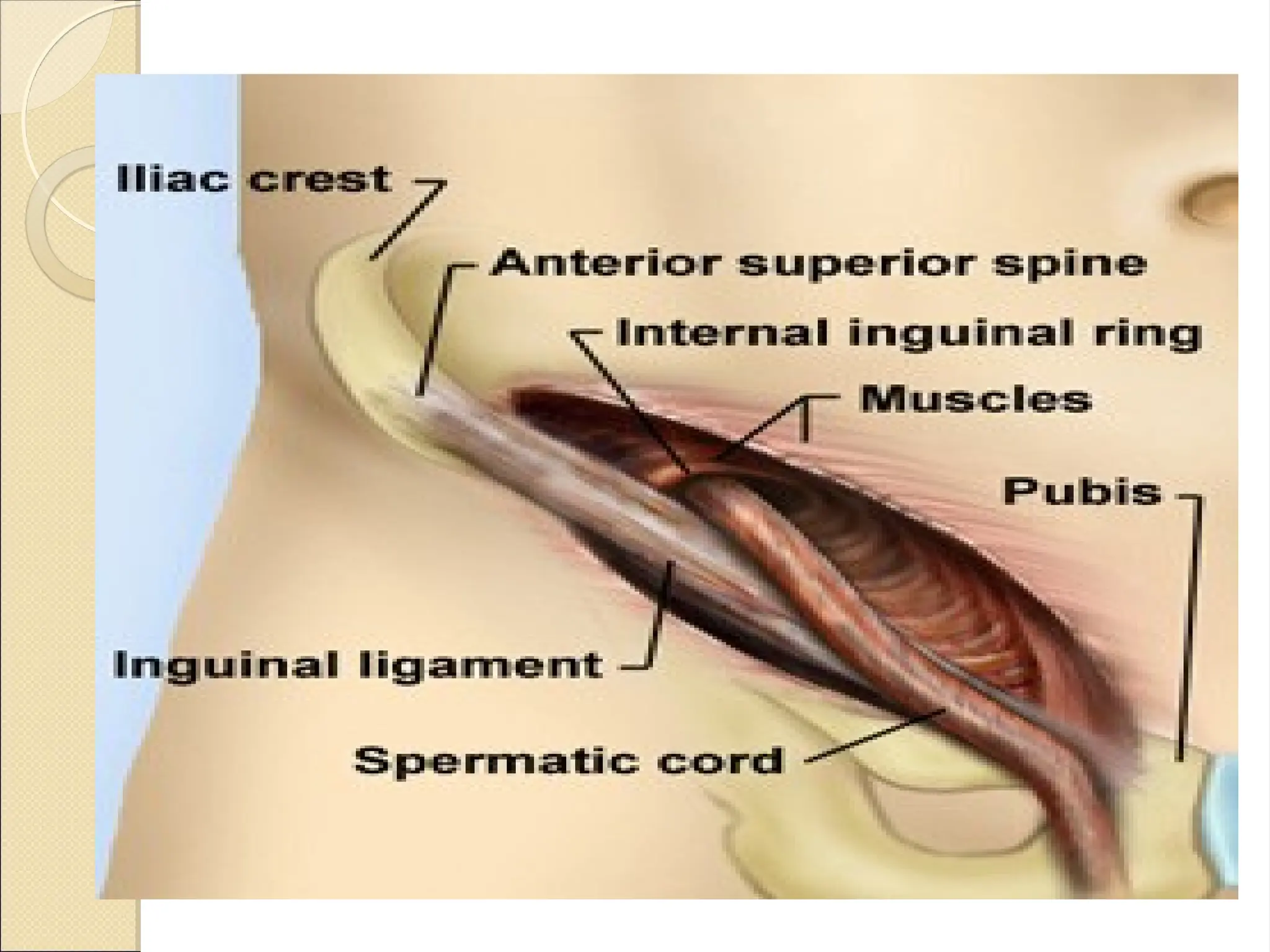
The inguinal canal is approximately 4 cm long and is directed obliquely
inferomedially through the inferior part of the anterolateral abdominal
wall. The canal lies parallel and 2-4 cm superior to the medial half of
the inguinal ligament.This ligament extends from the anterior
superior iliac spine to the pubic tubercle.
The inguinal canal has openings at either end : –
The deep (internal) inguinal ring is the entrance to the inguinal canal. It
is thesite of an outpouching of the transversalis fascia. This is
approximately 1.25 cm superior to the middle of the inguinal
ligament
The superficial, or external inguinal ring is the exit from the inguinal
canal. It is a slitlke opening between the diagonal fibres of the
aponeurosis of the external oblique
5.
Inguinal canal
Inguinal canal
walls of The inguinal canal :-
The anterior wall is formed mainly by the aponeurosis of the
external Oblique
. The posterior wall is formed mainly by transversalis fascia
The roof is formed by the arching fibres of the internal oblique and
transverse abdominal muscles.
The floor is formed by the inguinal ligament, which forms a shallow
trough. It is
reinforced in its most medial part by the lacunar ligament.
7.
Content :-
1. Spermaticcord ( round ligament of the uterus in female )
The Cord Itself.—The contents of the spermatic cord are
(a) the ductus (vas) deferens and its artery .
(b) the testicular artery and venous (pampiniform) plexus.
(c) the genital branch of the genitofemoral nerve.
(d) lymphatic vessels and sympathetic nerve fibers.
(e) fat and connective tissue surrounding the cord and its coverings
in various amounts
2. Ilioinguinal nerve .
3. Ilioinguinal lymph node .
8.
Femoral Canal
Femoral Canal
Themajor feature of the femoral canal is the femoral sheath. This
sheath is a condensation of the deep fascia (fascia lata) of the thigh
and contains, from lateral to medial, the femoral artery, femoral
vein, and femoral canal. The femoral canal is a space medial to the
vein that allows for venous expansion and contains a lymph node
(node of Cloquet). Other features of the femoral triangle include
the femoral nerve, which lies lateral to the sheath,
Wall of The Femoral canal
anterior is the inguinal ligament
posterior is the iliopsoas, pectineal, and long adductor muscles
(floor).
Medial is lacunar ligament
Lateral is femoral vessle
repeated INCREASE in
repeatedINCREASE in
abdominal pressure is usually
abdominal pressure is usually
due to
due to
Chronic cough
Straining
Bladder neck or urethral obstruction
Pregnancy
Vomiting
Sever muscular effort
Ascetic fluid
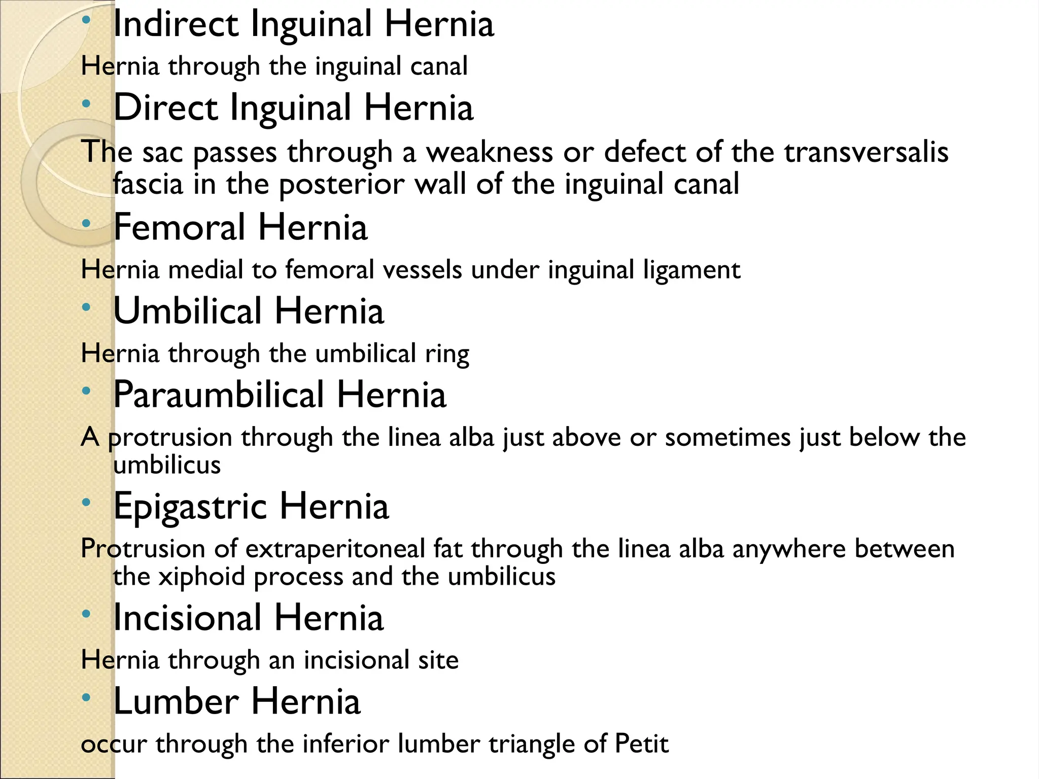
• Indirect InguinalHernia
Hernia through the inguinal canal
• Direct Inguinal Hernia
The sac passes through a weakness or defect of the transversalis
fascia in the posterior wall of the inguinal canal
• Femoral Hernia
Hernia medial to femoral vessels under inguinal ligament
• Umbilical Hernia
Hernia through the umbilical ring
• Paraumbilical Hernia
A protrusion through the linea alba just above or sometimes just below the
umbilicus
• Epigastric Hernia
Protrusion of extraperitoneal fat through the linea alba anywhere between
the xiphoid process and the umbilicus
• Incisional Hernia
Hernia through an incisional site
• Lumber Hernia
occur through the inferior lumber triangle of Petit
13.
Inguinal hernia
Inguinal hernia
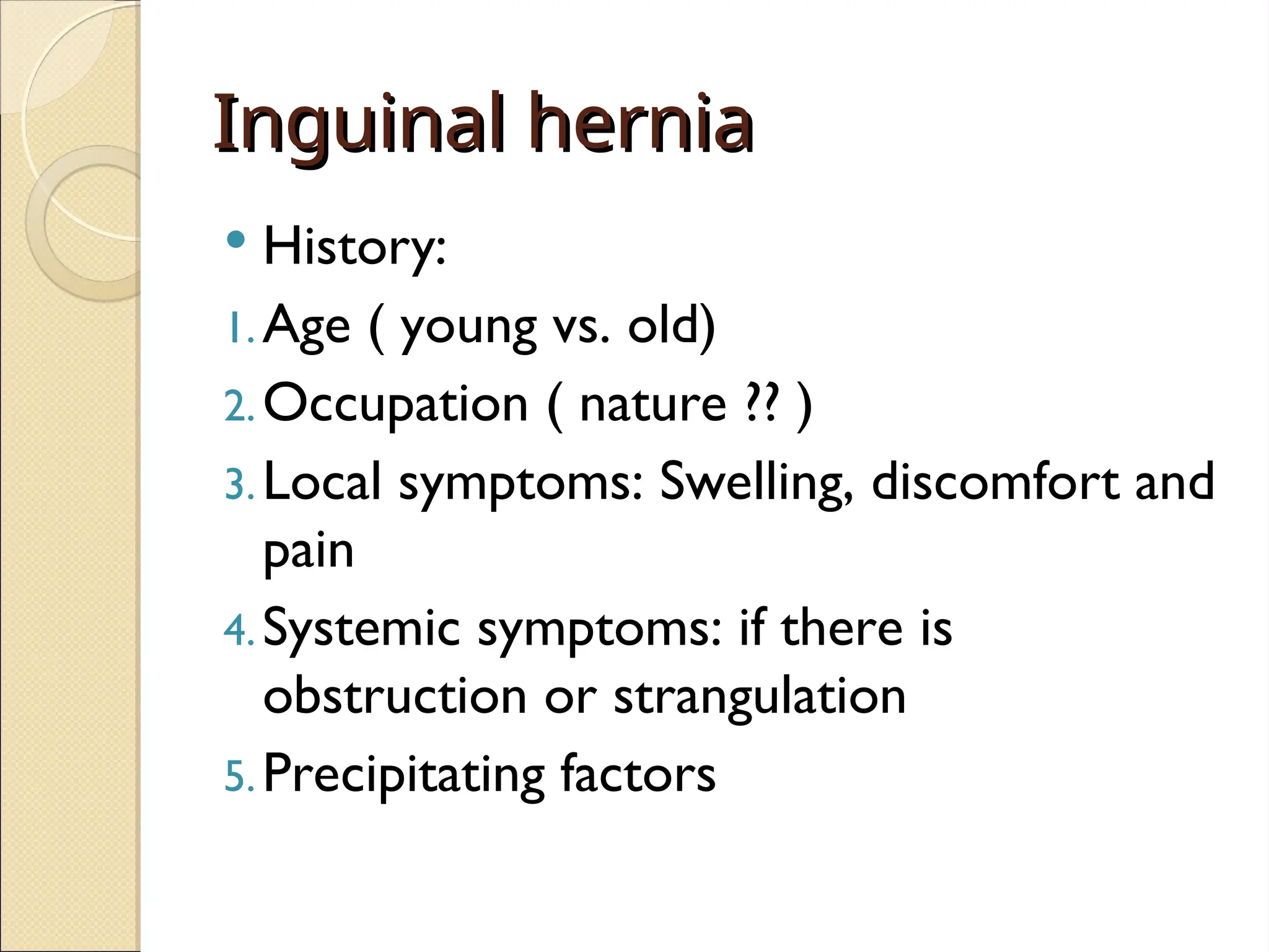
History:
1.Age ( young vs. old)
2.Occupation ( nature ?? )
3.Local symptoms: Swelling, discomfort and
pain
4.Systemic symptoms: if there is
obstruction or strangulation
5.Precipitating factors
14.
Inguinal hernia
Inguinal hernia
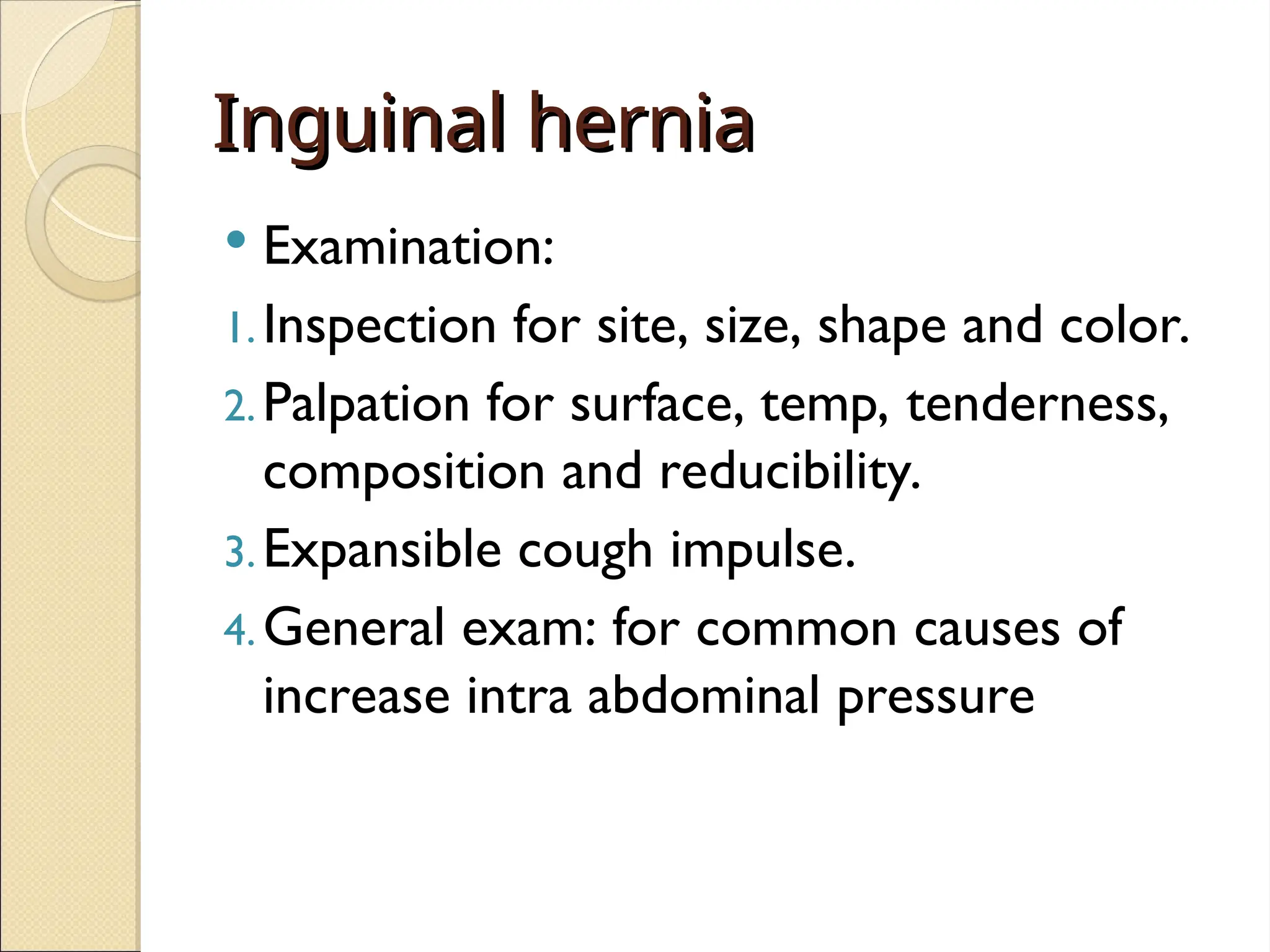
Examination:
1.Inspection for site, size, shape and color.
2.Palpation for surface, temp, tenderness,
composition and reducibility.
3.Expansible cough impulse.
4.General exam: for common causes of
increase intra abdominal pressure
15.
Indirect Versus Directinguinal
Indirect Versus Direct inguinal
hernias
hernias
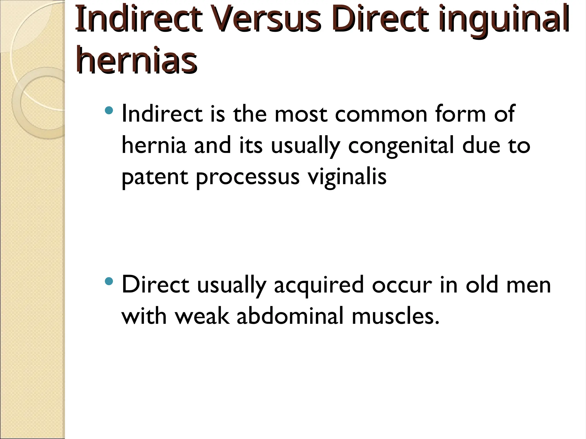
Indirect is the most common form of
hernia and its usually congenital due to
patent processus viginalis
Direct usually acquired occur in old men
with weak abdominal muscles.
16.
Indirect Versus Directinguinal hernias
Indirect Inguinal Hernia Direct Inguinal Hernia
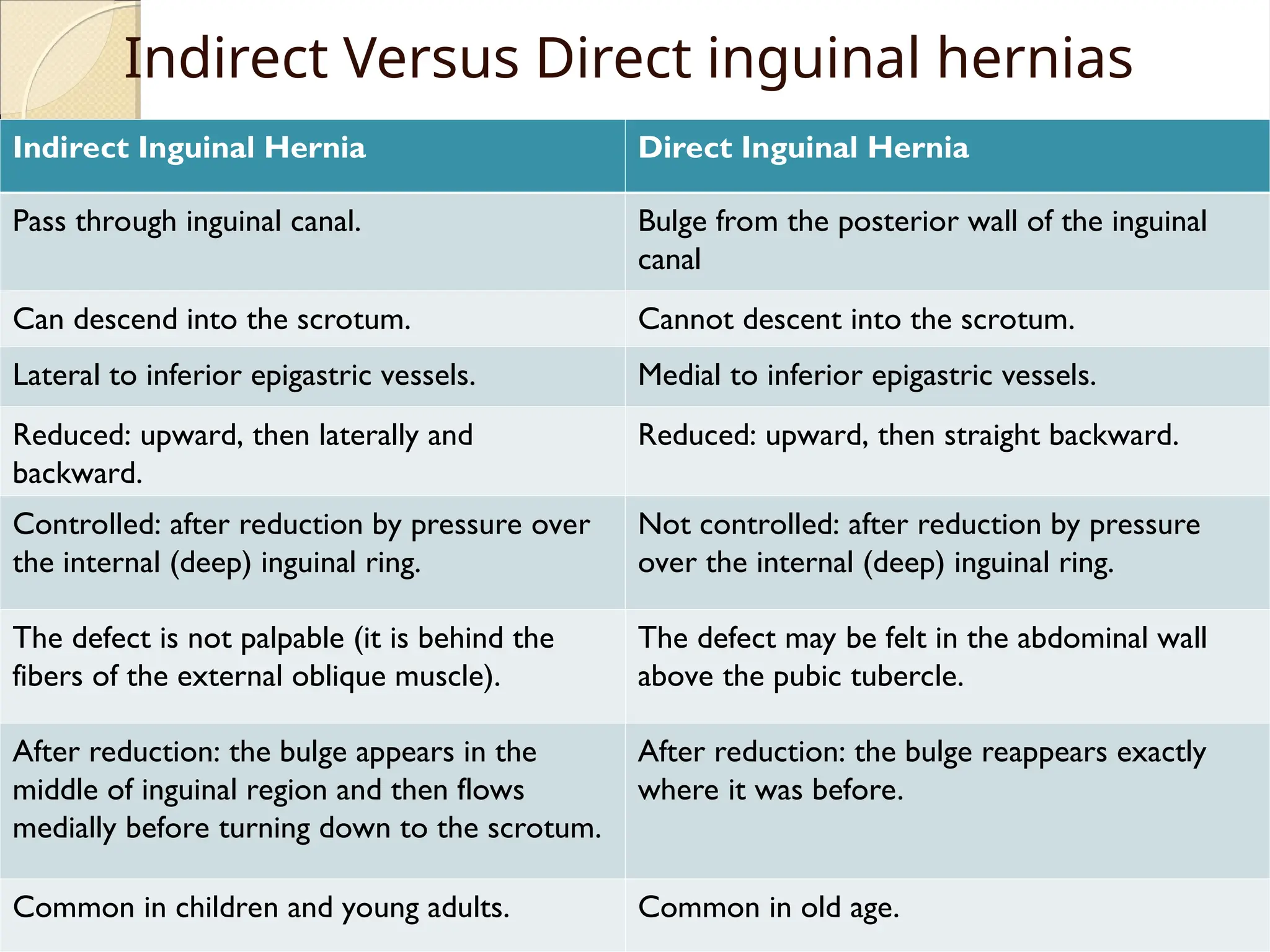
Pass through inguinal canal. Bulge from the posterior wall of the inguinal
canal
Can descend into the scrotum. Cannot descent into the scrotum.
Lateral to inferior epigastric vessels. Medial to inferior epigastric vessels.
Reduced: upward, then laterally and
backward.
Reduced: upward, then straight backward.
Controlled: after reduction by pressure over
the internal (deep) inguinal ring.
Not controlled: after reduction by pressure
over the internal (deep) inguinal ring.
The defect is not palpable (it is behind the
fibers of the external oblique muscle).
The defect may be felt in the abdominal wall
above the pubic tubercle.
After reduction: the bulge appears in the
middle of inguinal region and then flows
medially before turning down to the scrotum.
After reduction: the bulge reappears exactly
where it was before.
Common in children and young adults. Common in old age.
17.
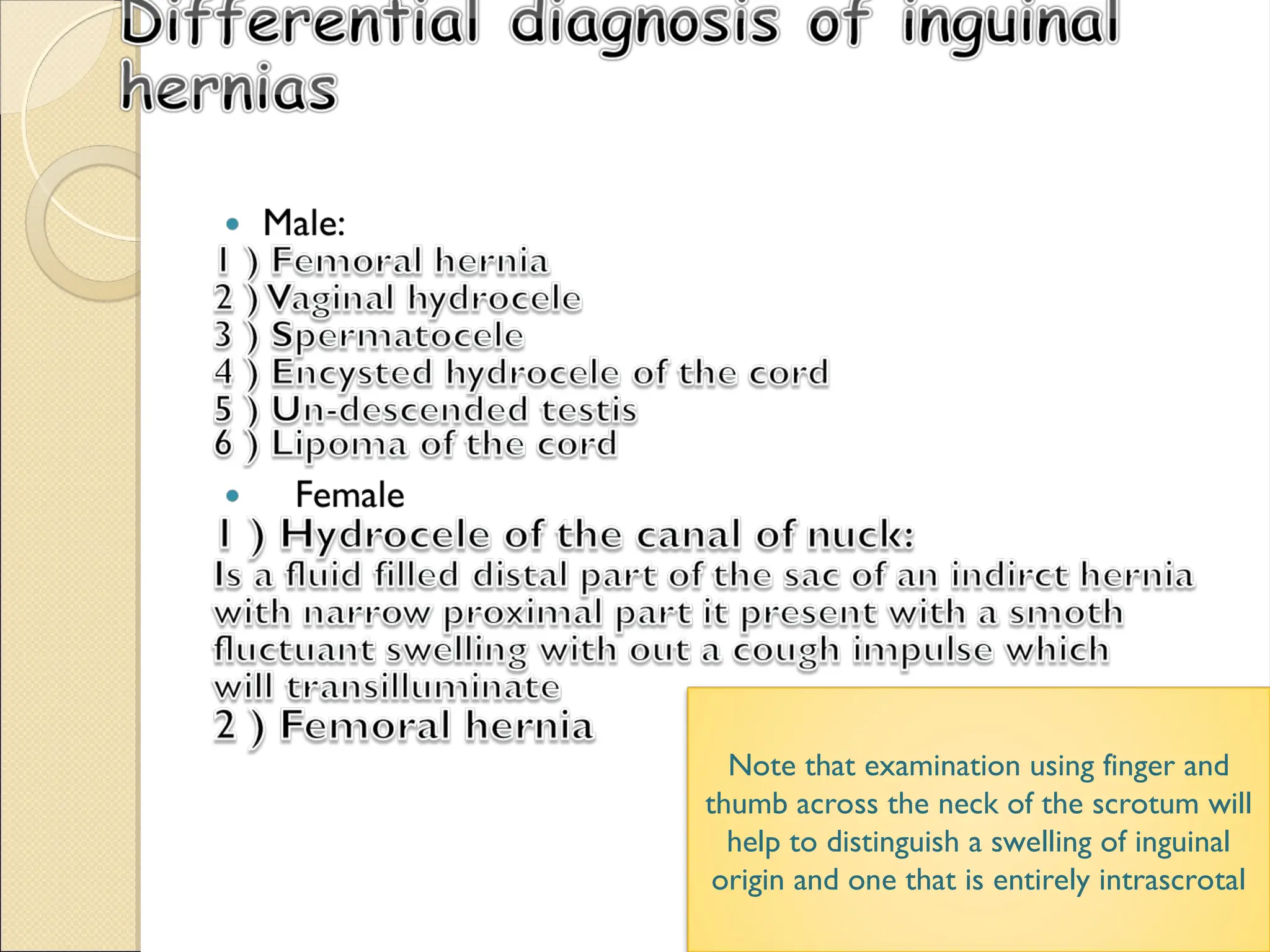
Note that examinationusing finger and
thumb across the neck of the scrotum will
help to distinguish a swelling of inguinal
origin and one that is entirely intrascrotal
18.
Femoral hernia
Femoral hernia
Smallfemoral hernia may be unnoticed by
the patient or disregarded for years
perhaps until the day it strangulates.
Adherence of the greater omentum
sometimes causes a dragging pain. Rarely
a large sac is present .
19.
Femoral hernia
Femoral hernia
History
Age ; uncommon in children , most common
in old age female .
Sex; women > men (but still commonest
hernia in women the inguinal hernia )
The patient came with local symptoms
1- discomfort and pain
2- swelling in the groin
General ; femoral hernia is more likely to be
strangulated than the inguinal hernia
Multiplicity ; often bilateral
20.
Femoral hernia versus
Femoralhernia versus
inguinal hernia
inguinal hernia
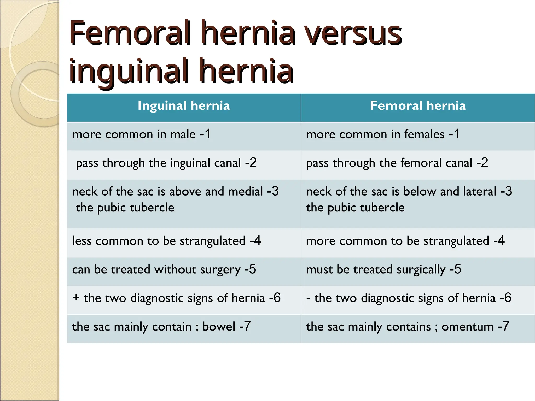
Inguinal hernia Femoral hernia
1
-
more common in male 1
-
more common in females
2
-
pass through the inguinal canal 2
-
pass through the femoral canal
3
-
neck of the sac is above and medial
the pubic tubercle
3
-
neck of the sac is below and lateral
the pubic tubercle
4
-
less common to be strangulated 4
-
more common to be strangulated
5
-
can be treated without surgery 5
-
must be treated surgically
6
-
the two diagnostic signs of hernia
+ 6
-
the two diagnostic signs of hernia
-
7
-
the sac mainly contain ; bowel 7
-
the sac mainly contains ; omentum
21.
Umbilical hernia
Umbilical hernia
Signs and symptoms
Age ; doesn’t appear until the umbilical
cord has separated and healed .
No specific symptoms
Have wide neck and reduce easily , rarely
give intestinal obstruction.
Nature history ; 90 % disappear
spontaneously during the first year.
22.
Examination
Inspection
Site ; in the center of the umbilicus
Size and shape ; size can vary from vary small to
very large . Shape is usually hemispherical.
Palpation
Composition ; contain bowel , which makes it
resonant to percussion . They reduce
spontaneously when the child lies down .
Reducibility ; easy
Cough impulse; invariably present .
23.
Incision hernia
Incision hernia
Signs and symptoms
Previous operation or accidental trauma
Age ; all ages , but more common in old age.
Symptom ; lump ,pain ,intestinal obstruction ( distention ,colic,
vomiting ,constipation , sever pain in the lump )
Examination
1- reducible lump
2- expansile cough impulse
3- if the lump dose not reduse and dose not have cough impulse ,
than it may be not a hernia
Ddx
Tumor
Chronic abscess
Hematoma
Foreign body granuloma
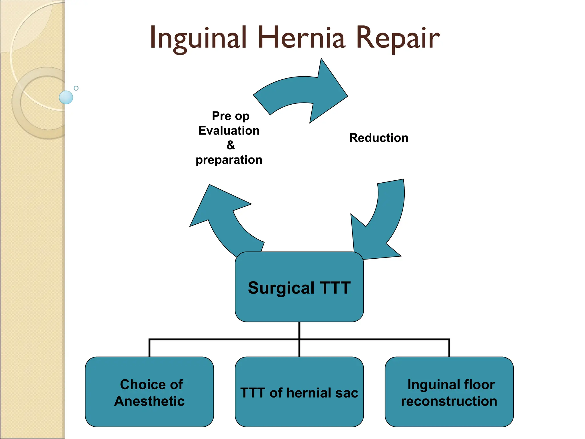
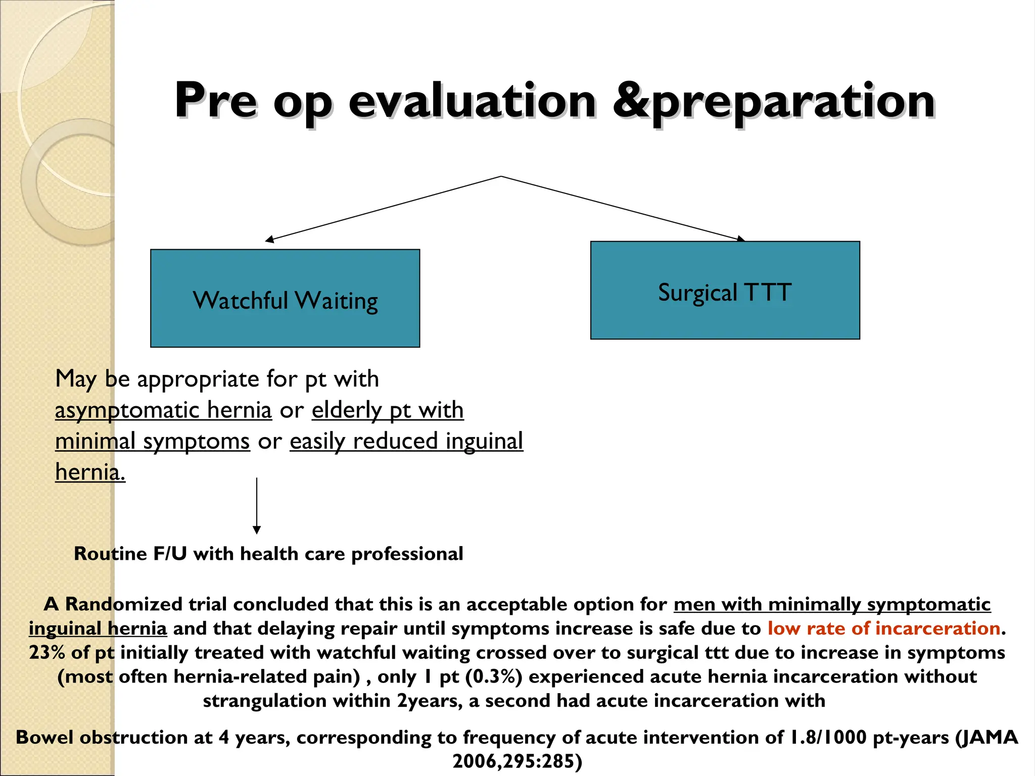
Pre op evaluation&preparation
Pre op evaluation &preparation
Watchful Waiting Surgical TTT
May be appropriate for pt with
asymptomatic hernia or elderly pt with
minimal symptoms or easily reduced inguinal
hernia.
Routine F/U with health care professional
A Randomized trial concluded that this is an acceptable option for men with minimally symptomatic
inguinal hernia and that delaying repair until symptoms increase is safe due to low rate of incarceration.
23% of pt initially treated with watchful waiting crossed over to surgical ttt due to increase in symptoms
(most often hernia-related pain) , only 1 pt (0.3%) experienced acute hernia incarceration without
strangulation within 2years, a second had acute incarceration with
Bowel obstruction at 4 years, corresponding to frequency of acute intervention of 1.8/1000 pt-years (JAMA
2006,295:285)
27.
Pre op
Pre oppreparation
preparation
Most pt are treated surgically
Increase IAP abnormalities (Chronic cough,
Constipation, Bladder outlet obstruction)
should be evaluated and remedied to extent
possible before elective herniorrhaphy.
In case of intestinal obstruction and possible
strangulation, Broad spectrum AB,NG suction
may be indicated, correction of volume status&
elctroyles.
28.
Reduction
Reduction
Uncomplicated:
ManualGentle pressure over hernia
Gentle traction over the mass sedation
and trendelenburg position.
Complicated (strangulated):
no attempt should be made to reduce the
hernia because of potential reduction of
gangrenous segment of bowel with the hernial
sac.
29.
Surgerical TTT
Surgerical TTT
1.choice of anesthetic:
elective open repair : Local is preferred
Laproscopic hernia repair: more
commonly under GA.
30.
2.TTT OF HERNIALSAC
2.TTT OF HERNIAL SAC
INDIRECT: sac is dissected free from the cord
structures and creamsteric fibers. Sac should be
open away from any herniated contents.
Contents are then reduced, and the sac is
ligated deep to inguinal ring with an absorbable
suture
DIRECT:
Too broadly based for ligation and should not
be opened, simple freed from transversalis
fibers and inverted.
31.
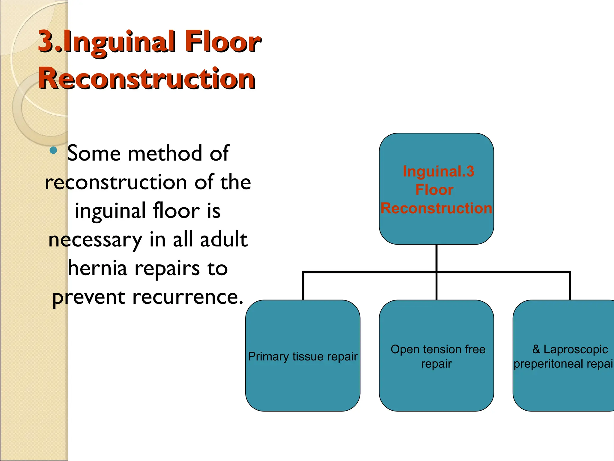
3.Inguinal Floor
3.Inguinal Floor
Reconstruction
Reconstruction
Some method of
reconstruction of the
inguinal floor is
necessary in all adult
hernia repairs to
prevent recurrence.
3
.
Inguinal
Floor
Reconstruction
Primary tissue repair
Open tension free
repair
Laproscopic
&
preperitoneal repair
32.
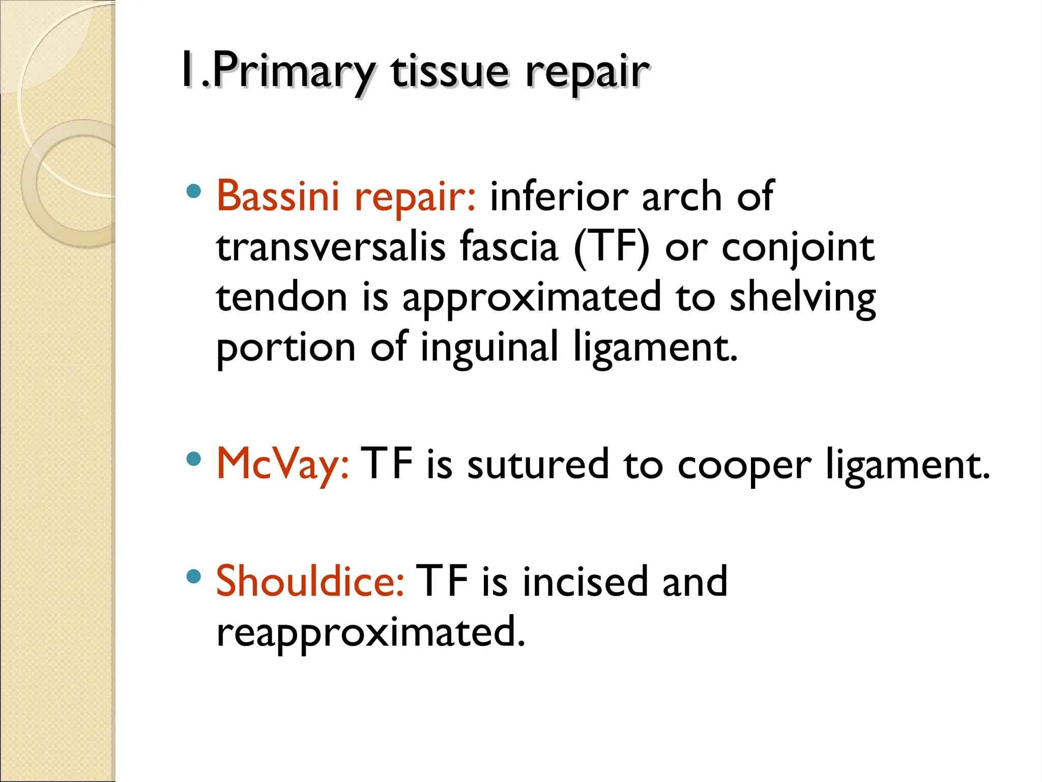
1.Primary tissue repair
1.Primarytissue repair
Bassini repair: inferior arch of
transversalis fascia (TF) or conjoint
tendon is approximated to shelving
portion of inguinal ligament.
McVay: TF is sutured to cooper ligament.
Shouldice: TF is incised and
reapproximated.
33.
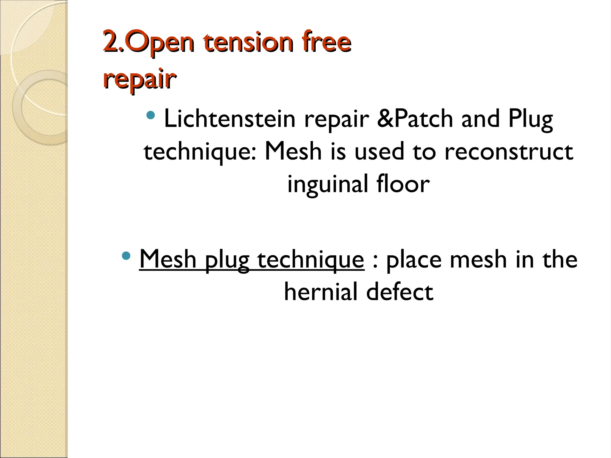
2.Open tension free
2.Opentension free
repair
repair
Lichtenstein repair &Patch and Plug
technique: Mesh is used to reconstruct
inguinal floor
Mesh plug technique : place mesh in the
hernial defect
34.
Laproscopic &
Laproscopic &
preperitonealrepairs
preperitoneal repairs
TAPP (transabdominal prepeitoneal procedure): peritoneal space
entered by conventional lap at umbilicus and peritoneum
overlaying inguinal floor is dissected away as flap.
TEP (Total extraperitoneal repair): preperitoneal space is
developed with a balloon inserted between posterior rectus
sheath and peritoneum balloon inflated to dissect the peritoneal
flaps awau from posterior abdomianl wall and the direct and
indirect spaces, other ports inserted into this preperitoneal space
without entering peritoneal cavity.
After lap. Dissection and reduction of hernia sac , a large piece of
mesh is placed over inguinal floor
35.
Femoral hernia repair
Femoralhernia repair
• Femoral hernias should be repaired very soon after the
diagnosis has been made because of the high risk of
strangulation.
• There is no place for a truss for a femoral hernia.
• Different approaches :
Open VS Laparoscopic
36.
Open surgery
Open surgery
Threeapproaches have been described for
open surgery :
1. Infra-inguinal approach (Lookwood)
2. Supra-inguinal approach ( McEvedy)
3. Trans-inguinal approach ( Lotheissen)
37.
Each techniquehas the principle of dissection
of the sac with reduction of its contents,
followed by ligation of the sac and closure
between the inguinal and pectineal ligaments.
38.
Lockwood’s infra-inguinal approach
Lockwood’sinfra-inguinal approach
The sac is dissected out below the
inguinal ligament via groin crease incision.
Then the sac is opened and the contents
are inspected and reduced into the
abdomen.
Then the neck of the sac is pulled down ,
ligated and allowed to retract through
femoral canal.
Then close the femoral canal by mesh
plug or non absorbable sutures.
39.
McEvedy’s high approach
McEvedy’shigh approach
Vertical incision is made over the femoral
canal and continued upwards above the
inguinal ligament.
This incision provides good access to the
preperitoneal space and then to the
peritoneum itself.
Use finger dissection to sweep peritoneum
from anterior abdominal wall , so the neck
of the sac can be identified.
Dissect the sac , reduce the contents and
repair the defect by mesh or sutures.