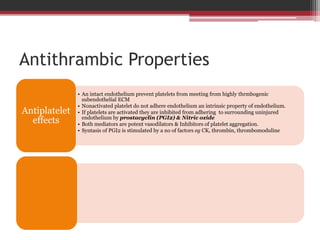
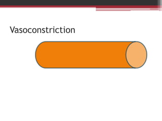
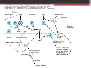
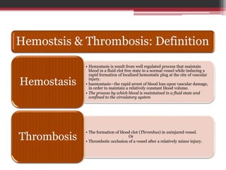
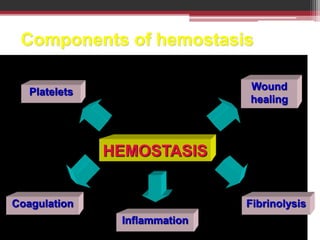
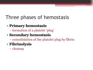
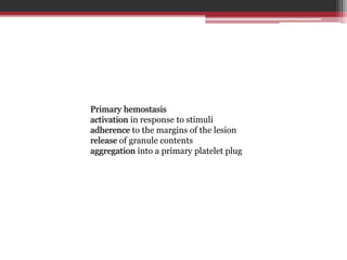
Hemostasis is the process by which blood is maintained in a fluid state within blood vessels, but forms clots to prevent blood loss when vessels are damaged. It involves three main components: primary hemostasis where platelets form a plug at the site of injury, secondary hemostasis where fibrin is formed to consolidate the platelet plug, and fibrinolysis which cleans up the clot once it is no longer needed. The process is tightly regulated to prevent inappropriate clot formation under normal conditions through properties of the endothelium like secretion of prostacyclin and nitric oxide. When vessels are damaged, factors like tissue factor activate the coagulation cascade leading to thrombin generation and fibrin clot formation to seal the break.
This seminar includes hemostasis,mechanism of blood clotting and associated blood dyscrasias commonly seen in children and their treatments with a note on antifibrinolytics
Hemostasis is the maintenance of blood flow is fluid state within the vascular system, the major components of hemostasis are vascular system, platelets, coagulation factors, inhibitors of coagulation and fibrinolytic system. details are given
Here's important & condensed ppt slides about hemostasis and its orchestrated steps and cogulation cascade, roles of endothelium,platelets and Coagulation protiens....!
Hemostasis and coagulation of blood For M.Sc & Basic Medical Students by Pand...Pandian M
Blood coagulation
Mechanism of coagulation
STAGES OF HEMOSTASIS
Coagulation of blood
Factors involved in blood clotting
Enzyme cascade theory
Mechanisms for formation of prothrombin activator
Fibrinolysis
Anticlotting mechanism in the body
Applied physiology
the objectives from this ppt :-
1.Define haemostasis.
2.Describe the main mechanisms that prevent blood loss after an injury.
3.Describe role of platelets in haemostasis.
4.Outline the mechanism of platelet plug formation.
5.Describe the mechanisms of blood coagulation.
Mechanisms of coagulation B.pharmacy 2 semesterKondal Reddy
Coagulation, also known as clotting, is the process by which
blood changes from a liquid to a gel, forming a blood clot.
It potentially results in haemostasis, the cessation of blood loss from a damaged vessel, followed by repair.
The mechanism of coagulation involves activation, adhesion and aggregation of platelets, as well as deposition and maturation of fibrin.
A detailed description of various stages in blood coagulation, clotting factors involved, the role of calcium, vitamin K, thrombin, phospholipids in blood coagulation, various tests for blood clotting, the significance of bleeding disorders in the treatment of periodontal disease and management.
Hemostasis definition, types and steps.
Hemostasis and coagulation physiology and pathology in steps and illustrated in simple way by diagrams.
Intrinsic and extrinsic pathways are mentioned in details.
Platelet function as a corner stone hemostasis in case of endothelial injury or another pathology taht affect endothelium or blood vessels.
Some pharmacological notes about drugs related to hemostasis and its clinical significance.
Indian Dental Academy: will be one of the most relevant and exciting training center with best faculty and flexible training programs for dental professionals who wish to advance in their dental practice,Offers certified courses in Dental implants,Orthodontics,Endodontics,Cosmetic Dentistry, Prosthetic Dentistry, Periodontics and General Dentistry.
Von Willebrand Disease is the most common hereditary bleeding disorder; roughly 1 in every 100 people suffers from the disease. People who suffer from VWD have blood that does not clot properly.
Normally when a person is injured and starts to bleed, the von Willebrand factor in the blood attaches to small blood cells called platelets. This helps the platelets stick together to form a clot at the site of the injury and stop bleeding. When a person has VWD, the clot might take longer to form or not form the way it should and bleeding might take longer to stop. This can lead to heavy, hard-to-stop bleeding. Although rare, the bleeding can be severe enough to damage joints or internal organs, or even be life-threatening.
http://www.nlm.nih.gov/medlineplus/plateletdisorders.html#cat1
http://www.cdc.gov/ncbddd/vwd/facts.html
Biochemistry of Hair fall, A complete review of hair fall cause, Types, Current methods of treatment, Natural methods of treatment,
for more detail text see :https://iiopinion.blogspot.in/2017/01/hair-fall-scientific-way-of-treatment.html
Non-Specific Immune Response, Innate immunity, inherent immunity, Role in overall immunity of individual, Significance, components involve in Non-Specific Immune Response,
- Video recording of this lecture in English language: https://youtu.be/lK81BzxMqdo
- Video recording of this lecture in Arabic language: https://youtu.be/Ve4P0COk9OI
- Link to download the book free: https://nephrotube.blogspot.com/p/nephrotube-nephrology-books.html
- Link to NephroTube website: www.NephroTube.com
- Link to NephroTube social media accounts: https://nephrotube.blogspot.com/p/join-nephrotube-on-social-media.html
Title: Sense of Smell
Presenter: Dr. Faiza, Assistant Professor of Physiology
Qualifications:
MBBS (Best Graduate, AIMC Lahore)
FCPS Physiology
ICMT, CHPE, DHPE (STMU)
MPH (GC University, Faisalabad)
MBA (Virtual University of Pakistan)
Learning Objectives:
Describe the primary categories of smells and the concept of odor blindness.
Explain the structure and location of the olfactory membrane and mucosa, including the types and roles of cells involved in olfaction.
Describe the pathway and mechanisms of olfactory signal transmission from the olfactory receptors to the brain.
Illustrate the biochemical cascade triggered by odorant binding to olfactory receptors, including the role of G-proteins and second messengers in generating an action potential.
Identify different types of olfactory disorders such as anosmia, hyposmia, hyperosmia, and dysosmia, including their potential causes.
Key Topics:
Olfactory Genes:
3% of the human genome accounts for olfactory genes.
400 genes for odorant receptors.
Olfactory Membrane:
Located in the superior part of the nasal cavity.
Medially: Folds downward along the superior septum.
Laterally: Folds over the superior turbinate and upper surface of the middle turbinate.
Total surface area: 5-10 square centimeters.
Olfactory Mucosa:
Olfactory Cells: Bipolar nerve cells derived from the CNS (100 million), with 4-25 olfactory cilia per cell.
Sustentacular Cells: Produce mucus and maintain ionic and molecular environment.
Basal Cells: Replace worn-out olfactory cells with an average lifespan of 1-2 months.
Bowman’s Gland: Secretes mucus.
Stimulation of Olfactory Cells:
Odorant dissolves in mucus and attaches to receptors on olfactory cilia.
Involves a cascade effect through G-proteins and second messengers, leading to depolarization and action potential generation in the olfactory nerve.
Quality of a Good Odorant:
Small (3-20 Carbon atoms), volatile, water-soluble, and lipid-soluble.
Facilitated by odorant-binding proteins in mucus.
Membrane Potential and Action Potential:
Resting membrane potential: -55mV.
Action potential frequency in the olfactory nerve increases with odorant strength.
Adaptation Towards the Sense of Smell:
Rapid adaptation within the first second, with further slow adaptation.
Psychological adaptation greater than receptor adaptation, involving feedback inhibition from the central nervous system.
Primary Sensations of Smell:
Camphoraceous, Musky, Floral, Pepperminty, Ethereal, Pungent, Putrid.
Odor Detection Threshold:
Examples: Hydrogen sulfide (0.0005 ppm), Methyl-mercaptan (0.002 ppm).
Some toxic substances are odorless at lethal concentrations.
Characteristics of Smell:
Odor blindness for single substances due to lack of appropriate receptor protein.
Behavioral and emotional influences of smell.
Transmission of Olfactory Signals:
From olfactory cells to glomeruli in the olfactory bulb, involving lateral inhibition.
Primitive, less old, and new olfactory systems with different path
Ozempic: Preoperative Management of Patients on GLP-1 Receptor Agonists Saeid Safari
Preoperative Management of Patients on GLP-1 Receptor Agonists like Ozempic and Semiglutide
ASA GUIDELINE
NYSORA Guideline
2 Case Reports of Gastric Ultrasound
Report Back from SGO 2024: What’s the Latest in Cervical Cancer?bkling
Are you curious about what’s new in cervical cancer research or unsure what the findings mean? Join Dr. Emily Ko, a gynecologic oncologist at Penn Medicine, to learn about the latest updates from the Society of Gynecologic Oncology (SGO) 2024 Annual Meeting on Women’s Cancer. Dr. Ko will discuss what the research presented at the conference means for you and answer your questions about the new developments.
These lecture slides, by Dr Sidra Arshad, offer a quick overview of physiological basis of a normal electrocardiogram.
Learning objectives:
1. Define an electrocardiogram (ECG) and electrocardiography
2. Describe how dipoles generated by the heart produce the waveforms of the ECG
3. Describe the components of a normal electrocardiogram of a typical bipolar leads (limb II)
4. Differentiate between intervals and segments
5. Enlist some common indications for obtaining an ECG
Study Resources:
1. Chapter 11, Guyton and Hall Textbook of Medical Physiology, 14th edition
2. Chapter 9, Human Physiology - From Cells to Systems, Lauralee Sherwood, 9th edition
3. Chapter 29, Ganong’s Review of Medical Physiology, 26th edition
4. Electrocardiogram, StatPearls - https://www.ncbi.nlm.nih.gov/books/NBK549803/
5. ECG in Medical Practice by ABM Abdullah, 4th edition
6. ECG Basics, http://www.nataliescasebook.com/tag/e-c-g-basics
Ethanol (CH3CH2OH), or beverage alcohol, is a two-carbon alcohol
that is rapidly distributed in the body and brain. Ethanol alters many
neurochemical systems and has rewarding and addictive properties. It
is the oldest recreational drug and likely contributes to more morbidity,
mortality, and public health costs than all illicit drugs combined. The
5th edition of the Diagnostic and Statistical Manual of Mental Disorders
(DSM-5) integrates alcohol abuse and alcohol dependence into a single
disorder called alcohol use disorder (AUD), with mild, moderate,
and severe subclassifications (American Psychiatric Association, 2013).
In the DSM-5, all types of substance abuse and dependence have been
combined into a single substance use disorder (SUD) on a continuum
from mild to severe. A diagnosis of AUD requires that at least two of
the 11 DSM-5 behaviors be present within a 12-month period (mild
AUD: 2–3 criteria; moderate AUD: 4–5 criteria; severe AUD: 6–11 criteria).
The four main behavioral effects of AUD are impaired control over
drinking, negative social consequences, risky use, and altered physiological
effects (tolerance, withdrawal). This chapter presents an overview
of the prevalence and harmful consequences of AUD in the U.S.,
the systemic nature of the disease, neurocircuitry and stages of AUD,
comorbidities, fetal alcohol spectrum disorders, genetic risk factors, and
pharmacotherapies for AUD.
micro teaching on communication m.sc nursing.pdfAnurag Sharma
Microteaching is a unique model of practice teaching. It is a viable instrument for the. desired change in the teaching behavior or the behavior potential which, in specified types of real. classroom situations, tends to facilitate the achievement of specified types of objectives.
Couples presenting to the infertility clinic- Do they really have infertility...Sujoy Dasgupta
Dr Sujoy Dasgupta presented the study on "Couples presenting to the infertility clinic- Do they really have infertility? – The unexplored stories of non-consummation" in the 13th Congress of the Asia Pacific Initiative on Reproduction (ASPIRE 2024) at Manila on 24 May, 2024.
Title: Sense of Taste
Presenter: Dr. Faiza, Assistant Professor of Physiology
Qualifications:
MBBS (Best Graduate, AIMC Lahore)
FCPS Physiology
ICMT, CHPE, DHPE (STMU)
MPH (GC University, Faisalabad)
MBA (Virtual University of Pakistan)
Learning Objectives:
Describe the structure and function of taste buds.
Describe the relationship between the taste threshold and taste index of common substances.
Explain the chemical basis and signal transduction of taste perception for each type of primary taste sensation.
Recognize different abnormalities of taste perception and their causes.
Key Topics:
Significance of Taste Sensation:
Differentiation between pleasant and harmful food
Influence on behavior
Selection of food based on metabolic needs
Receptors of Taste:
Taste buds on the tongue
Influence of sense of smell, texture of food, and pain stimulation (e.g., by pepper)
Primary and Secondary Taste Sensations:
Primary taste sensations: Sweet, Sour, Salty, Bitter, Umami
Chemical basis and signal transduction mechanisms for each taste
Taste Threshold and Index:
Taste threshold values for Sweet (sucrose), Salty (NaCl), Sour (HCl), and Bitter (Quinine)
Taste index relationship: Inversely proportional to taste threshold
Taste Blindness:
Inability to taste certain substances, particularly thiourea compounds
Example: Phenylthiocarbamide
Structure and Function of Taste Buds:
Composition: Epithelial cells, Sustentacular/Supporting cells, Taste cells, Basal cells
Features: Taste pores, Taste hairs/microvilli, and Taste nerve fibers
Location of Taste Buds:
Found in papillae of the tongue (Fungiform, Circumvallate, Foliate)
Also present on the palate, tonsillar pillars, epiglottis, and proximal esophagus
Mechanism of Taste Stimulation:
Interaction of taste substances with receptors on microvilli
Signal transduction pathways for Umami, Sweet, Bitter, Sour, and Salty tastes
Taste Sensitivity and Adaptation:
Decrease in sensitivity with age
Rapid adaptation of taste sensation
Role of Saliva in Taste:
Dissolution of tastants to reach receptors
Washing away the stimulus
Taste Preferences and Aversions:
Mechanisms behind taste preference and aversion
Influence of receptors and neural pathways
Impact of Sensory Nerve Damage:
Degeneration of taste buds if the sensory nerve fiber is cut
Abnormalities of Taste Detection:
Conditions: Ageusia, Hypogeusia, Dysgeusia (parageusia)
Causes: Nerve damage, neurological disorders, infections, poor oral hygiene, adverse drug effects, deficiencies, aging, tobacco use, altered neurotransmitter levels
Neurotransmitters and Taste Threshold:
Effects of serotonin (5-HT) and norepinephrine (NE) on taste sensitivity
Supertasters:
25% of the population with heightened sensitivity to taste, especially bitterness
Increased number of fungiform papillae
Explore natural remedies for syphilis treatment in Singapore. Discover alternative therapies, herbal remedies, and lifestyle changes that may complement conventional treatments. Learn about holistic approaches to managing syphilis symptoms and supporting overall health.
2. Hemostsis & Thrombosis: Definition
• Hemostasis is result from well regulated process that maintain
blood in a fluid clot free state in a normal vessel while inducing a
rapid formation of localized hemostatic plug at the site of vascular
injury.
• haemostasis—the rapid arrest of blood loss upon vascular damage,
in order to maintain a relatively constant blood volume.
• The process by which blood is maintained in a fluid state and
confined to the circulatory system
Hemostasis
• The formation of blood clot (Thrombus) in uninjured vessel.
Or
• Thrombotic occlusion of a vessel after a relatively minor injury.
Thrombosis
3. Components of hemostasis
Hemostasis is a complex process and actually comprises a number of distinct, but
closely linked biochemical systems. The blood platelets are responsible for
primary hemostasis and for providing the framework for subsequent coagulation
and wound healing. Fibrinolysis is required for the removal of fibrin that is no
longer needed and for the eventual remodeling of the injured area. All these
systems are closely linked to inflammation, especially through contact activation
of FXII.
Platelets
Coagulation
Inflammation
Wound
healing
HEMOSTASIS
Fibrinolysis
4. Three phases of hemostasis
• Primary hemostasis
▫ formation of a platelet ‘plug’
• Secondary hemostasis
▫ consolidation of the platelet plug by fibrin
• Fibrinolysis
▫ cleanup
5. Primary hemostasis
activation in response to stimuli
adherence to the margins of the lesion
release of granule contents
aggregation into a primary platelet plug
9. Fig. 1. Wound healing is a
complex process
encompassing a number of
overlapping phases,
including inflammation,
epithelialization,
angiogenesis and matrix
deposition. During
inflammation, the
formation of a blood clot
re-establishes hemostasis
and provides a provisional
matrix for cell migration.
Cytokines play an
important role in the
evolution of granulation
tissue through recruitment
of inflammatory
leukocytes and stimulation
of fibroblasts and
epithelial cells. [Note:
figure is adapted from
reference 1.]
10. Normal Hemostasis: 1
General Sequence of Events are:
Endothelial injury : it is also exposed highly thrmbogenic
subendothelial extracellular matrix (ECM) which allow platelets to adhere
and activated
ie. a shape change
Release of secretory
granules
Within a minute secretory
product recruits several
platelets to form hemostatic
plug.
Vasoconstriction : After a initial injury there is a brief period of
arteriolar vasoconstriction due to
Reflex neurogenic mechanism
Local secretion of the factors such as
endotheline
11. three main mechanisms underline the
haemostatic process
The congregation and clumping of blood platelets at the site of vascular
injury, thus effectively plugging the site of blood leakage.
Localized constriction of the blood vessel, which minimizes further blood
flow through the area.
Induction of the blood coagulation cascade. This culminates in the
conversion of a soluble serum protein, fibrinogen, into insoluble fibrin.
Fibrin monomers then aggregate at the site of damage, thus forming a clot
(thrombus), which seals it off. These mechanisms are effective in dealing
with small vessel injuries, e.g. in capillaries and arterioles, although they
are ineffective when the damage relates to large veins/arteries.
12. Normal Hemostasis:2
General Sequence of Events are:
Polymerized fibrin & platelet aggregates form a permanent plug to
prevent any further hemorrhage
Tissue Factors: a membrane bound procougulant factors is
synthesized by the endothelium, also released at the site of injury.
It act in conjugation with secreted
platelet factor actor to activate the
coagulation cascades in activation
of thrombin
In turn thrombin cleave fibrinogen
into insoluble fibrine, creating a
fibrin meshwork
Thrombin also induced further
platelet requirement & granules
released.
13. Endothelium
Modulate several
aspect of anti-
cougulating
properties
Anti-platelet ,Anti-
couagulent &
fibrinolytic
Exerts pro-
couagulent
functions
It may activated by infectious
agents, hemodynamic factors
plasma mediators & CK
14. Antithrambic Properties
• An intact endothelium prevent platelets from meeting from highly thrmbogenic
subendothelial ECM
• Nonactivated platelet do not adhere endothelium an intrinsic property of endothelium.
• If platelets are activated they are inhibited from adhering to surrounding uninjured
endothelium by prostacyclin (PGI2) & Nitric oxide
• Both mediators are potent vasodilators & Inhibitors of platelet aggregation.
• Syntasis of PGI2 is stimulated by a no of factors eg CK, thrombin, thrombomoduline
Antiplatelet
effects
16. Figure 51–8. Diagrammatic representation of platelet activation. The external environment,
the plasma membrane, and the inside of a platelet are depicted from top to bottom.
Thrombin and collagen are the two most important platelet activators. ADP is considered
a weak agonist; it causes aggregation but not granule release. (GP, glycoprotein; R1–R5,
various receptors; AC, adenylyl cyclase; PLA2, phospholipase A2; PL, phospholipids; PLCβ,
phospholipase Cβ; PIP2, phosphatidylinositol 4,5-bisphosphate; cAMP, cyclic AMP; PKC,
protein kinase C; TxA2, thromboxane A2; IP3, inositol 1,4,5-trisphosphate; DAG, 1,2-diacylglycerol.
The G proteins that are involved are not shown.)