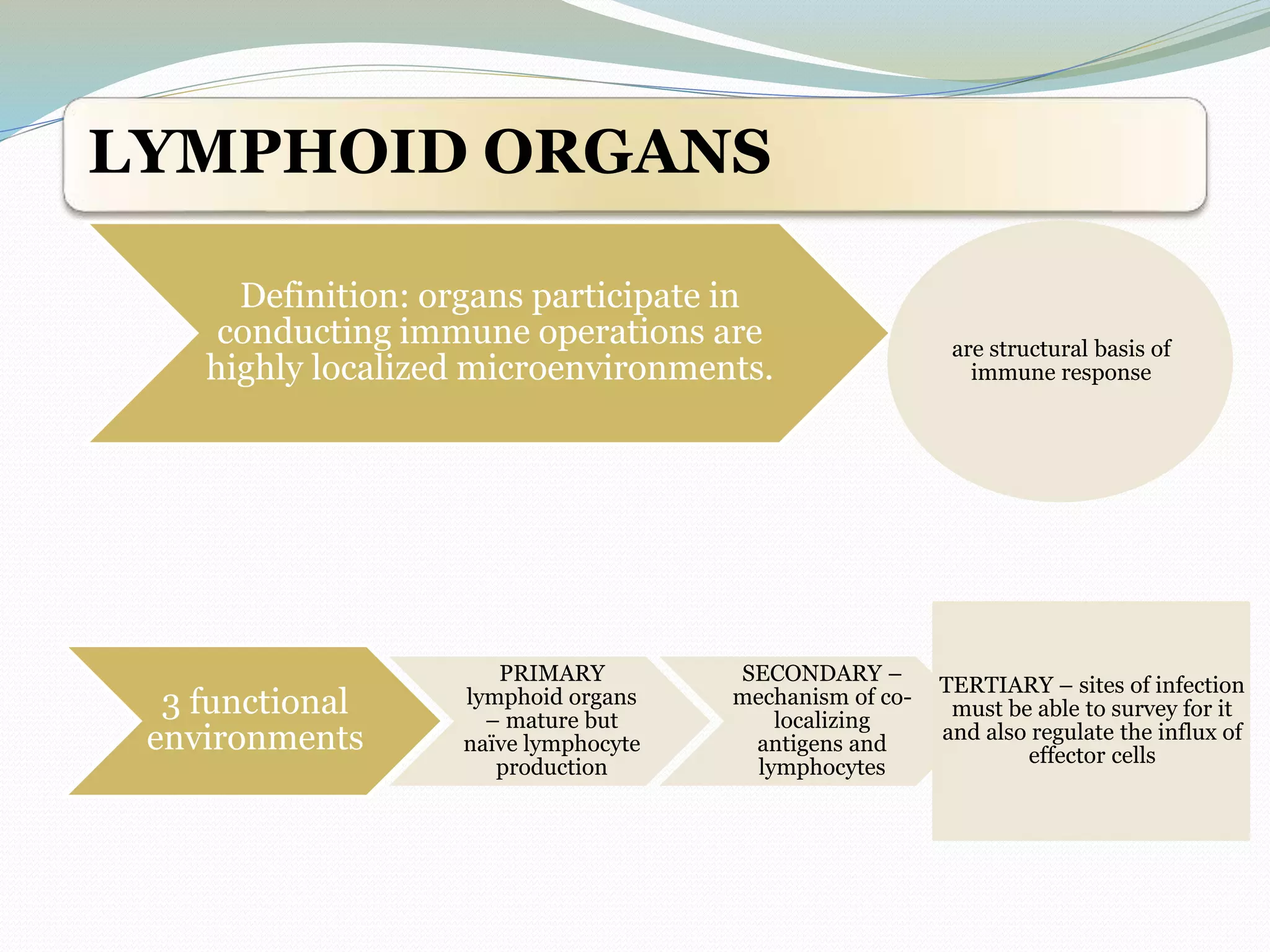
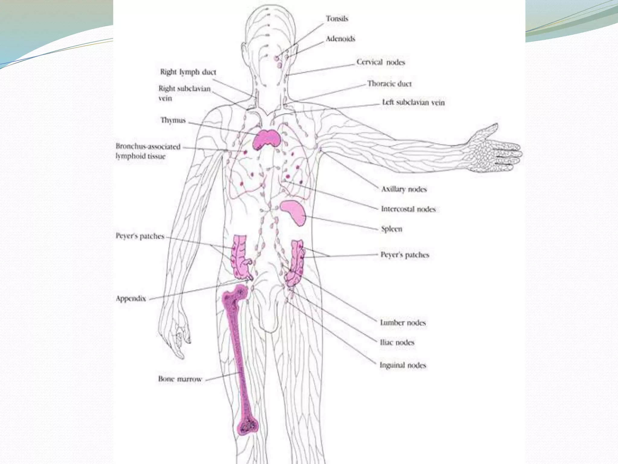
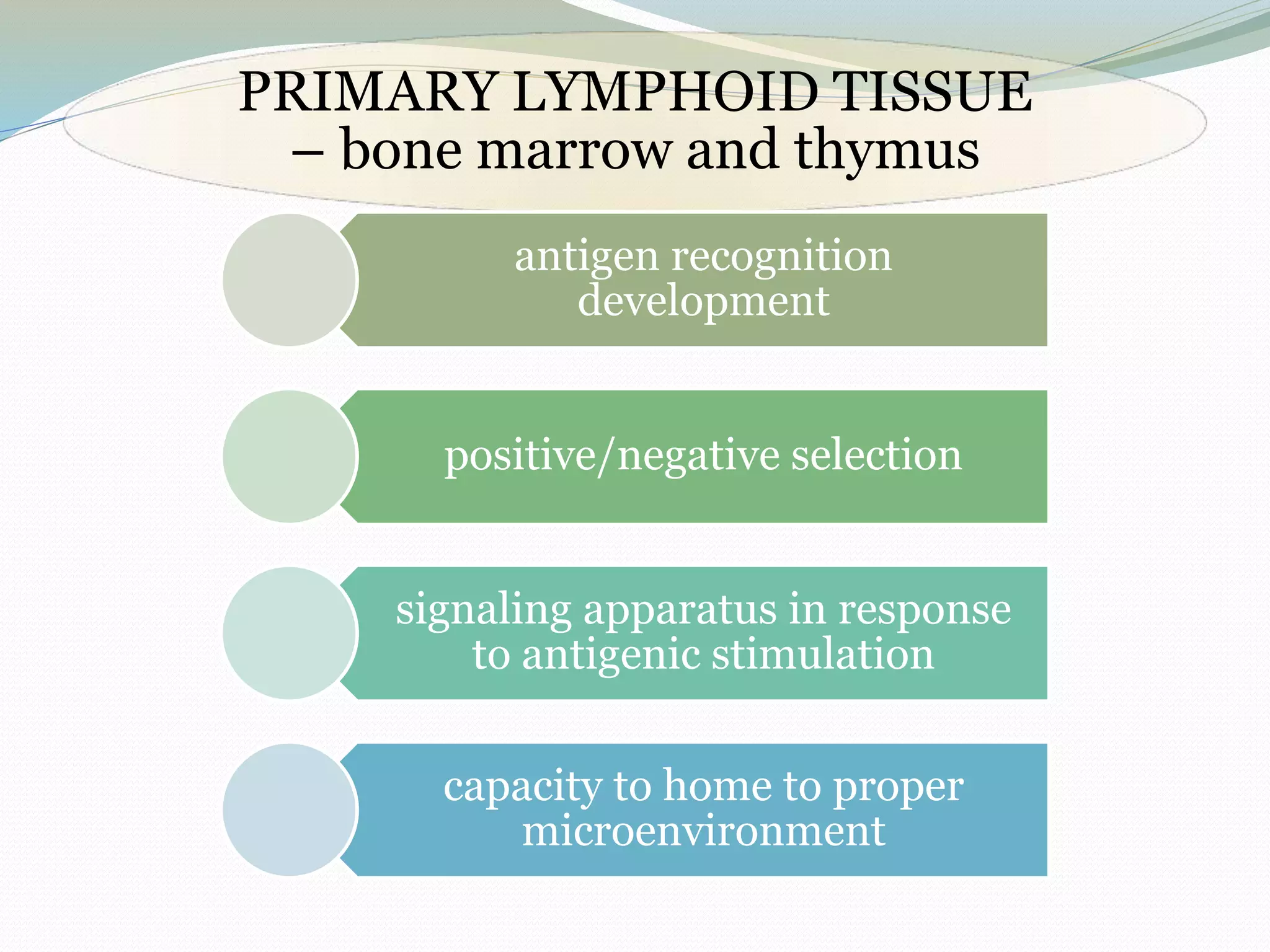
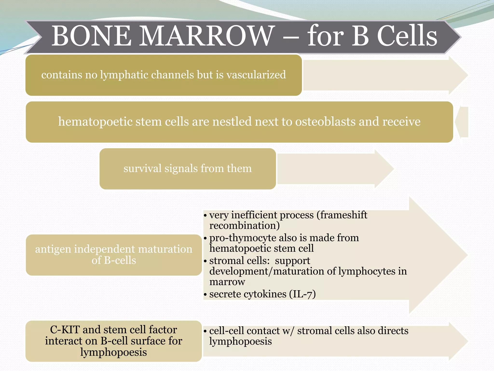
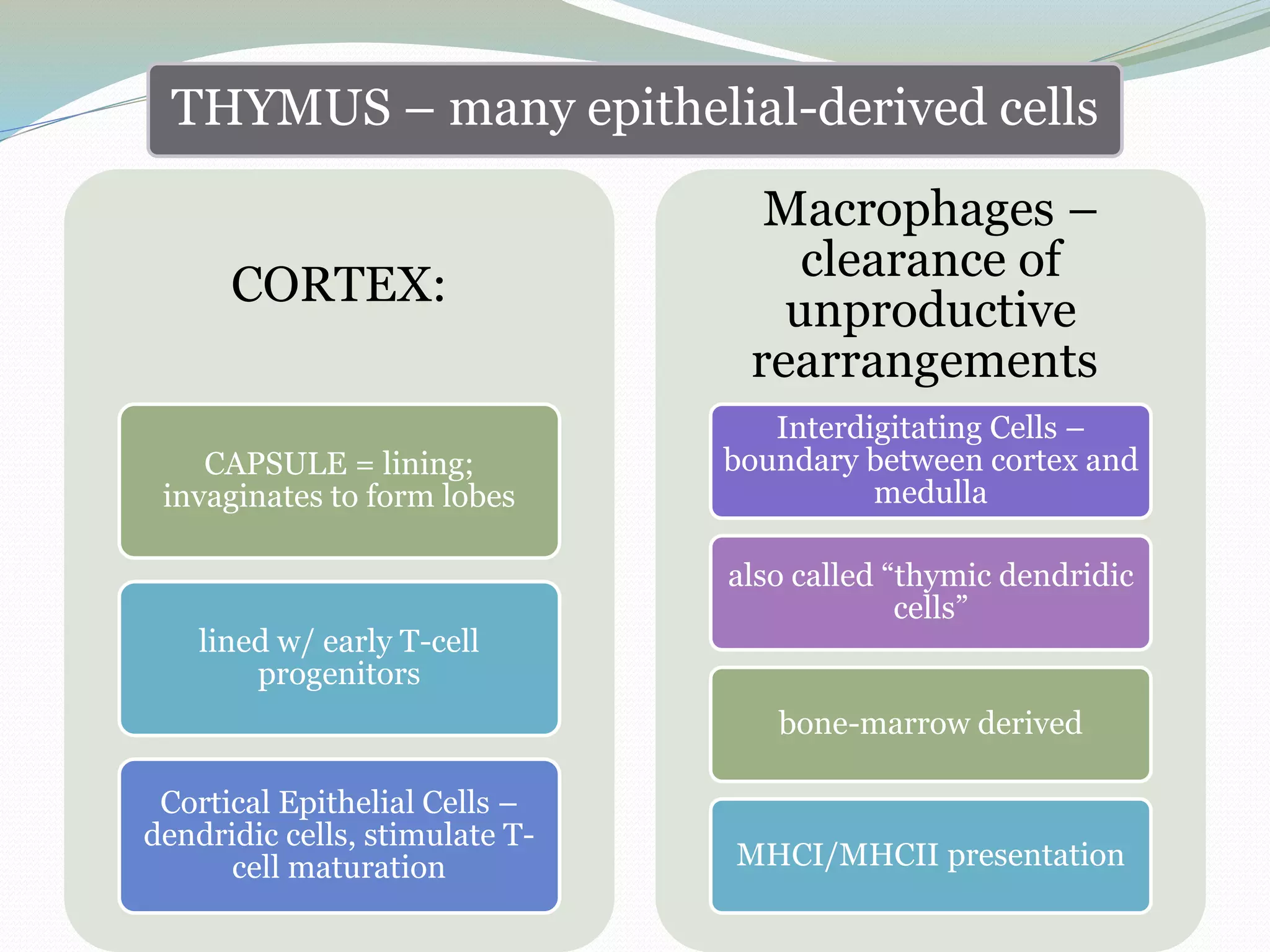
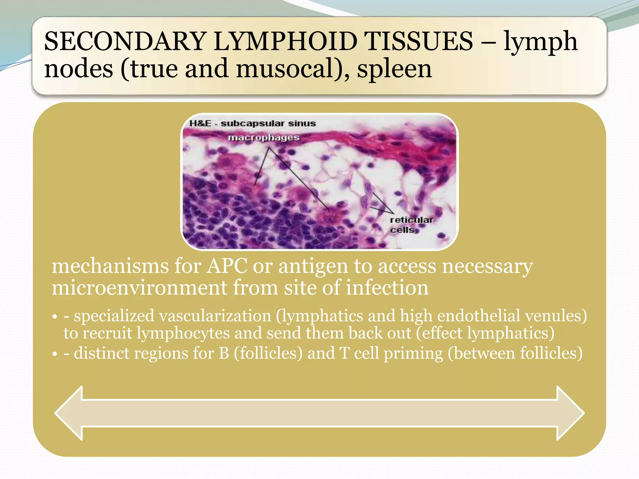
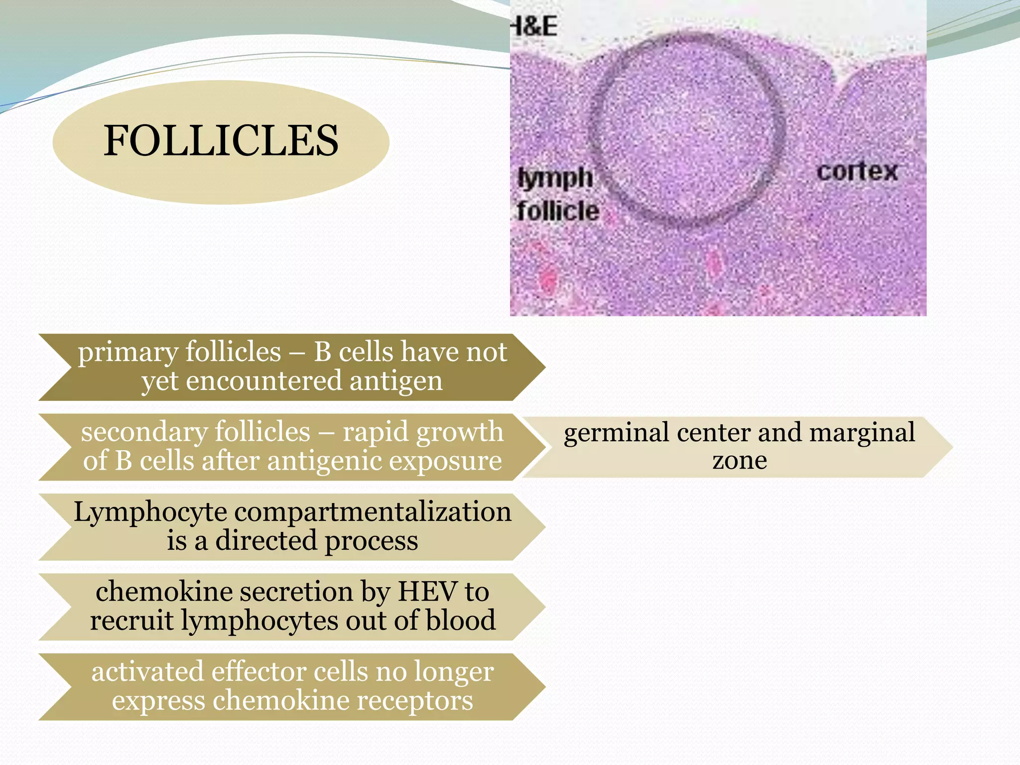
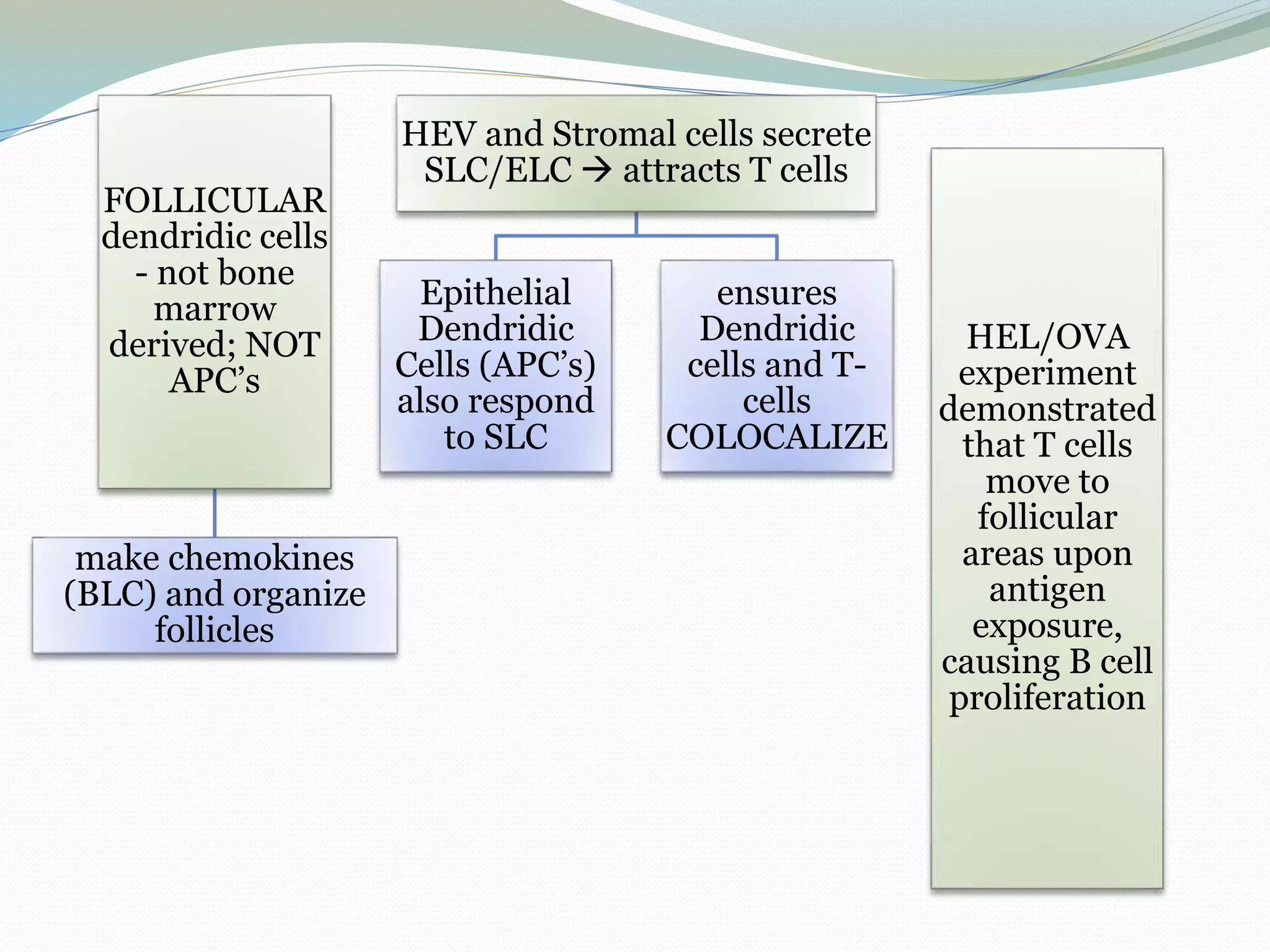
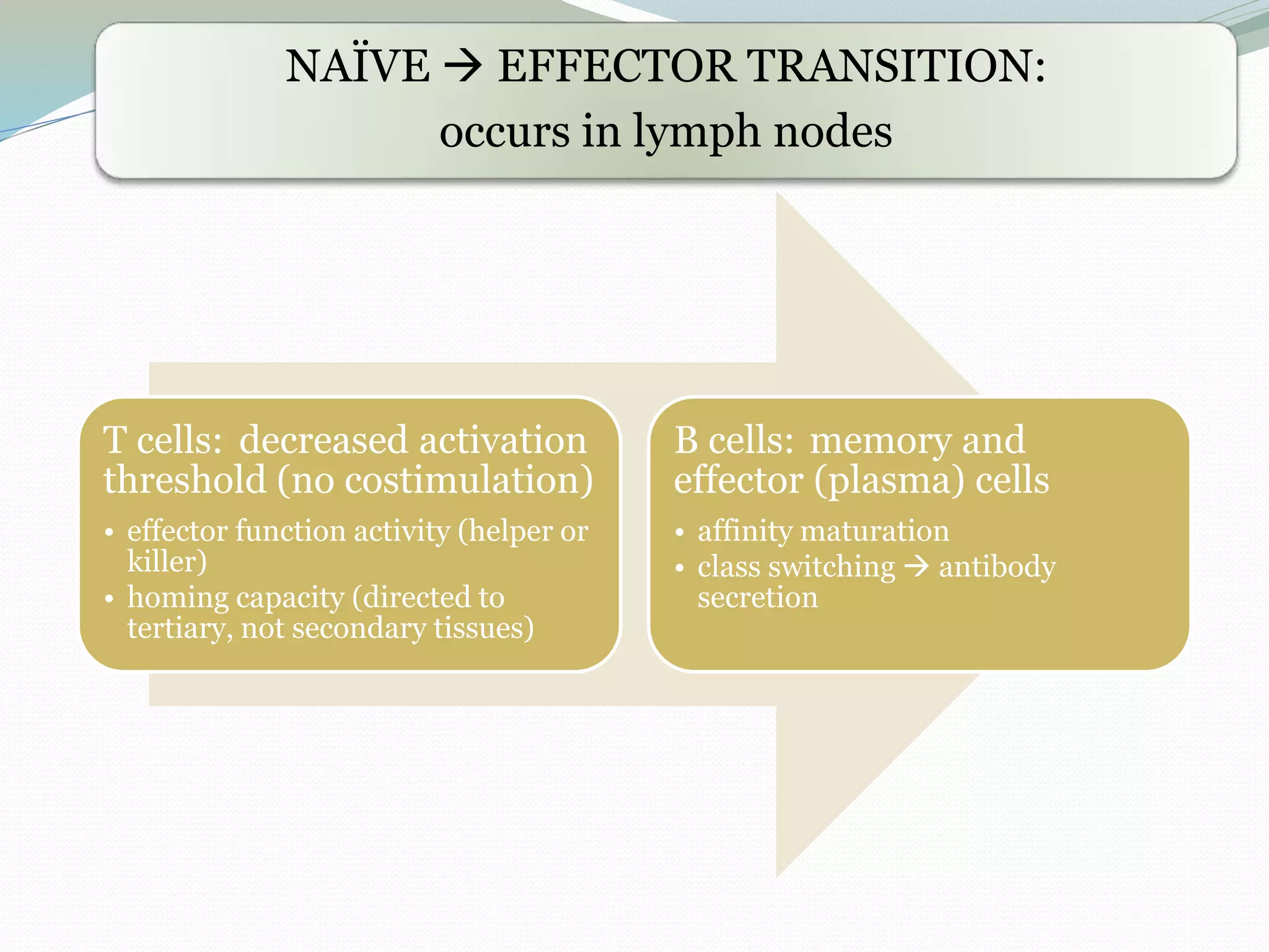
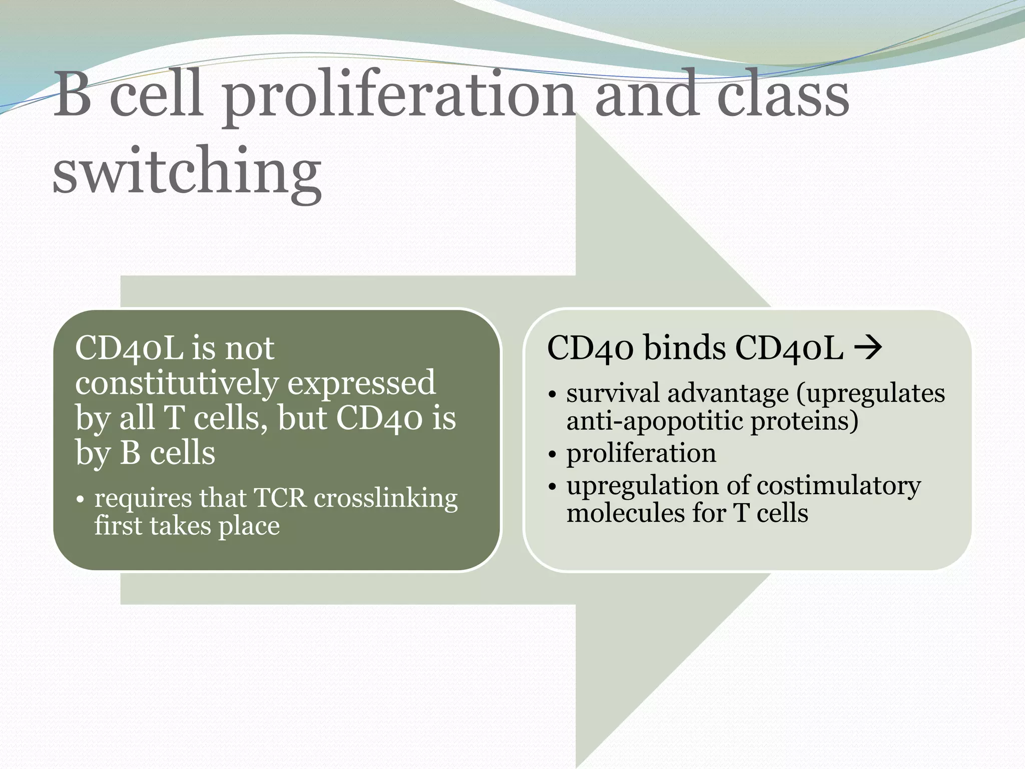
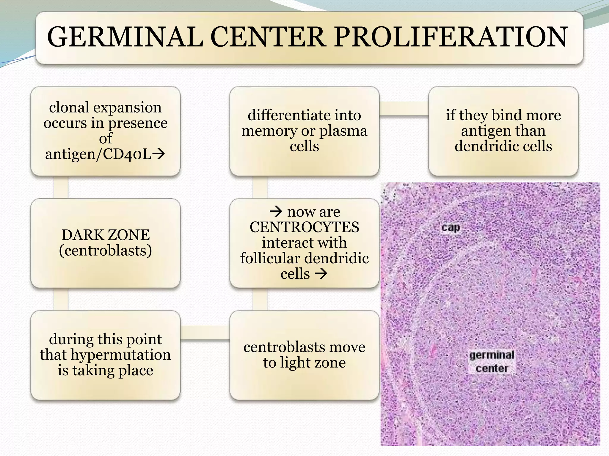
SlideShare a Scribd company logo
Dr Alok Tripathi
Department of Biotechnology,
Saaii College of Medical Science & Technology, Kanpur
aquaimmuno@yahoo.co.in
09451662231
LYMPHOID ORGANS
Definition: organs participate in
conducting immune operations are
highly localized microenvironments.
are structural basis of
immune response
3 functional
environments
PRIMARY
lymphoid organs
– mature but
naïve lymphocyte
production
SECONDARY –
mechanism of co-
localizing
antigens and
lymphocytes
TERTIARY – sites of infection
must be able to survey for it
and also regulate the influx of
effector cells
PRIMARY LYMPHOID TISSUE
– bone marrow and thymus
antigen recognition
development
positive/negative selection
signaling apparatus in response
to antigenic stimulation
capacity to home to proper
microenvironment
BONE MARROW – for B Cells
contains no lymphatic channels but is vascularized
hematopoetic stem cells are nestled next to osteoblasts and receive
survival signals from them
• very inefficient process (frameshift
recombination)
• pro-thymocyte also is made from
hematopoetic stem cell
• stromal cells: support
development/maturation of lymphocytes in
marrow
• secrete cytokines (IL-7)
antigen independent maturation
of B-cells
• cell-cell contact w/ stromal cells also directs
lymphopoesis
C-KIT and stem cell factor
interact on B-cell surface for
lymphopoesis
THYMUS – many epithelial-derived cells
CORTEX:
CAPSULE = lining;
invaginates to form lobes
lined w/ early T-cell
progenitors
Cortical Epithelial Cells –
dendridic cells, stimulate T-
cell maturation
Macrophages –
clearance of
unproductive
rearrangements
Interdigitating Cells –
boundary between cortex and
medulla
also called “thymic dendridic
cells”
bone-marrow derived
MHCI/MHCII presentation
The thymus is enclosed by a
thin connective tissue capsule
from which numerous septa
extend into the thymus
subdividing the two lobes into
numerous lobules (about 0.5 -
2 mm in diameter).
Blood vessels enter and leave
the thymus via the connective
tissue septa.
Each lobulus is divided into a
darker peripheral zone, the
cortex, and a lighter, central
zone, the medulla.
Medullary tissue is
continuous from lobule to
lobule throughout each lobe.
Thymus: Cortex+ Medulla
Cells in Thymus
Reticular cells
Reticular cells sheathe
the cortical capillaries
• are quite abundant.
• cytoplasm eosinophilic, with large,
ovoid and light nuclei may contain 1
or 2 nucleoli.
• cells branched, and their slender
processes are connected with the
processes of other reticular cells to
form a cellular reticulum
• This cellular network (reticular fibres
are scant in the thymus) provides
support for other cells of the thymus.
• form an epitheloid layer which
delimits the cortical tissue from
the connective tissue
• and secrete substances
important for thymic function.
• → create and maintain the
microenvironment necessary
for the development of T-
lymphocytes in the cortex.
Macrophages occur in both
cortex and medulla. They are
difficult to distinguish from the
reticular cells in H&E stained
sections.
Lymphocytes are present in
both cortex and medulla,
• more numerous (denser) in the cortex.
• sizes are variable (5 - 15 µm) in the
cortex but generally small in the
medulla.
• The vast majority of them will be
developing T-lymphocytes.
• They are also called thymic lymphocytes
or thymocytes.
Function of the Thymus
necessary for the development of the recirculating pool of small,
long-lived lymphocytes, the T-lymphocytes.
T cells mainly responsible for the cell-mediated immune response.
Stem cells invade the cortical regions of the thymus, where they
divide to form lymphocytes.
Only a small fraction (10-30%) of the cells generated in the cortex
leave the thymus.
They migrate via the medulla into the blood stream to populate
the T-lymphocyte areas of other lymphoid tissues and organs.
Cells which do not express the necessary receptors to recognize AP to them or which react
incorrectly towards "self-antigens" die and are removed by cortical macrophages.
Since the function of the thymus is to produce lymphocytes for the
other lymphoid tissues it is a primary lymphoid organ.
SECONDARY LYMPHOID TISSUES – lymph
nodes (true and musocal), spleen
mechanisms for APC or antigen to access necessary
microenvironment from site of infection
• - specialized vascularization (lymphatics and high endothelial venules)
to recruit lymphocytes and send them back out (effect lymphatics)
• - distinct regions for B (follicles) and T cell priming (between follicles)
FOLLICLES
primary follicles – B cells have not
yet encountered antigen
secondary follicles – rapid growth
of B cells after antigenic exposure
germinal center and marginal
zone
Lymphocyte compartmentalization
is a directed process
chemokine secretion by HEV to
recruit lymphocytes out of blood
activated effector cells no longer
express chemokine receptors
FOLLICULAR
dendridic cells
- not bone
marrow
derived; NOT
APC’s
make chemokines
(BLC) and organize
follicles
HEV and Stromal cells secrete
SLC/ELC  attracts T cells
Epithelial
Dendridic
Cells (APC’s)
also respond
to SLC
ensures
Dendridic
cells and T-
cells
COLOCALIZE
HEL/OVA
experiment
demonstrated
that T cells
move to
follicular
areas upon
antigen
exposure,
causing B cell
proliferation
NAÏVE  EFFECTOR TRANSITION:
occurs in lymph nodes
T cells: decreased activation
threshold (no costimulation)
• effector function activity (helper or
killer)
• homing capacity (directed to
tertiary, not secondary tissues)
B cells: memory and
effector (plasma) cells
• affinity maturation
• class switching  antibody
secretion
OVERVIEW
no antigen  B and T
cells come in from high
endothelial venule migrate
to follicle/dendridic cell
So they LEAVE via the
efferent lympathics
antigen present 
B cell enters and remains in
follicle (binds soluble
antigen) antigen on DC
causes CD4 TH2
differentiation
CD4 proliferates and
releases cytokines
(IL4/5/6)
CD4 follows SLC and
moves to follicular
margin for T/B
interaction
•CD40L----CD40
•TCR/CD4 -----MHC: peptide
•CD28 ----B7 (stim)
•CTLA4 --- B7 (inhib)
migration occurs in the
absence of CCR7
receptors
**localization is
very
important**
B cell proliferation and class
switching
CD40L is not
constitutively expressed
by all T cells, but CD40 is
by B cells
• requires that TCR crosslinking
first takes place
CD40 binds CD40L 
• survival advantage (upregulates
anti-apopotitic proteins)
• proliferation
• upregulation of costimulatory
molecules for T cells
GERMINAL CENTER PROLIFERATION
clonal expansion
occurs in presence
of
antigen/CD40L
DARK ZONE
(centroblasts)
during this point
that hypermutation
is taking place
centroblasts move
to light zone
 now are
CENTROCYTES
interact with
follicular dendridic
cells 
differentiate into
memory or plasma
cells
if they bind more
antigen than
dendridic cells
Phagocyte:
Macrophage (MФ) & Neutrophils (PMN)
A phagocyte is a cell that ingests
(and destroys) foreign matter, such
as microorganisms or debris via a
process known as phagocytosis,
• in which these cells ingest and kill offending
cells by cellular digestion.
These phagocytes are extremely
useful as an initial immune system
response to tissue damage.
Blood Cells
Granulocytes
Cells with various types of granules
Agranulocytes
Cells without granules
Eosinophils
Stain with acidic dyes
Lymphocytes
Basophils
Satin with basic dyes
Monocytes
Neutrophils
Stain with neutral dyes
MФ Macrophages adapted specially for sustained
battles against foreign agents.
In addition, they help to
clean up and remove
damaged tissues.
Immature macrophages which are
circulating in the bloodstream are
called monocytes.
These macrophages cannot react
immediately, but once they have
developed, they are often referred to as
'killing machines' they act by
phagocytizing and destroying anything
that isn't recognized as belonging to the
body.
A number
of cell types
are closely
related to
MФ -
Dendritic cells
(including
Langerhans
cells)
Microglia Kupffer cells
Osteoclasts
Neutrophil
• express receptors for
immunoglobulin and
complement and are
involved in the acute
inflammatory response.
Morphology
• Large mononuclear cells with granular cytoplasm
• Smaller cells with multi-lobed nucleus and neutral cytoplasmic
granules
Location
• Often resident in tissues
(remove routine cell debris)
• Blood – requires recruitment to
site of infection
Killing
ability
After killing
Antigen
presentation
• Require activation by
bacterial molecules ±IFNg
• Activated during
recruitment, then able to kill
internalised bacteria
automatically
• Migrate to local lymph
nodes
• Die at site by apoptosis
(then taken up by
macrophages)
• Can present antigen (Class
II upregulated by IFNg)
• Cannot present antigen
(don't normally express
Class II)
Neutrophil
Eosinophil granulocyte
Eosinophil granulocytes, commonly referred to as eosinophils (or less
commonly as acidophils), are white blood cells that are responsible for
combating infection by parasites in the body.
Transparent in vivo, these cells appear brick-red when stained with eosin
using the Romanowsky method (and are thus, 'eosin (or acid)-loving' cells,
hence the name).
The red color is visible as small granules within the cellular cytoplasm.
These granules contain histamine and proteins such as eosinophil peroxidase,
RNase, DNases, lipase, plasminogen, and Major Basic Protein that are toxic
to both parasites and the host's tissues.
Eosinophils make up about 2.3% of the all white blood cells, and are about
10-12 micrometres in size.
A key mediator in eosinophil activation is interleukin 5.
Functions
of
eosinophils
• Eosinophils play a role in fighting
viral infections which is evident
from the abundance of RNAses
they contain within their granules.
• Eosinophils also play a role in the
allergic response, and in fibrin
removal in inflammation.
• Eosinophils are considered the
main effector cells in asthma
pathogenesis and are associated
with disease severity.
• Eosinophils fight helminth (worm)
colonization.
Basophil
granulocyte
 least common of the granulocytes, representing about 1% of
circulating leukocytes.
 They contain large cytoplasmic granules which obscure the
cell nucleus under the microscope.
 when unstained, the nucleus is visible and it usually has 2
lobes.
 A cell in tissues, the mast cell, has many similar
characteristics.
 For example, both cell types store histamine, a chemical that
is secreted by the cells when stimulated in certain ways
(histamine causes some of the symptoms of an allergic
reaction).
 Like all circulating granulocytes, basophils can be recruited
out of the blood into a tissue when needed.
Function
Basophils tend to appear in specific kinds of inflammatory
reactions, particularly those that cause allergic symptoms.
While the exact purpose of basophils has never been proven,
they appear often in tissues where parasites are found.
They can be found in unusually high numbers at sites of
exoparasite infection, e.g., ticks.
also appear in tissues where allergic reactions are occurring
and probably contribute to the severity of these reactions.
Basophils have protein receptors on their cell surface that
bind IgE antibody very tightly.
It is the bound IgE antibody that confers a selective
response of these cells to environmental substances, for
example, pollen proteins.
Secretions
When activated, basophils secrete histamine, several proteoglycans, lipid
mediators like leukotrienes, and several cytokines.
Histamine and proteoglycans are pre-stored in the cell's granules while the
other secreted substances are newly generated.
Each of these substances contributes to inflammation.
Recent evidence suggests that basophils are an important source of the
cytokine, IL-4, perhaps more important than T cells.
Interleukin-4 is considered one of the critical cytokines in the development of
allergies and the production of IgE antibody by the immune system.
other substances that can activate basophils to secrete which suggests that
these cells have other roles in inflammation.
Lymphocyte
A lymphocyte is a
type of white blood
cell involved in the
human body's
immune system.
There are two broad
categories of
lymphocytes, namely
T cells and B cells.
Lymphocytes play an
important and
integral part of the
body's defenses.
T cells are chiefly responsible for cell-
mediated immunity whereas B cells are
primarily responsible for humoral
immunity (relating to antibodies).
T cells are named such
because these lymphocytes
mature in the thymus; B
cells, named for the bursa of
Fabricius in which they
mature in bird species, are
thought to mature in the
bone marrow in humans.
In the presence of an
antigen, B cells can
become much more
metabolically active and
differentiate into plasma
cells, which secrete large
quantities of antibodies.
Microscopically, in a Wright's stained peripheral blood smear, a normal
lymphocyte has a large, dark-staining nucleus with little to no basophilic
cytoplasm.
In normal situations, the coarse, dense nucleus of a lymphocyte is
approximately the size of a red blood cell (about 7 micrometres in diameter).
Some lymphocytes show a clear perinuclear zone (or halo) around the nucleus
or could exhibit a small clear zone to one side of the nucleus.
It is impossible to distinguish between T cells and B cells in a peripheral blood
smear.
Normally, flow cytometry testing is used for specific lymphocyte population
counts.
When one must specifically determine the percentage of lymphocytes that
produce a particular secretion (say, a specific antibody or cytokine), the
ELISPOT or secretion assay techniques can be used instead.
The HIV hijacks and destroys T cells (specifically, CD4+
lymphocytes).
Without this key defense, the body is susceptible to
opportunistic diseases that otherwise would not kill healthy
people.
A lymphocyte count is part of a peripheral complete blood
cell count and is expressed as percentage of lymphocytes to
total WBC counted.
An increase in lymphocytes is usually a sign of a viral
infection
A general increase in the number of lymphocytes is known
as lymphocytosis whereas a decrease is lymphocytopenia.
Monocyte
A monocyte is a
leukocyte, part of the
human body's immune
system that protect
against blood-borne
pathogens and move
quickly to sites of
infection in the tissues.
It is one of the five
major types of white
blood cell, based on the
appearance of white
blood cells in stained
smears as viewed under
a light microscope.
Microscopic
anatomy
• Monocytes are 13 to 25 μm
in diameter. On a Wright's
stained peripheral blood
smear they appear larger
than red blood cells and
have a bluish-grey
cytoplasm with a large
cytoplasm to nucleus ratio.
Monocytes are typically
identified by flow
cytometry by surface
expression of the protein
CD14, a receptor for
bacterial endotoxin that
gives rise to septicaemia.
Physiology
Monocytes are
produced by
the bone
marrow from
haematopoietic
stem cell
precursors,
circulate in the
blood stream
for about one
to three days
and then
typically move
into tissues
throughout the
body. In the
tissues
monocytes
mature into
different types
of
macrophages
at different
anatomical
locations.
Monocytes are
responsible for
phagocytosis
Monocytes can
perform
phagocytosis
using
intermediary
(opsonising)
proteins such
as antibodies
or
complement.
Monocytes are
also capable of
killing infected
host cells via
antibody,
termed
antibody-
mediated
cellular
cytotoxicity.
Vacuolization
may be present
in a cell that
has recently
phagocytized
foreign matter.
Monocytes
which migrate
from the blood
stream to other
tissues are
called
macrophages.
Macrophages
are responsible
for protecting
tissues from
foreign
substances but
are also the
predominant
cells involved
in
atherosclerosis.
 Diagnostic use
 A monocyte count is part of a complete blood count
and is expressed either as a ratio of monocytes to the
total number of white blood cells counted, or by
absolute numbers. Both may be useful. Monocytosis
is the state of excess monocytes in the peripheral
blood. It may be indicative of various disease states.

More Related Content

What's hot

clonal selection theory.PPTX
clonal selection theory.PPTXclonal selection theory.PPTX
clonal selection theory.PPTX
akshyhari
 
5 immune defense against bacterial pathogens
5 immune defense against bacterial  pathogens5 immune defense against bacterial  pathogens
5 immune defense against bacterial pathogens
Prabesh Raj Jamkatel
 
Organs of the immune system
Organs of the immune systemOrgans of the immune system
Cells & organs of immune system
Cells & organs of immune systemCells & organs of immune system
Cells & organs of immune system
ArchanaSoni3
 
Cytokines
Cytokines Cytokines
Cytokines
Juliet Abisha
 
Cytokines
CytokinesCytokines
B cell(Immunology)
B cell(Immunology)B cell(Immunology)
B cell(Immunology)
Caroline Karunya
 
Natural killer cells
Natural killer cellsNatural killer cells
Natural killer cells
Kanchan Rawat
 
T-cell activation
T-cell activationT-cell activation
T-cell activation
Microbiology
 
T CELL RECEPTOR.pptx
T CELL RECEPTOR.pptxT CELL RECEPTOR.pptx
T CELL RECEPTOR.pptx
BinteHawah1
 
INNATE IMMUNITY
INNATE IMMUNITYINNATE IMMUNITY
INNATE IMMUNITY
akshyhari
 
Immunology (Innate and adaptive immune systems) (ANTIGENS (Ag))
Immunology (Innate and adaptive immune systems) (ANTIGENS (Ag)) Immunology (Innate and adaptive immune systems) (ANTIGENS (Ag))
Immunology (Innate and adaptive immune systems) (ANTIGENS (Ag))
Amany Elsayed
 
Haptens
HaptensHaptens
T cell development, maturation, activation and differentiation
T cell development, maturation, activation and differentiationT cell development, maturation, activation and differentiation
T cell development, maturation, activation and differentiation
Kamaraj College of Engineering & Technology, Virudhunagar
 
T-cell
T-cellT-cell
Cells of immune system
Cells of immune systemCells of immune system
Cells of immune system
Saranraj P
 
Adaptive immunity
Adaptive immunityAdaptive immunity
Adaptive immunity
Hari Sharan Makaju
 
Organs of the Immune system
Organs of the Immune systemOrgans of the Immune system
Organs of the Immune system
Dr. Amer Ali Khaleel /HMU
 
B cell generation-activation_and_differentiation
B cell generation-activation_and_differentiationB cell generation-activation_and_differentiation
B cell generation-activation_and_differentiation
DUSHYANT KUMAR
 

What's hot (20)

clonal selection theory.PPTX
clonal selection theory.PPTXclonal selection theory.PPTX
clonal selection theory.PPTX
 
5 immune defense against bacterial pathogens
5 immune defense against bacterial  pathogens5 immune defense against bacterial  pathogens
5 immune defense against bacterial pathogens
 
Organs of the immune system
Organs of the immune systemOrgans of the immune system
Organs of the immune system
 
Cytokines
CytokinesCytokines
Cytokines
 
Cells & organs of immune system
Cells & organs of immune systemCells & organs of immune system
Cells & organs of immune system
 
Cytokines
Cytokines Cytokines
Cytokines
 
Cytokines
CytokinesCytokines
Cytokines
 
B cell(Immunology)
B cell(Immunology)B cell(Immunology)
B cell(Immunology)
 
Natural killer cells
Natural killer cellsNatural killer cells
Natural killer cells
 
T-cell activation
T-cell activationT-cell activation
T-cell activation
 
T CELL RECEPTOR.pptx
T CELL RECEPTOR.pptxT CELL RECEPTOR.pptx
T CELL RECEPTOR.pptx
 
INNATE IMMUNITY
INNATE IMMUNITYINNATE IMMUNITY
INNATE IMMUNITY
 
Immunology (Innate and adaptive immune systems) (ANTIGENS (Ag))
Immunology (Innate and adaptive immune systems) (ANTIGENS (Ag)) Immunology (Innate and adaptive immune systems) (ANTIGENS (Ag))
Immunology (Innate and adaptive immune systems) (ANTIGENS (Ag))
 
Haptens
HaptensHaptens
Haptens
 
T cell development, maturation, activation and differentiation
T cell development, maturation, activation and differentiationT cell development, maturation, activation and differentiation
T cell development, maturation, activation and differentiation
 
T-cell
T-cellT-cell
T-cell
 
Cells of immune system
Cells of immune systemCells of immune system
Cells of immune system
 
Adaptive immunity
Adaptive immunityAdaptive immunity
Adaptive immunity
 
Organs of the Immune system
Organs of the Immune systemOrgans of the Immune system
Organs of the Immune system
 
B cell generation-activation_and_differentiation
B cell generation-activation_and_differentiationB cell generation-activation_and_differentiation
B cell generation-activation_and_differentiation
 

Viewers also liked

Basic immunology from the dermatologic point of view(innate)
Basic immunology from the dermatologic point of view(innate)Basic immunology from the dermatologic point of view(innate)
Basic immunology from the dermatologic point of view(innate)
Hima Farag
 
Primary and Secondary Lymphoid Organs
Primary and Secondary Lymphoid OrgansPrimary and Secondary Lymphoid Organs
Primary and Secondary Lymphoid OrgansAngelica Nhoj Gemora
 
Biology for Computer Engineers, Part 2: The Cell
Biology for Computer Engineers, Part 2: The CellBiology for Computer Engineers, Part 2: The Cell
Biology for Computer Engineers, Part 2: The Cell
ubio Biotechnology Systems Pvt Ltd
 
Nutritional importance of proteins (biochemistry)
Nutritional importance of proteins (biochemistry)Nutritional importance of proteins (biochemistry)
Nutritional importance of proteins (biochemistry)
autumnpianist
 
Slideshare biological actions of endothelium aj
Slideshare  biological actions of endothelium aj Slideshare  biological actions of endothelium aj
Slideshare biological actions of endothelium aj
Anu Priya
 
Cell-mediated immune responses
Cell-mediated immune responsesCell-mediated immune responses
NUTRITIONAL IMPORTANCE OF PROTEINS
NUTRITIONAL IMPORTANCE OF PROTEINSNUTRITIONAL IMPORTANCE OF PROTEINS
NUTRITIONAL IMPORTANCE OF PROTEINS
YESANNA
 
section 1, chapter 16: lymphatic system
section 1, chapter 16: lymphatic systemsection 1, chapter 16: lymphatic system
section 1, chapter 16: lymphatic system
Michael Walls
 
The immune system
The immune systemThe immune system
The immune systempiskopos
 
Chapter 3: the composition and structure of the earth
Chapter 3: the composition and structure of the earthChapter 3: the composition and structure of the earth
Chapter 3: the composition and structure of the earthChristine Joy Tonquin
 
Organs of the immune system
Organs of the immune systemOrgans of the immune system
Organs of the immune system
National Academy of Young Scientists
 
Lesson 1 - 'Layers of the Earth' PowerPoint
Lesson 1 - 'Layers of the Earth' PowerPointLesson 1 - 'Layers of the Earth' PowerPoint
Lesson 1 - 'Layers of the Earth' PowerPointKarlee K
 
Immune system
Immune systemImmune system
Immune system
Carmela Domocmat
 

Viewers also liked (13)

Basic immunology from the dermatologic point of view(innate)
Basic immunology from the dermatologic point of view(innate)Basic immunology from the dermatologic point of view(innate)
Basic immunology from the dermatologic point of view(innate)
 
Primary and Secondary Lymphoid Organs
Primary and Secondary Lymphoid OrgansPrimary and Secondary Lymphoid Organs
Primary and Secondary Lymphoid Organs
 
Biology for Computer Engineers, Part 2: The Cell
Biology for Computer Engineers, Part 2: The CellBiology for Computer Engineers, Part 2: The Cell
Biology for Computer Engineers, Part 2: The Cell
 
Nutritional importance of proteins (biochemistry)
Nutritional importance of proteins (biochemistry)Nutritional importance of proteins (biochemistry)
Nutritional importance of proteins (biochemistry)
 
Slideshare biological actions of endothelium aj
Slideshare  biological actions of endothelium aj Slideshare  biological actions of endothelium aj
Slideshare biological actions of endothelium aj
 
Cell-mediated immune responses
Cell-mediated immune responsesCell-mediated immune responses
Cell-mediated immune responses
 
NUTRITIONAL IMPORTANCE OF PROTEINS
NUTRITIONAL IMPORTANCE OF PROTEINSNUTRITIONAL IMPORTANCE OF PROTEINS
NUTRITIONAL IMPORTANCE OF PROTEINS
 
section 1, chapter 16: lymphatic system
section 1, chapter 16: lymphatic systemsection 1, chapter 16: lymphatic system
section 1, chapter 16: lymphatic system
 
The immune system
The immune systemThe immune system
The immune system
 
Chapter 3: the composition and structure of the earth
Chapter 3: the composition and structure of the earthChapter 3: the composition and structure of the earth
Chapter 3: the composition and structure of the earth
 
Organs of the immune system
Organs of the immune systemOrgans of the immune system
Organs of the immune system
 
Lesson 1 - 'Layers of the Earth' PowerPoint
Lesson 1 - 'Layers of the Earth' PowerPointLesson 1 - 'Layers of the Earth' PowerPoint
Lesson 1 - 'Layers of the Earth' PowerPoint
 
Immune system
Immune systemImmune system
Immune system
 

Similar to Cell & tissue of Immune System

Cells of immune
Cells of immuneCells of immune
Cells of immune
AakifahAmreen
 
cells of immune system.pdf
cells of immune system.pdfcells of immune system.pdf
cells of immune system.pdf
Anjalirawat415746
 
EMH and Bubbleology.pptx
EMH and Bubbleology.pptxEMH and Bubbleology.pptx
EMH and Bubbleology.pptx
OsmanHassan35
 
Structure and functions of immune system
Structure and functions of immune systemStructure and functions of immune system
Structure and functions of immune system
Guddeti Prashanth Kumar
 
Cells and organs of the immune system.pptx
Cells and organs of the immune system.pptxCells and organs of the immune system.pptx
Cells and organs of the immune system.pptx
GirjaPrasad
 
structureandfunctionsofimmunesystem-lecture 2.pptx
structureandfunctionsofimmunesystem-lecture 2.pptxstructureandfunctionsofimmunesystem-lecture 2.pptx
structureandfunctionsofimmunesystem-lecture 2.pptx
OsmanHassan35
 
Cells of the Immune System
Cells of the Immune SystemCells of the Immune System
Cells of the Immune System
Dr. Amer Ali Khaleel /HMU
 
Cells And Organ of Immune System and Their Functions
Cells And Organ of Immune System and Their FunctionsCells And Organ of Immune System and Their Functions
Cells And Organ of Immune System and Their Functions
Kuldeep Gauliya
 
Lymphoid organs
Lymphoid organsLymphoid organs
Lymphoid organs
renubiokem
 
Lymphoid organs
Lymphoid organsLymphoid organs
Lymphoid organs
RenukaR17
 
Lymphoid organs
Lymphoid organsLymphoid organs
Lymphoid organs
renubiokem
 
Immunology [autosaved]
Immunology [autosaved]Immunology [autosaved]
Immunology [autosaved]
DrsmritiGupta1
 
structureandfunctionsofimmunesystem- lecture 2.pptx
structureandfunctionsofimmunesystem- lecture 2.pptxstructureandfunctionsofimmunesystem- lecture 2.pptx
structureandfunctionsofimmunesystem- lecture 2.pptx
OsmanHassan35
 
Cells of immune system by pranzly.ppt
Cells of immune system by pranzly.pptCells of immune system by pranzly.ppt
Cells of immune system by pranzly.ppt
Pranzly Rajput
 
24. blood 2-07-08
24. blood 2-07-0824. blood 2-07-08
24. blood 2-07-08Nasir Koko
 
lymphoid organs.
lymphoid organs.lymphoid organs.
lymphoid organs.
GanapathiM10
 
Antigen processing-and-presentation-09
Antigen processing-and-presentation-09Antigen processing-and-presentation-09
Antigen processing-and-presentation-09
Dr.Dinesh Jain
 
Anatomy and biology of immune response
Anatomy and biology of immune responseAnatomy and biology of immune response
Anatomy and biology of immune responseBruno Mmassy
 
leukocyte disorders.pptx
leukocyte disorders.pptxleukocyte disorders.pptx
leukocyte disorders.pptx
Askin Kaplan
 

Similar to Cell & tissue of Immune System (20)

Cells of immune
Cells of immuneCells of immune
Cells of immune
 
cells of immune system.pdf
cells of immune system.pdfcells of immune system.pdf
cells of immune system.pdf
 
EMH and Bubbleology.pptx
EMH and Bubbleology.pptxEMH and Bubbleology.pptx
EMH and Bubbleology.pptx
 
Structure and functions of immune system
Structure and functions of immune systemStructure and functions of immune system
Structure and functions of immune system
 
Cells and organs of the immune system.pptx
Cells and organs of the immune system.pptxCells and organs of the immune system.pptx
Cells and organs of the immune system.pptx
 
structureandfunctionsofimmunesystem-lecture 2.pptx
structureandfunctionsofimmunesystem-lecture 2.pptxstructureandfunctionsofimmunesystem-lecture 2.pptx
structureandfunctionsofimmunesystem-lecture 2.pptx
 
Cells of the Immune System
Cells of the Immune SystemCells of the Immune System
Cells of the Immune System
 
Cells And Organ of Immune System and Their Functions
Cells And Organ of Immune System and Their FunctionsCells And Organ of Immune System and Their Functions
Cells And Organ of Immune System and Their Functions
 
13 lymphoid organs dr kirti 21 dec (2)
13 lymphoid organs dr kirti  21 dec (2)13 lymphoid organs dr kirti  21 dec (2)
13 lymphoid organs dr kirti 21 dec (2)
 
Lymphoid organs
Lymphoid organsLymphoid organs
Lymphoid organs
 
Lymphoid organs
Lymphoid organsLymphoid organs
Lymphoid organs
 
Lymphoid organs
Lymphoid organsLymphoid organs
Lymphoid organs
 
Immunology [autosaved]
Immunology [autosaved]Immunology [autosaved]
Immunology [autosaved]
 
structureandfunctionsofimmunesystem- lecture 2.pptx
structureandfunctionsofimmunesystem- lecture 2.pptxstructureandfunctionsofimmunesystem- lecture 2.pptx
structureandfunctionsofimmunesystem- lecture 2.pptx
 
Cells of immune system by pranzly.ppt
Cells of immune system by pranzly.pptCells of immune system by pranzly.ppt
Cells of immune system by pranzly.ppt
 
24. blood 2-07-08
24. blood 2-07-0824. blood 2-07-08
24. blood 2-07-08
 
lymphoid organs.
lymphoid organs.lymphoid organs.
lymphoid organs.
 
Antigen processing-and-presentation-09
Antigen processing-and-presentation-09Antigen processing-and-presentation-09
Antigen processing-and-presentation-09
 
Anatomy and biology of immune response
Anatomy and biology of immune responseAnatomy and biology of immune response
Anatomy and biology of immune response
 
leukocyte disorders.pptx
leukocyte disorders.pptxleukocyte disorders.pptx
leukocyte disorders.pptx
 

More from Dr Alok Tripathi

Hair fall: Cause & Solution
Hair fall: Cause & SolutionHair fall: Cause & Solution
Hair fall: Cause & Solution
Dr Alok Tripathi
 
Vaccines & immunomodulation
Vaccines & immunomodulationVaccines & immunomodulation
Vaccines & immunomodulation
Dr Alok Tripathi
 
T-Cell Mediated Immunity
T-Cell Mediated ImmunityT-Cell Mediated Immunity
T-Cell Mediated Immunity
Dr Alok Tripathi
 
Phagocytosis: Physiology and Biochemistry
Phagocytosis: Physiology and Biochemistry Phagocytosis: Physiology and Biochemistry
Phagocytosis: Physiology and Biochemistry
Dr Alok Tripathi
 
Pain: Biochemistry and Physiology
Pain: Biochemistry and PhysiologyPain: Biochemistry and Physiology
Pain: Biochemistry and Physiology
Dr Alok Tripathi
 
Non-Specific Immune Response
Non-Specific Immune ResponseNon-Specific Immune Response
Non-Specific Immune Response
Dr Alok Tripathi
 
MHC: Major Histocompetability Complex
MHC: Major Histocompetability ComplexMHC: Major Histocompetability Complex
MHC: Major Histocompetability Complex
Dr Alok Tripathi
 
Lymphocytes : Structure & immunological Function
Lymphocytes : Structure & immunological Function Lymphocytes : Structure & immunological Function
Lymphocytes : Structure & immunological Function
Dr Alok Tripathi
 
Key concept of immunology
Key concept of immunologyKey concept of immunology
Key concept of immunology
Dr Alok Tripathi
 
Introduction to immunology
Introduction to immunologyIntroduction to immunology
Introduction to immunology
Dr Alok Tripathi
 
Inflammation: Introduction
Inflammation: Introduction Inflammation: Introduction
Inflammation: Introduction
Dr Alok Tripathi
 
Immunotolerance: mechanism and consequence
Immunotolerance: mechanism and consequence Immunotolerance: mechanism and consequence
Immunotolerance: mechanism and consequence
Dr Alok Tripathi
 
Introduction to Hypersensitivity & Allergy
Introduction to Hypersensitivity & Allergy Introduction to Hypersensitivity & Allergy
Introduction to Hypersensitivity & Allergy
Dr Alok Tripathi
 
Hemostsis & thrombosis
Hemostsis & thrombosisHemostsis & thrombosis
Hemostsis & thrombosis
Dr Alok Tripathi
 
Hemostasis : Blood clotting
Hemostasis : Blood clottingHemostasis : Blood clotting
Hemostasis : Blood clotting
Dr Alok Tripathi
 
Introductions to immunology
Introductions to immunologyIntroductions to immunology
Introductions to immunology
Dr Alok Tripathi
 
Complement:
Complement: Complement:
Complement:
Dr Alok Tripathi
 
Blood
BloodBlood
Autoimmune
AutoimmuneAutoimmune
Autoimmune
Dr Alok Tripathi
 
Antigen processing & presentation
Antigen processing & presentationAntigen processing & presentation
Antigen processing & presentation
Dr Alok Tripathi
 

More from Dr Alok Tripathi (20)

Hair fall: Cause & Solution
Hair fall: Cause & SolutionHair fall: Cause & Solution
Hair fall: Cause & Solution
 
Vaccines & immunomodulation
Vaccines & immunomodulationVaccines & immunomodulation
Vaccines & immunomodulation
 
T-Cell Mediated Immunity
T-Cell Mediated ImmunityT-Cell Mediated Immunity
T-Cell Mediated Immunity
 
Phagocytosis: Physiology and Biochemistry
Phagocytosis: Physiology and Biochemistry Phagocytosis: Physiology and Biochemistry
Phagocytosis: Physiology and Biochemistry
 
Pain: Biochemistry and Physiology
Pain: Biochemistry and PhysiologyPain: Biochemistry and Physiology
Pain: Biochemistry and Physiology
 
Non-Specific Immune Response
Non-Specific Immune ResponseNon-Specific Immune Response
Non-Specific Immune Response
 
MHC: Major Histocompetability Complex
MHC: Major Histocompetability ComplexMHC: Major Histocompetability Complex
MHC: Major Histocompetability Complex
 
Lymphocytes : Structure & immunological Function
Lymphocytes : Structure & immunological Function Lymphocytes : Structure & immunological Function
Lymphocytes : Structure & immunological Function
 
Key concept of immunology
Key concept of immunologyKey concept of immunology
Key concept of immunology
 
Introduction to immunology
Introduction to immunologyIntroduction to immunology
Introduction to immunology
 
Inflammation: Introduction
Inflammation: Introduction Inflammation: Introduction
Inflammation: Introduction
 
Immunotolerance: mechanism and consequence
Immunotolerance: mechanism and consequence Immunotolerance: mechanism and consequence
Immunotolerance: mechanism and consequence
 
Introduction to Hypersensitivity & Allergy
Introduction to Hypersensitivity & Allergy Introduction to Hypersensitivity & Allergy
Introduction to Hypersensitivity & Allergy
 
Hemostsis & thrombosis
Hemostsis & thrombosisHemostsis & thrombosis
Hemostsis & thrombosis
 
Hemostasis : Blood clotting
Hemostasis : Blood clottingHemostasis : Blood clotting
Hemostasis : Blood clotting
 
Introductions to immunology
Introductions to immunologyIntroductions to immunology
Introductions to immunology
 
Complement:
Complement: Complement:
Complement:
 
Blood
BloodBlood
Blood
 
Autoimmune
AutoimmuneAutoimmune
Autoimmune
 
Antigen processing & presentation
Antigen processing & presentationAntigen processing & presentation
Antigen processing & presentation
 

Recently uploaded

Charaka Samhita Sutra Sthana 9 Chapter khuddakachatuspadadhyaya
Charaka Samhita Sutra Sthana 9 Chapter khuddakachatuspadadhyayaCharaka Samhita Sutra Sthana 9 Chapter khuddakachatuspadadhyaya
Charaka Samhita Sutra Sthana 9 Chapter khuddakachatuspadadhyaya
Dr KHALID B.M
 
TEST BANK for Operations Management, 14th Edition by William J. Stevenson, Ve...
TEST BANK for Operations Management, 14th Edition by William J. Stevenson, Ve...TEST BANK for Operations Management, 14th Edition by William J. Stevenson, Ve...
TEST BANK for Operations Management, 14th Edition by William J. Stevenson, Ve...
kevinkariuki227
 
Surgical Site Infections, pathophysiology, and prevention.pptx
Surgical Site Infections, pathophysiology, and prevention.pptxSurgical Site Infections, pathophysiology, and prevention.pptx
Surgical Site Infections, pathophysiology, and prevention.pptx
jval Landero
 
The Normal Electrocardiogram - Part I of II
The Normal Electrocardiogram - Part I of IIThe Normal Electrocardiogram - Part I of II
The Normal Electrocardiogram - Part I of II
MedicoseAcademics
 
Phone Us ❤85270-49040❤ #ℂall #gIRLS In Surat By Surat @ℂall @Girls Hotel With...
Phone Us ❤85270-49040❤ #ℂall #gIRLS In Surat By Surat @ℂall @Girls Hotel With...Phone Us ❤85270-49040❤ #ℂall #gIRLS In Surat By Surat @ℂall @Girls Hotel With...
Phone Us ❤85270-49040❤ #ℂall #gIRLS In Surat By Surat @ℂall @Girls Hotel With...
Savita Shen $i11
 
Are There Any Natural Remedies To Treat Syphilis.pdf
Are There Any Natural Remedies To Treat Syphilis.pdfAre There Any Natural Remedies To Treat Syphilis.pdf
Are There Any Natural Remedies To Treat Syphilis.pdf
Little Cross Family Clinic
 
Couples presenting to the infertility clinic- Do they really have infertility...
Couples presenting to the infertility clinic- Do they really have infertility...Couples presenting to the infertility clinic- Do they really have infertility...
Couples presenting to the infertility clinic- Do they really have infertility...
Sujoy Dasgupta
 
BENIGN PROSTATIC HYPERPLASIA.BPH. BPHpdf
BENIGN PROSTATIC HYPERPLASIA.BPH. BPHpdfBENIGN PROSTATIC HYPERPLASIA.BPH. BPHpdf
BENIGN PROSTATIC HYPERPLASIA.BPH. BPHpdf
DR SETH JOTHAM
 
Ocular injury ppt Upendra pal optometrist upums saifai etawah
Ocular injury  ppt  Upendra pal  optometrist upums saifai etawahOcular injury  ppt  Upendra pal  optometrist upums saifai etawah
Ocular injury ppt Upendra pal optometrist upums saifai etawah
pal078100
 
Antiulcer drugs Advance Pharmacology .pptx
Antiulcer drugs Advance Pharmacology .pptxAntiulcer drugs Advance Pharmacology .pptx
Antiulcer drugs Advance Pharmacology .pptx
Rohit chaurpagar
 
HOT NEW PRODUCT! BIG SALES FAST SHIPPING NOW FROM CHINA!! EU KU DB BK substit...
HOT NEW PRODUCT! BIG SALES FAST SHIPPING NOW FROM CHINA!! EU KU DB BK substit...HOT NEW PRODUCT! BIG SALES FAST SHIPPING NOW FROM CHINA!! EU KU DB BK substit...
HOT NEW PRODUCT! BIG SALES FAST SHIPPING NOW FROM CHINA!! EU KU DB BK substit...
GL Anaacs
 
Flu Vaccine Alert in Bangalore Karnataka
Flu Vaccine Alert in Bangalore KarnatakaFlu Vaccine Alert in Bangalore Karnataka
Flu Vaccine Alert in Bangalore Karnataka
addon Scans
 
Evaluation of antidepressant activity of clitoris ternatea in animals
Evaluation of antidepressant activity of clitoris ternatea in animalsEvaluation of antidepressant activity of clitoris ternatea in animals
Evaluation of antidepressant activity of clitoris ternatea in animals
Shweta
 
BRACHYTHERAPY OVERVIEW AND APPLICATORS
BRACHYTHERAPY OVERVIEW  AND  APPLICATORSBRACHYTHERAPY OVERVIEW  AND  APPLICATORS
BRACHYTHERAPY OVERVIEW AND APPLICATORS
Krishan Murari
 
The hemodynamic and autonomic determinants of elevated blood pressure in obes...
The hemodynamic and autonomic determinants of elevated blood pressure in obes...The hemodynamic and autonomic determinants of elevated blood pressure in obes...
The hemodynamic and autonomic determinants of elevated blood pressure in obes...
Catherine Liao
 
Alcohol_Dr. Jeenal Mistry MD Pharmacology.pdf
Alcohol_Dr. Jeenal Mistry MD Pharmacology.pdfAlcohol_Dr. Jeenal Mistry MD Pharmacology.pdf
Alcohol_Dr. Jeenal Mistry MD Pharmacology.pdf
Dr Jeenal Mistry
 
Lung Cancer: Artificial Intelligence, Synergetics, Complex System Analysis, S...
Lung Cancer: Artificial Intelligence, Synergetics, Complex System Analysis, S...Lung Cancer: Artificial Intelligence, Synergetics, Complex System Analysis, S...
Lung Cancer: Artificial Intelligence, Synergetics, Complex System Analysis, S...
Oleg Kshivets
 
Report Back from SGO 2024: What’s the Latest in Cervical Cancer?
Report Back from SGO 2024: What’s the Latest in Cervical Cancer?Report Back from SGO 2024: What’s the Latest in Cervical Cancer?
Report Back from SGO 2024: What’s the Latest in Cervical Cancer?
bkling
 
Physiology of Chemical Sensation of smell.pdf
Physiology of Chemical Sensation of smell.pdfPhysiology of Chemical Sensation of smell.pdf
Physiology of Chemical Sensation of smell.pdf
MedicoseAcademics
 
ARTIFICIAL INTELLIGENCE IN HEALTHCARE.pdf
ARTIFICIAL INTELLIGENCE IN  HEALTHCARE.pdfARTIFICIAL INTELLIGENCE IN  HEALTHCARE.pdf
ARTIFICIAL INTELLIGENCE IN HEALTHCARE.pdf
Anujkumaranit
 

Recently uploaded (20)

Charaka Samhita Sutra Sthana 9 Chapter khuddakachatuspadadhyaya
Charaka Samhita Sutra Sthana 9 Chapter khuddakachatuspadadhyayaCharaka Samhita Sutra Sthana 9 Chapter khuddakachatuspadadhyaya
Charaka Samhita Sutra Sthana 9 Chapter khuddakachatuspadadhyaya
 
TEST BANK for Operations Management, 14th Edition by William J. Stevenson, Ve...
TEST BANK for Operations Management, 14th Edition by William J. Stevenson, Ve...TEST BANK for Operations Management, 14th Edition by William J. Stevenson, Ve...
TEST BANK for Operations Management, 14th Edition by William J. Stevenson, Ve...
 
Surgical Site Infections, pathophysiology, and prevention.pptx
Surgical Site Infections, pathophysiology, and prevention.pptxSurgical Site Infections, pathophysiology, and prevention.pptx
Surgical Site Infections, pathophysiology, and prevention.pptx
 
The Normal Electrocardiogram - Part I of II
The Normal Electrocardiogram - Part I of IIThe Normal Electrocardiogram - Part I of II
The Normal Electrocardiogram - Part I of II
 
Phone Us ❤85270-49040❤ #ℂall #gIRLS In Surat By Surat @ℂall @Girls Hotel With...
Phone Us ❤85270-49040❤ #ℂall #gIRLS In Surat By Surat @ℂall @Girls Hotel With...Phone Us ❤85270-49040❤ #ℂall #gIRLS In Surat By Surat @ℂall @Girls Hotel With...
Phone Us ❤85270-49040❤ #ℂall #gIRLS In Surat By Surat @ℂall @Girls Hotel With...
 
Are There Any Natural Remedies To Treat Syphilis.pdf
Are There Any Natural Remedies To Treat Syphilis.pdfAre There Any Natural Remedies To Treat Syphilis.pdf
Are There Any Natural Remedies To Treat Syphilis.pdf
 
Couples presenting to the infertility clinic- Do they really have infertility...
Couples presenting to the infertility clinic- Do they really have infertility...Couples presenting to the infertility clinic- Do they really have infertility...
Couples presenting to the infertility clinic- Do they really have infertility...
 
BENIGN PROSTATIC HYPERPLASIA.BPH. BPHpdf
BENIGN PROSTATIC HYPERPLASIA.BPH. BPHpdfBENIGN PROSTATIC HYPERPLASIA.BPH. BPHpdf
BENIGN PROSTATIC HYPERPLASIA.BPH. BPHpdf
 
Ocular injury ppt Upendra pal optometrist upums saifai etawah
Ocular injury  ppt  Upendra pal  optometrist upums saifai etawahOcular injury  ppt  Upendra pal  optometrist upums saifai etawah
Ocular injury ppt Upendra pal optometrist upums saifai etawah
 
Antiulcer drugs Advance Pharmacology .pptx
Antiulcer drugs Advance Pharmacology .pptxAntiulcer drugs Advance Pharmacology .pptx
Antiulcer drugs Advance Pharmacology .pptx
 
HOT NEW PRODUCT! BIG SALES FAST SHIPPING NOW FROM CHINA!! EU KU DB BK substit...
HOT NEW PRODUCT! BIG SALES FAST SHIPPING NOW FROM CHINA!! EU KU DB BK substit...HOT NEW PRODUCT! BIG SALES FAST SHIPPING NOW FROM CHINA!! EU KU DB BK substit...
HOT NEW PRODUCT! BIG SALES FAST SHIPPING NOW FROM CHINA!! EU KU DB BK substit...
 
Flu Vaccine Alert in Bangalore Karnataka
Flu Vaccine Alert in Bangalore KarnatakaFlu Vaccine Alert in Bangalore Karnataka
Flu Vaccine Alert in Bangalore Karnataka
 
Evaluation of antidepressant activity of clitoris ternatea in animals
Evaluation of antidepressant activity of clitoris ternatea in animalsEvaluation of antidepressant activity of clitoris ternatea in animals
Evaluation of antidepressant activity of clitoris ternatea in animals
 
BRACHYTHERAPY OVERVIEW AND APPLICATORS
BRACHYTHERAPY OVERVIEW  AND  APPLICATORSBRACHYTHERAPY OVERVIEW  AND  APPLICATORS
BRACHYTHERAPY OVERVIEW AND APPLICATORS
 
The hemodynamic and autonomic determinants of elevated blood pressure in obes...
The hemodynamic and autonomic determinants of elevated blood pressure in obes...The hemodynamic and autonomic determinants of elevated blood pressure in obes...
The hemodynamic and autonomic determinants of elevated blood pressure in obes...
 
Alcohol_Dr. Jeenal Mistry MD Pharmacology.pdf
Alcohol_Dr. Jeenal Mistry MD Pharmacology.pdfAlcohol_Dr. Jeenal Mistry MD Pharmacology.pdf
Alcohol_Dr. Jeenal Mistry MD Pharmacology.pdf
 
Lung Cancer: Artificial Intelligence, Synergetics, Complex System Analysis, S...
Lung Cancer: Artificial Intelligence, Synergetics, Complex System Analysis, S...Lung Cancer: Artificial Intelligence, Synergetics, Complex System Analysis, S...
Lung Cancer: Artificial Intelligence, Synergetics, Complex System Analysis, S...
 
Report Back from SGO 2024: What’s the Latest in Cervical Cancer?
Report Back from SGO 2024: What’s the Latest in Cervical Cancer?Report Back from SGO 2024: What’s the Latest in Cervical Cancer?
Report Back from SGO 2024: What’s the Latest in Cervical Cancer?
 
Physiology of Chemical Sensation of smell.pdf
Physiology of Chemical Sensation of smell.pdfPhysiology of Chemical Sensation of smell.pdf
Physiology of Chemical Sensation of smell.pdf
 
ARTIFICIAL INTELLIGENCE IN HEALTHCARE.pdf
ARTIFICIAL INTELLIGENCE IN  HEALTHCARE.pdfARTIFICIAL INTELLIGENCE IN  HEALTHCARE.pdf
ARTIFICIAL INTELLIGENCE IN HEALTHCARE.pdf
 

Cell & tissue of Immune System

  • 1. Dr Alok Tripathi Department of Biotechnology, Saaii College of Medical Science & Technology, Kanpur aquaimmuno@yahoo.co.in 09451662231
  • 2. LYMPHOID ORGANS Definition: organs participate in conducting immune operations are highly localized microenvironments. are structural basis of immune response 3 functional environments PRIMARY lymphoid organs – mature but naïve lymphocyte production SECONDARY – mechanism of co- localizing antigens and lymphocytes TERTIARY – sites of infection must be able to survey for it and also regulate the influx of effector cells
  • 3.
  • 4. PRIMARY LYMPHOID TISSUE – bone marrow and thymus antigen recognition development positive/negative selection signaling apparatus in response to antigenic stimulation capacity to home to proper microenvironment
  • 5. BONE MARROW – for B Cells contains no lymphatic channels but is vascularized hematopoetic stem cells are nestled next to osteoblasts and receive survival signals from them • very inefficient process (frameshift recombination) • pro-thymocyte also is made from hematopoetic stem cell • stromal cells: support development/maturation of lymphocytes in marrow • secrete cytokines (IL-7) antigen independent maturation of B-cells • cell-cell contact w/ stromal cells also directs lymphopoesis C-KIT and stem cell factor interact on B-cell surface for lymphopoesis
  • 6.
  • 7. THYMUS – many epithelial-derived cells CORTEX: CAPSULE = lining; invaginates to form lobes lined w/ early T-cell progenitors Cortical Epithelial Cells – dendridic cells, stimulate T- cell maturation Macrophages – clearance of unproductive rearrangements Interdigitating Cells – boundary between cortex and medulla also called “thymic dendridic cells” bone-marrow derived MHCI/MHCII presentation
  • 8. The thymus is enclosed by a thin connective tissue capsule from which numerous septa extend into the thymus subdividing the two lobes into numerous lobules (about 0.5 - 2 mm in diameter). Blood vessels enter and leave the thymus via the connective tissue septa. Each lobulus is divided into a darker peripheral zone, the cortex, and a lighter, central zone, the medulla. Medullary tissue is continuous from lobule to lobule throughout each lobe.
  • 10. Cells in Thymus Reticular cells Reticular cells sheathe the cortical capillaries • are quite abundant. • cytoplasm eosinophilic, with large, ovoid and light nuclei may contain 1 or 2 nucleoli. • cells branched, and their slender processes are connected with the processes of other reticular cells to form a cellular reticulum • This cellular network (reticular fibres are scant in the thymus) provides support for other cells of the thymus. • form an epitheloid layer which delimits the cortical tissue from the connective tissue • and secrete substances important for thymic function. • → create and maintain the microenvironment necessary for the development of T- lymphocytes in the cortex.
  • 11.
  • 12. Macrophages occur in both cortex and medulla. They are difficult to distinguish from the reticular cells in H&E stained sections. Lymphocytes are present in both cortex and medulla, • more numerous (denser) in the cortex. • sizes are variable (5 - 15 µm) in the cortex but generally small in the medulla. • The vast majority of them will be developing T-lymphocytes. • They are also called thymic lymphocytes or thymocytes.
  • 13. Function of the Thymus necessary for the development of the recirculating pool of small, long-lived lymphocytes, the T-lymphocytes. T cells mainly responsible for the cell-mediated immune response. Stem cells invade the cortical regions of the thymus, where they divide to form lymphocytes. Only a small fraction (10-30%) of the cells generated in the cortex leave the thymus. They migrate via the medulla into the blood stream to populate the T-lymphocyte areas of other lymphoid tissues and organs. Cells which do not express the necessary receptors to recognize AP to them or which react incorrectly towards "self-antigens" die and are removed by cortical macrophages. Since the function of the thymus is to produce lymphocytes for the other lymphoid tissues it is a primary lymphoid organ.
  • 14. SECONDARY LYMPHOID TISSUES – lymph nodes (true and musocal), spleen mechanisms for APC or antigen to access necessary microenvironment from site of infection • - specialized vascularization (lymphatics and high endothelial venules) to recruit lymphocytes and send them back out (effect lymphatics) • - distinct regions for B (follicles) and T cell priming (between follicles)
  • 15. FOLLICLES primary follicles – B cells have not yet encountered antigen secondary follicles – rapid growth of B cells after antigenic exposure germinal center and marginal zone Lymphocyte compartmentalization is a directed process chemokine secretion by HEV to recruit lymphocytes out of blood activated effector cells no longer express chemokine receptors
  • 16. FOLLICULAR dendridic cells - not bone marrow derived; NOT APC’s make chemokines (BLC) and organize follicles HEV and Stromal cells secrete SLC/ELC  attracts T cells Epithelial Dendridic Cells (APC’s) also respond to SLC ensures Dendridic cells and T- cells COLOCALIZE HEL/OVA experiment demonstrated that T cells move to follicular areas upon antigen exposure, causing B cell proliferation
  • 17. NAÏVE  EFFECTOR TRANSITION: occurs in lymph nodes T cells: decreased activation threshold (no costimulation) • effector function activity (helper or killer) • homing capacity (directed to tertiary, not secondary tissues) B cells: memory and effector (plasma) cells • affinity maturation • class switching  antibody secretion
  • 18. OVERVIEW no antigen  B and T cells come in from high endothelial venule migrate to follicle/dendridic cell So they LEAVE via the efferent lympathics antigen present  B cell enters and remains in follicle (binds soluble antigen) antigen on DC causes CD4 TH2 differentiation CD4 proliferates and releases cytokines (IL4/5/6) CD4 follows SLC and moves to follicular margin for T/B interaction •CD40L----CD40 •TCR/CD4 -----MHC: peptide •CD28 ----B7 (stim) •CTLA4 --- B7 (inhib) migration occurs in the absence of CCR7 receptors **localization is very important**
  • 19. B cell proliferation and class switching CD40L is not constitutively expressed by all T cells, but CD40 is by B cells • requires that TCR crosslinking first takes place CD40 binds CD40L  • survival advantage (upregulates anti-apopotitic proteins) • proliferation • upregulation of costimulatory molecules for T cells
  • 20. GERMINAL CENTER PROLIFERATION clonal expansion occurs in presence of antigen/CD40L DARK ZONE (centroblasts) during this point that hypermutation is taking place centroblasts move to light zone  now are CENTROCYTES interact with follicular dendridic cells  differentiate into memory or plasma cells if they bind more antigen than dendridic cells
  • 21. Phagocyte: Macrophage (MФ) & Neutrophils (PMN) A phagocyte is a cell that ingests (and destroys) foreign matter, such as microorganisms or debris via a process known as phagocytosis, • in which these cells ingest and kill offending cells by cellular digestion. These phagocytes are extremely useful as an initial immune system response to tissue damage.
  • 22. Blood Cells Granulocytes Cells with various types of granules Agranulocytes Cells without granules Eosinophils Stain with acidic dyes Lymphocytes Basophils Satin with basic dyes Monocytes Neutrophils Stain with neutral dyes
  • 23. MФ Macrophages adapted specially for sustained battles against foreign agents. In addition, they help to clean up and remove damaged tissues. Immature macrophages which are circulating in the bloodstream are called monocytes. These macrophages cannot react immediately, but once they have developed, they are often referred to as 'killing machines' they act by phagocytizing and destroying anything that isn't recognized as belonging to the body.
  • 24. A number of cell types are closely related to MФ - Dendritic cells (including Langerhans cells) Microglia Kupffer cells Osteoclasts
  • 25. Neutrophil • express receptors for immunoglobulin and complement and are involved in the acute inflammatory response. Morphology • Large mononuclear cells with granular cytoplasm • Smaller cells with multi-lobed nucleus and neutral cytoplasmic granules Location • Often resident in tissues (remove routine cell debris) • Blood – requires recruitment to site of infection
  • 26. Killing ability After killing Antigen presentation • Require activation by bacterial molecules ±IFNg • Activated during recruitment, then able to kill internalised bacteria automatically • Migrate to local lymph nodes • Die at site by apoptosis (then taken up by macrophages) • Can present antigen (Class II upregulated by IFNg) • Cannot present antigen (don't normally express Class II) Neutrophil
  • 27. Eosinophil granulocyte Eosinophil granulocytes, commonly referred to as eosinophils (or less commonly as acidophils), are white blood cells that are responsible for combating infection by parasites in the body. Transparent in vivo, these cells appear brick-red when stained with eosin using the Romanowsky method (and are thus, 'eosin (or acid)-loving' cells, hence the name). The red color is visible as small granules within the cellular cytoplasm. These granules contain histamine and proteins such as eosinophil peroxidase, RNase, DNases, lipase, plasminogen, and Major Basic Protein that are toxic to both parasites and the host's tissues. Eosinophils make up about 2.3% of the all white blood cells, and are about 10-12 micrometres in size. A key mediator in eosinophil activation is interleukin 5.
  • 28. Functions of eosinophils • Eosinophils play a role in fighting viral infections which is evident from the abundance of RNAses they contain within their granules. • Eosinophils also play a role in the allergic response, and in fibrin removal in inflammation. • Eosinophils are considered the main effector cells in asthma pathogenesis and are associated with disease severity. • Eosinophils fight helminth (worm) colonization.
  • 29. Basophil granulocyte  least common of the granulocytes, representing about 1% of circulating leukocytes.  They contain large cytoplasmic granules which obscure the cell nucleus under the microscope.  when unstained, the nucleus is visible and it usually has 2 lobes.  A cell in tissues, the mast cell, has many similar characteristics.  For example, both cell types store histamine, a chemical that is secreted by the cells when stimulated in certain ways (histamine causes some of the symptoms of an allergic reaction).  Like all circulating granulocytes, basophils can be recruited out of the blood into a tissue when needed.
  • 30. Function Basophils tend to appear in specific kinds of inflammatory reactions, particularly those that cause allergic symptoms. While the exact purpose of basophils has never been proven, they appear often in tissues where parasites are found. They can be found in unusually high numbers at sites of exoparasite infection, e.g., ticks. also appear in tissues where allergic reactions are occurring and probably contribute to the severity of these reactions. Basophils have protein receptors on their cell surface that bind IgE antibody very tightly. It is the bound IgE antibody that confers a selective response of these cells to environmental substances, for example, pollen proteins.
  • 31. Secretions When activated, basophils secrete histamine, several proteoglycans, lipid mediators like leukotrienes, and several cytokines. Histamine and proteoglycans are pre-stored in the cell's granules while the other secreted substances are newly generated. Each of these substances contributes to inflammation. Recent evidence suggests that basophils are an important source of the cytokine, IL-4, perhaps more important than T cells. Interleukin-4 is considered one of the critical cytokines in the development of allergies and the production of IgE antibody by the immune system. other substances that can activate basophils to secrete which suggests that these cells have other roles in inflammation.
  • 32. Lymphocyte A lymphocyte is a type of white blood cell involved in the human body's immune system. There are two broad categories of lymphocytes, namely T cells and B cells. Lymphocytes play an important and integral part of the body's defenses. T cells are chiefly responsible for cell- mediated immunity whereas B cells are primarily responsible for humoral immunity (relating to antibodies). T cells are named such because these lymphocytes mature in the thymus; B cells, named for the bursa of Fabricius in which they mature in bird species, are thought to mature in the bone marrow in humans. In the presence of an antigen, B cells can become much more metabolically active and differentiate into plasma cells, which secrete large quantities of antibodies.
  • 33. Microscopically, in a Wright's stained peripheral blood smear, a normal lymphocyte has a large, dark-staining nucleus with little to no basophilic cytoplasm. In normal situations, the coarse, dense nucleus of a lymphocyte is approximately the size of a red blood cell (about 7 micrometres in diameter). Some lymphocytes show a clear perinuclear zone (or halo) around the nucleus or could exhibit a small clear zone to one side of the nucleus. It is impossible to distinguish between T cells and B cells in a peripheral blood smear. Normally, flow cytometry testing is used for specific lymphocyte population counts. When one must specifically determine the percentage of lymphocytes that produce a particular secretion (say, a specific antibody or cytokine), the ELISPOT or secretion assay techniques can be used instead.
  • 34. The HIV hijacks and destroys T cells (specifically, CD4+ lymphocytes). Without this key defense, the body is susceptible to opportunistic diseases that otherwise would not kill healthy people. A lymphocyte count is part of a peripheral complete blood cell count and is expressed as percentage of lymphocytes to total WBC counted. An increase in lymphocytes is usually a sign of a viral infection A general increase in the number of lymphocytes is known as lymphocytosis whereas a decrease is lymphocytopenia.
  • 35. Monocyte A monocyte is a leukocyte, part of the human body's immune system that protect against blood-borne pathogens and move quickly to sites of infection in the tissues. It is one of the five major types of white blood cell, based on the appearance of white blood cells in stained smears as viewed under a light microscope. Microscopic anatomy • Monocytes are 13 to 25 μm in diameter. On a Wright's stained peripheral blood smear they appear larger than red blood cells and have a bluish-grey cytoplasm with a large cytoplasm to nucleus ratio. Monocytes are typically identified by flow cytometry by surface expression of the protein CD14, a receptor for bacterial endotoxin that gives rise to septicaemia.
  • 36. Physiology Monocytes are produced by the bone marrow from haematopoietic stem cell precursors, circulate in the blood stream for about one to three days and then typically move into tissues throughout the body. In the tissues monocytes mature into different types of macrophages at different anatomical locations. Monocytes are responsible for phagocytosis Monocytes can perform phagocytosis using intermediary (opsonising) proteins such as antibodies or complement. Monocytes are also capable of killing infected host cells via antibody, termed antibody- mediated cellular cytotoxicity. Vacuolization may be present in a cell that has recently phagocytized foreign matter. Monocytes which migrate from the blood stream to other tissues are called macrophages. Macrophages are responsible for protecting tissues from foreign substances but are also the predominant cells involved in atherosclerosis.
  • 37.  Diagnostic use  A monocyte count is part of a complete blood count and is expressed either as a ratio of monocytes to the total number of white blood cells counted, or by absolute numbers. Both may be useful. Monocytosis is the state of excess monocytes in the peripheral blood. It may be indicative of various disease states.