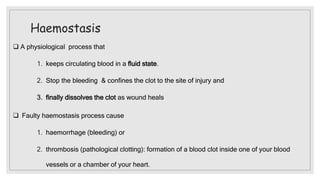
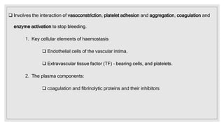
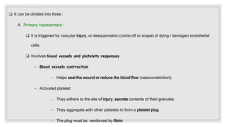
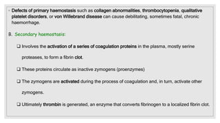
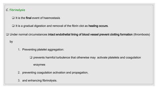
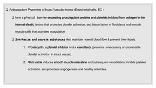
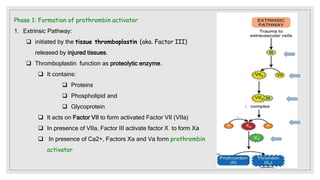
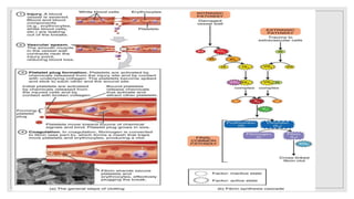
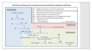
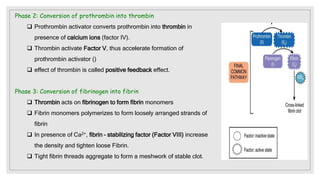
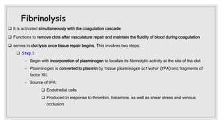
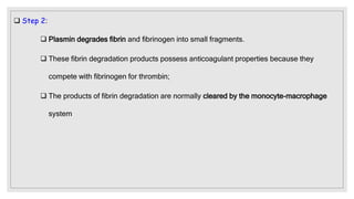
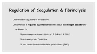
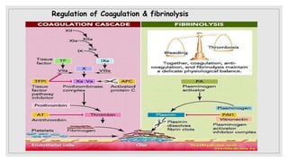
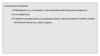
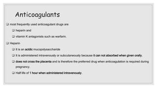
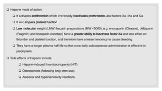
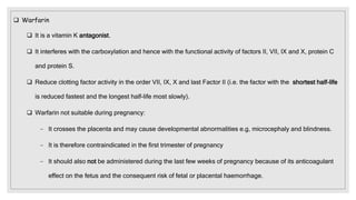
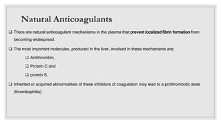
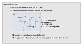
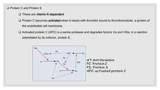
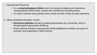
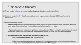
HAEMOSTASIS
Mirriam Mbilu
◦ Lecture Outline
1. Haemostasis:
a. Coagulation and fibrinolysis
b. vascular reconstruction
2. Disorders of coagulation and thrombosis
3. Anticoagulants
4. fibrinolytic and anti-platelet therapy
Haemostasis
 A physiological process that
1. keeps circulating blood in a fluid state.
2. Stop the bleeding & confines the clot to the site of injury and
3. finally dissolves the clot as wound heals
 Faulty haemostasis process cause
1. haemorrhage (bleeding) or
2. thrombosis (pathological clotting): formation of a blood clot inside one of your blood
vessels or a chamber of your heart.
 Involves the interaction of vasoconstriction, platelet adhesion and aggregation, coagulation and
enzyme activation to stop bleeding.
1. Key cellular elements of haemostasis
 Endothelial cells of the vascular intima,
 Extravascular tissue factor (TF) – bearing cells, and platelets.
2. The plasma components:
 coagulation and fibrinolytic proteins and their inhibitors
 It can be divided into three :
A. Primary haemostasis :
 It is triggered by vascular injury, or desquamation (come off or scape) of dying / damaged endothelial
cells.
 Involves blood vessels and platelets responses
− Blood vessels contraction
− Helps seal the wound or reduce the blood flow (vasoconstriction).
− Activated platelet:
− They adhere to the site of injury, secrete contents of their granules
− They aggregate with other platelets to form a platelet plug
− The plug must be reinforced by fibrin
◦ Defects of primary haemostasis such as collagen abnormalities, thrombocytopenia, qualitative
platelet disorders, or von Willebrand disease can cause debilitating, sometimes fatal, chronic
haemorrhage.
B. Secondary haemostasis:
 Involves the activation of a series of coagulation proteins in the plasma, mostly serine
proteases, to form a fibrin clot.
 These proteins circulate as inactive zymogens (proenzymes)
 The zymogens are activated during the process of coagulation and, in turn, activate other
zymogens.
 Ultimately thrombin is generated, an enzyme that converts fibrinogen to a localized fibrin clot.
C. fibrinolysis
 It is the final event of haemostasis
 It is a gradual digestion and removal of the fibrin clot as healing occurs.
 Under normal circumstances intact endothelial lining of blood vessel prevent clotting formation (thrombosis)
by
1. Preventing platelet aggregation:
 prevents harmful turbulence that otherwise may activate platelets and coagulation
enzymes
2. preventing coagulation activation and propagation,
3. and enhancing fibrinolysis.
 Anticoagulant Properties of Intact Vascular Intima (Endothelial cells, EC )
 form a physical barrier separating procoagulant proteins and platelets in blood from collagen in the
internal elastic lamina that promotes platelet adhesion, and tissue factor in fibroblasts and smooth
muscle cells that activates coagulation
 Synthesize and secrete substances that maintain normal blood flow & prevent thrombosis.
1. Prostacyclin, a platelet inhibitor and a vasodilator (prevents unnecessary or undesirable
platelet activation in intact vessel).
2. Nitric oxide induces smooth muscle relaxation and subsequent vasodilation, inhibits platelet
activation, and promotes angiogenesis and healthy arterioles.
 Synthesize and secrete a variety of substances that maintain normal blood flow & prevent thrombosis.
3. Tissue factor pathway inhibitor (TFPI), which controls activation of the tissue factor pathway, also
called the extrinsic coagulation pathway.
4. Production of inhibitors of thrombin formation such as thrombomodulin, facilitated by endothelial
protein C receptor (EPCR), and heparan sulfate.
PGI2: prostacyclin
TPA: Tissue Plasminogen
Activator
◦ Steps of haemostasis after Any harmful local stimulus either mechanical or chemical signal:
 First, it induces vasoconstriction, the vascular lumen narrows or closes, & blood flow to the injured site
is minimized.
 Second, exposed collagen of subendothelial connective tissues binds and activates platelets
 Third, ECs secrete molecules that promote platelet and Leukocytes adhesion to the exposed
subendothelial collagen in arterioles e.g.
 von Willebrand factor (VWF). VWF are necessary for platelets to adhere to.
 P-selectin
 intercellular adhesion molecules (ICAMs) and platelet endothelial cell adhesion molecules
(PECAMs)
 Finally, subendothelial smooth muscle cells and fibroblasts support the constitutive membrane protein
tissue factor which bind clotting factor & activate lead to lot formation and vascular sealing
◦ EC disruption exposes tissue factor in subendothelial cells and activates the coagulation system through
contact with plasma factor VI.
 Vascular Intima (endothelial cells) also express molecules fibrinolytic properties:
 Tissue plasminogen activator (TPA):
 TPA activates fibrinolysis by converting plasminogen to plasmin, which gradually digests fibrin
and restores blood flow
 Both TPA and plasminogen bind to polymerized fibrin during thrombus formation:.
 inhibitors to prevent excessive plasmin generation:
 Plasminogen activator inhibitor 1 (PAI-1), a TPA control protein that inhibits plasmin generation
and fibrinolysis.
 thrombin-activatable fibrinolysis inhibitor (TAFI), is activated by thrombin bound to EC
membrane thrombomodulin.
◦ NB: Elevations in PAI-1 or TAFI can slow fibrinolysis and increase the tendency for thrombosis
Coagulation
 This involves a series of concerted activation steps where inactive proteases (zymogens) are activated in a
cascade of reactions.
 principal enzyme complexes are composed of a vitamin K–dependent enzyme and a non-enzyme cofactor
assembled on anionic phospholipid membranes in a calcium-dependent fashion
 Because of the cascade nature of the process, a small stimulus produce a robust response.
 This process require activation of zymogen
 Coagulation is divided into three phases:
1. Formation of Prothrombin activator (initiated by intrinsic mechanism like trauma to the blood vessel)
2. Conversion of prothrombin into thrombin
3. Conversion of fibrinogen into fibrin
Phase 1: Formation of prothrombin activator
 Blood clotting commences with the formation of a substance called prothrombin activator, which converts
prothrombin into thrombin.
 Its formation is initiated by substances either within the blood or outside the blood.
 Formation of prothrombin activator occurs through two pathways:
1. Intrinsic Pathway (initiated by damage blood vessels)
2. Extrinsic Pathway (initiated by damage tissues)
◦ Phase 1: Formation of prothrombin activator
1. Intrinsic Pathway:
 Initiated when certain elements come into
contact with exposed collagen of damaged
blood wall
− Factor XII is activated to the Factor XIIa
− Platelets are activated to release
phospholipids
 Factor V is also activated by positive feedback
by thrombin
 Calcium ions also known as factor IV
◦ Phase 1: Formation of prothrombin activator
 In this pathway, the formation of prothrombin activator is initiated by platelets, which are within the
blood itself. Sequence of Events in Intrinsic Pathway During the injury, the blood vessel is ruptured;
 Endothelium is damaged and collagen beneath the endothelium is exposed.
 When factor XII (Hageman factor) comes in contact with collagen, it is converted into activated factor
XII in the presence of Kallikrein and Kinogen.
 The activated factor XII Converts factor XI into activated XI in the presence of high molecular weight
Kinogen.
 The activated factor XI activates factor IX in the presence of factor IV (Calcium).
 Activated factor IX activates factor X in the presence of factor VIII and Calcium. When platelet comes in
contact with collagen of damaged blood vessel, it gets activated and releases phospholipids. Now the
activated factor X reacts with platelet phospholipid and factor V to form Prothrombin activator. This
needs the presence of calcium ions, Factor V is also activated by the positive feedback effect of
thrombin.
Phase 1: Formation of prothrombin activator
1. Extrinsic Pathway:
 initiated by the tissue thromboplastin (aka. Factor III)
released by injured tissues.
 Thromboplastin function as proteolytic enzyme.
 It contains:
 Proteins
 Phospholipid and
 Glycoprotein
 It acts on Factor VII to form activated Factor VII (VIIa)
 In presence of VIIa, Factor III activate factor X to form Xa
 In presence of Ca2+, Factors Xa and Va form prothrombin
activator
Phase 2: Conversion of prothrombin into thrombin
 Prothrombin activator converts prothrombin into thrombin in
presence of calcium ions (factor IV).
 Thrombin activate Factor V, thus accelerate formation of
prothrombin activator ()
 effect of thrombin is called positive feedback effect.
Phase 3: Conversion of fibrinogen into fibrin
 Thrombin acts on fibrinogen to form fibrin monomers
 Fibrin monomers polymerizes to form loosely arranged strands of
fibrin
 In presence of Ca2+, fibrin – stabilizing factor (Factor VIII) increase
the density and tighten loose Fibrin.
 Tight fibrin threads aggregate to form a meshwork of stable clot.
Fibrinolysis
 It is activated simultaneously with the coagulation cascade.
 Functions to remove clots after vasculature repair and maintain the fluidity of blood during coagulation
 serves in clot lysis once tissue repair begins. This involves two steps:
 Step 1:
− Begin with incorporation of plasminogen to localize its fibrinolytic activity at the site of the clot
− Plasminogen is converted to plasmin by tissue plasminogen activator (tPA) and fragments of
factor XII.
− Source of tPA:
 Endothelial cells
 Produced in response to thrombin, histamine, as well as shear stress and venous
occlusion
 Step 2:
 Plasmin degrades fibrin and fibrinogen into small fragments.
 These fibrin degradation products possess anticoagulant properties because they
compete with fibrinogen for thrombin;
 The products of fibrin degradation are normally cleared by the monocyte-macrophage
system
Regulation of Coagulation & fibrinolysis
 Inhibited at Key points of the cascade
 Fibrinolysis is regulated by proteins that inhibit tissue plasminogen activator and
urokinase .i.e
 plasminogen activator inhibitors 1 & 2 (PAI-1 & PAI-2),
 activated protein C-inhibitor
 and thrombin activatable fibrinolysis inhibitor (TAFI).
Regulation of Coagulation & fibrinolysis
Disorders of coagulation
 This are conditions whereby there is abnormal bleeding due to impairment of haemostasis
(cessation of bleeding)
 classified into:
1. Inherited disorders
2. Acquired disorders
Inherited disorders of coagulation
 Occur as a result of an inherited defect of one or other protein involved in
coagulation.
 Examples:
 Hemophilia A
 haemophilia B, Christmas disease
 Von Willebrand disease
Hemophilia A
 is the most common inherited coagulation disorder
 Caused by factor VIII deficiency
 Attributed to inherited genetic mutation on the X chromosome
such as deletions, insertions, point mutations and a common
intra-gene inversion
 Thus it is sex-linked. Severe disease occur in males
 Clinical features
 Range from severe spontaneous bleeding, especially into joints
(haemarthroses) and muscles, to mild symptoms,
 Symptom on the factor VIII level deficiency
 Onset in early childhood (e.g. post-circumcision).
 Individual have Increased risk of post-operative or post-traumatic
haemorrhage
 Chronic debilitating joint disease caused by repeated bleeds.
 Hemophilia A: Laboratory features
 Plasma factor VIII activity reduced to:
 <1% of normal = severe cases,
 1–5% of normal = moderate cases and
 5–40% of normal = in mild cases.
 Carriers have factor VIII levels in plasma approximately 50% of normal.
 DNA analysis is helpful in carrier detection and antenatal diagnosis.
 Von Willebrand factor (vWF) level is normal (vWF is procoagulant released by endothelial cells)
◦ Treatment of hemophilia A
 Infusions of factor VIII:
− Using either recombinant or concentrate from normal donated plasma
− Aimed at elevating patient’s Factor VIII level:
 to 20–50% of normal in cases of severe bleeding.
 Level is raised to and maintained at 80–100% for elective surgery
 Desmopressin, an analogue of vasopressin, leads to a modest rise in endogenous factor VIII
which is useful in mild cases.
 Avoid aspirin, other antiplatelet drugs and intramuscular injections.
B. Haemophilia B,.
 Aka Christmas disease , named after first person
(Stephen Christmas) diagnosed with the condition in 1952.
 Caused by Factor IX deficiency
 has similar clinical features to haemophilia A
 It is X-linked
 Less common than hemophilia A
 Diagnosis and treatment are similar to haemophilia A,
except that factor IX concentrate is infused
C. Von Willebrand disease
 Caused by mutations in the vWF gene.
 VWF (Von Willebrand factor) is a large multimeric protein produced by endothelial cells.
 VWF carries factor VIII in plasma and mediates platelet adhesion to endothelium
 The disease is more frequent than haemophilia A; males and females are affected equally.
 Clinical features •
− Bleeding, typically from mucous membranes (mouth, epistaxis, menorrhagia).
− Excess blood loss following trauma or surgery
◦ Treatment of Von Willebrand disease
 Intermediate purity factor VIII concentrate (contains both vWF and factor VIII) for bleeding.
High purity vWF concentrates for severe bleeding.
 Desmopressin is helpful for mild bleeding
 Fibrinolytic inhibitors (e.g. tranexamic acid) are helpful.
 Carrier detection and antenatal diagnosis based on fetal DNA analysis is available.
Other Inherited disorders of coagulation
 Factor XI deficiency
 Congenital deficiencies of factors II, V, VII, X and XIII are rare and usually cause mild bleeding
disorder.
 Fibrinogen deficiency occurs as a moderately severe autosomal recessive disorder.
 Dysfibrinogenaemia (presence of a functionally abnormal molecule) is both a rare autosomal dominant
disorder and a more common acquired disorder (liver disease, malignancy and systemic lupus
erythematosus).
Acquired disorders of coagulation
◦ Liver disease
 This leads to defects of coagulation, platelets and fibrinolysis;
− Reduced synthesis of vitamin K-dependent factors (II, VII,IX, X, proteins C and
S) due to impaired vitamin K absorption (biliary obstruction).
− Impaired synthesis of other coagulation proteins (factors I and V).
− Thrombocytopenia (hypersplenism) and abnormal platelet function (cirrhosis).
− Impaired fibrinolysis due to reduced levels of proteins C and S, antithrombin and α2-
antiplasmin lead to susceptibility to disseminated intravascular coagulation (DIC).
◦ Abnormal fibrinogen (dysfibrinogenaemia) may lead to haemorrhage or thrombosis due to
formation abnormal clots
Vitamin K deficiency
◦ This vitamin is necessary for synthesis of factors II, VII, IX, & X and proteins C and S
Causes
 Inadequate body stores e.g. in malnutrition, heamorrhrage, of the new bone.
 Mal-absorption of vitamin k. often occurs in obstructive jaundice.
 The billiary duct becomes obstructed and bile is not poured in the GIT.
 Bile emulsifies fat and since vitamin k is fat soluble, It can not be absorbed into blood stream.
◦ Clinical features
 Bleeding tendencies.
 Cerebral bleeding
◦ Treatment
 Replace vitamin k injection intramascular vitamin k 10mg OR.
 Identify and treat the cause Vitamin K deficiency
THROMBOSIS
 A pathological process where platelets and fibrin interact within the vessel wall to form a haemostatic plug to
cause vascular obstruction
 Can be either
 Arterial thrombosis
 venous thrombosis
 Arterial thrombosis
 This occurs in relation to damaged endothelium, e.g. atherosclerotic plaques.
 Exposed collagen and released tissue factor cause platelet aggregation and fibrin formation
 It causes ischaemia (restriction of blood supply to tissue),
 Risk factor include:
− Hypertension
− Smoking
− Diabetes
− Hyperlipidaemia
− ↑ Homocysteine
− Polycythaemia / thrombocythaemia
− ↑ Factor VIII
− ↑ Fibrinogen
− Lupus anticoagulant (one of malfunctioning antibodies)
− Heparin therapy
Venous thrombosis
 Is a blood clot (thrombus) that forms within a vein
 It leads to stasis (slow blood flow in veins)
 Risk of venous thrombosis increased by
1. Factors affecting blood flow e.g. stasis,
obesity),
2. alterations in blood constituents and damage to
vascular endothelium e.g. caused by sepsis,
surgery or indwelling catheters
Thrombophilia
 is a congenital or acquired predisposition to venous thrombosis
 It should be suspected and screened for in patients with thrombosis who are young, have a positive family
history, have thrombosis
 Inherited thrombophilia:
 Caused by Inherited genetic mutations that may predispose to venous and, more rarely, arterial
thrombosis
 inheritance of a variant form of factor V (factor V Leiden) is the most common
 Acquired thrombophilia
 Pathogenesis, e.g. in pregnancy, oral contraceptive pill therapy and malignancy,
 It is multifactorial
 It relates to elevated levels of procoagulant factors, depressed levels of inhibitor proteins
and physical factors (e.g. stasis, surgery).
Anticoagulants
 most frequently used anticoagulant drugs are
 heparin and
 vitamin K antagonists such as warfarin.
 Heparin
 It is an acidic mucopolysaccharide
 It is administered intravenously or subcutaneously because it can not absorbed when given orally.
 does not cross the placenta and is therefore the preferred drug when anticoagulation is required during
pregnancy.
 Half life of 1 hour when administered intravenously.
 Heparin mode of action:
 It activates antithrombin which irreversibly inactivates prothrombin, and factors Xa, IXa and Xia
 It also impairs platelet function
 Low molecular weight (LMW) heparin preparations (MW <5000), e.g. enoxaparin (Clexane), deltaparin
(Fragmin) and tinzaparin (Innohep) have a greater ability to inactivate factor Xa and less effect on
thrombin and platelet function, and therefore have a lesser tendency to cause bleeding.
 They have a longer plasma half-life so that once daily subcutaneous administration is effective in
prophylaxis.
 Side effects of Heparin include;
 Heparin-induced thrombocytopenia (HIT)
 Osteoporosis (following long-term use),
 Alopecia and hypersensitivity reactions.
 Warfarin
 It is a vitamin K antagonist.
 It interferes with the carboxylation and hence with the functional activity of factors II, VII, IX and X, protein C
and protein S.
 Reduce clotting factor activity in the order VII, IX, X and last Factor II (i.e. the factor with the shortest half-life
is reduced fastest and the longest half-life most slowly).
 Warfarin not suitable during pregnancy:
− It crosses the placenta and may cause developmental abnormalities e.g, microcephaly and blindness.
− It is therefore contraindicated in the first trimester of pregnancy
− It should also not be administered during the last few weeks of pregnancy because of its anticoagulant
effect on the fetus and the consequent risk of fetal or placental haemorrhage.
Natural Anticoagulants
 There are natural anticoagulant mechanisms in the plasma that prevent localized fibrin formation from
becoming widespread.
 The most important molecules, produced in the liver, involved in these mechanisms are;
 Antithrombin,
 Protein C and
 protein S.
 Inherited or acquired abnormalities of these inhibitors of coagulation may lead to a prothrombotic state
(thrombophilia).
 Antithrombin (AT)
 Mainly an inhibitor of thrombin and factor Xa
 It also inhibits factors IXa and XIa and the TF–VIIa complex
 its action is markedly potentiated by heparin
 Congenital AT deficiency is inherited as an autosomal dominant character trait
 Protein C and Protein S
 These are vitamin K-dependent
 Protein C becomes activated when it reacts with thrombin bound to thrombomodulin, a protein of
the endothelial cell membrane.
 Activated protein C (APC) is a serine protease and degrades factors Va and VIIIa, in a reaction
potentiated by its cofactor, protein S.
fibrinolytic and anti-platelet therapy
Antiplatelet therapy
1. Aspirin (75 mg/day and 300 mg post-myocardial infarction)
 Most widely used to prevent arterial thrombosis.
 It inhibits platelet function by inhibiting cyclo-oxygenase, thus reducing thromboxane A2 production
2. Clopidogrel is an adenosine diphosphate (ADP) receptor antagonist.
 At 75 mg/day it is widely used with aspirin or alone.
 It is less likely than aspirin to cause gastrointestinal haemorrhage.
3. Prasugrel
 Mode of action to similar clopidogrel.
1. Dipyridamole (Persantin)
 It is a phosphodiesterase inhibitor which, by raising the platelet cyclic adenosine
monophosphate (AMP) levels, reduces their sensitivity to activating stimuli.
 It is used in patients with prosthetic heart valves and after cardiac by-pass operations.
2. Others antiplatelet therapies include:
 Monoclonal antibodies directed to platelet glycoproteins (e.g. abciximab, which is
directed against glycoprotein IIb/IIIa) or
 Small molecule inhibitors of glycoprotein IIb/IIIa eptifibatide or tirofiban are used, for
example, post-angioplasty or stent insertion.
Fibrinolytic therapy
 This is used to enhance conversion of plasminogen to plasmin which degrades fibrin.
 It must be used within 5–7 days for venous thrombosis or pulmonary embolus and 5–7 hours for arterial
thrombosis•
 Fibrinolytic therapy include:
− Streptokinase directly activates plasminogen. Most individuals have antistreptococcal antibodies; a loading
dose is therefore required and treatment becomes ineffective after 4–10 days.
− Urokinase has a similar action as streptokinase but may be used if there are high levels of antistreptococcal
antibodies. Single-chain urokinase-type plasminogen activator (SCU-PA) has also been developed.
− Acylated plasminogen streptokinase activator complex (APSAC)
 activates streptokinase bound to plasminogen.
 Recombinant tissue plasminogen activation (TPA) causes activation of fibrin-bound plasminogen
only, and is associated with less systemic activation of fibrinolysis.

HEMOSTASIS & ANTICOAGULANTS document pptx

  • 1.
  • 2.
    ◦ Lecture Outline 1.Haemostasis: a. Coagulation and fibrinolysis b. vascular reconstruction 2. Disorders of coagulation and thrombosis 3. Anticoagulants 4. fibrinolytic and anti-platelet therapy
  • 3.
    Haemostasis  A physiologicalprocess that 1. keeps circulating blood in a fluid state. 2. Stop the bleeding & confines the clot to the site of injury and 3. finally dissolves the clot as wound heals  Faulty haemostasis process cause 1. haemorrhage (bleeding) or 2. thrombosis (pathological clotting): formation of a blood clot inside one of your blood vessels or a chamber of your heart.
  • 4.
     Involves theinteraction of vasoconstriction, platelet adhesion and aggregation, coagulation and enzyme activation to stop bleeding. 1. Key cellular elements of haemostasis  Endothelial cells of the vascular intima,  Extravascular tissue factor (TF) – bearing cells, and platelets. 2. The plasma components:  coagulation and fibrinolytic proteins and their inhibitors
  • 5.
     It canbe divided into three : A. Primary haemostasis :  It is triggered by vascular injury, or desquamation (come off or scape) of dying / damaged endothelial cells.  Involves blood vessels and platelets responses − Blood vessels contraction − Helps seal the wound or reduce the blood flow (vasoconstriction). − Activated platelet: − They adhere to the site of injury, secrete contents of their granules − They aggregate with other platelets to form a platelet plug − The plug must be reinforced by fibrin
  • 6.
    ◦ Defects ofprimary haemostasis such as collagen abnormalities, thrombocytopenia, qualitative platelet disorders, or von Willebrand disease can cause debilitating, sometimes fatal, chronic haemorrhage. B. Secondary haemostasis:  Involves the activation of a series of coagulation proteins in the plasma, mostly serine proteases, to form a fibrin clot.  These proteins circulate as inactive zymogens (proenzymes)  The zymogens are activated during the process of coagulation and, in turn, activate other zymogens.  Ultimately thrombin is generated, an enzyme that converts fibrinogen to a localized fibrin clot.
  • 7.
    C. fibrinolysis  Itis the final event of haemostasis  It is a gradual digestion and removal of the fibrin clot as healing occurs.  Under normal circumstances intact endothelial lining of blood vessel prevent clotting formation (thrombosis) by 1. Preventing platelet aggregation:  prevents harmful turbulence that otherwise may activate platelets and coagulation enzymes 2. preventing coagulation activation and propagation, 3. and enhancing fibrinolysis.
  • 8.
     Anticoagulant Propertiesof Intact Vascular Intima (Endothelial cells, EC )  form a physical barrier separating procoagulant proteins and platelets in blood from collagen in the internal elastic lamina that promotes platelet adhesion, and tissue factor in fibroblasts and smooth muscle cells that activates coagulation  Synthesize and secrete substances that maintain normal blood flow & prevent thrombosis. 1. Prostacyclin, a platelet inhibitor and a vasodilator (prevents unnecessary or undesirable platelet activation in intact vessel). 2. Nitric oxide induces smooth muscle relaxation and subsequent vasodilation, inhibits platelet activation, and promotes angiogenesis and healthy arterioles.
  • 9.
     Synthesize andsecrete a variety of substances that maintain normal blood flow & prevent thrombosis. 3. Tissue factor pathway inhibitor (TFPI), which controls activation of the tissue factor pathway, also called the extrinsic coagulation pathway. 4. Production of inhibitors of thrombin formation such as thrombomodulin, facilitated by endothelial protein C receptor (EPCR), and heparan sulfate. PGI2: prostacyclin TPA: Tissue Plasminogen Activator
  • 10.
    ◦ Steps ofhaemostasis after Any harmful local stimulus either mechanical or chemical signal:  First, it induces vasoconstriction, the vascular lumen narrows or closes, & blood flow to the injured site is minimized.  Second, exposed collagen of subendothelial connective tissues binds and activates platelets  Third, ECs secrete molecules that promote platelet and Leukocytes adhesion to the exposed subendothelial collagen in arterioles e.g.  von Willebrand factor (VWF). VWF are necessary for platelets to adhere to.  P-selectin  intercellular adhesion molecules (ICAMs) and platelet endothelial cell adhesion molecules (PECAMs)  Finally, subendothelial smooth muscle cells and fibroblasts support the constitutive membrane protein tissue factor which bind clotting factor & activate lead to lot formation and vascular sealing
  • 11.
    ◦ EC disruptionexposes tissue factor in subendothelial cells and activates the coagulation system through contact with plasma factor VI.
  • 12.
     Vascular Intima(endothelial cells) also express molecules fibrinolytic properties:  Tissue plasminogen activator (TPA):  TPA activates fibrinolysis by converting plasminogen to plasmin, which gradually digests fibrin and restores blood flow  Both TPA and plasminogen bind to polymerized fibrin during thrombus formation:.  inhibitors to prevent excessive plasmin generation:  Plasminogen activator inhibitor 1 (PAI-1), a TPA control protein that inhibits plasmin generation and fibrinolysis.  thrombin-activatable fibrinolysis inhibitor (TAFI), is activated by thrombin bound to EC membrane thrombomodulin. ◦ NB: Elevations in PAI-1 or TAFI can slow fibrinolysis and increase the tendency for thrombosis
  • 13.
    Coagulation  This involvesa series of concerted activation steps where inactive proteases (zymogens) are activated in a cascade of reactions.  principal enzyme complexes are composed of a vitamin K–dependent enzyme and a non-enzyme cofactor assembled on anionic phospholipid membranes in a calcium-dependent fashion  Because of the cascade nature of the process, a small stimulus produce a robust response.  This process require activation of zymogen
  • 14.
     Coagulation isdivided into three phases: 1. Formation of Prothrombin activator (initiated by intrinsic mechanism like trauma to the blood vessel) 2. Conversion of prothrombin into thrombin 3. Conversion of fibrinogen into fibrin Phase 1: Formation of prothrombin activator  Blood clotting commences with the formation of a substance called prothrombin activator, which converts prothrombin into thrombin.  Its formation is initiated by substances either within the blood or outside the blood.  Formation of prothrombin activator occurs through two pathways: 1. Intrinsic Pathway (initiated by damage blood vessels) 2. Extrinsic Pathway (initiated by damage tissues)
  • 15.
    ◦ Phase 1:Formation of prothrombin activator 1. Intrinsic Pathway:  Initiated when certain elements come into contact with exposed collagen of damaged blood wall − Factor XII is activated to the Factor XIIa − Platelets are activated to release phospholipids  Factor V is also activated by positive feedback by thrombin  Calcium ions also known as factor IV
  • 16.
    ◦ Phase 1:Formation of prothrombin activator  In this pathway, the formation of prothrombin activator is initiated by platelets, which are within the blood itself. Sequence of Events in Intrinsic Pathway During the injury, the blood vessel is ruptured;  Endothelium is damaged and collagen beneath the endothelium is exposed.  When factor XII (Hageman factor) comes in contact with collagen, it is converted into activated factor XII in the presence of Kallikrein and Kinogen.  The activated factor XII Converts factor XI into activated XI in the presence of high molecular weight Kinogen.  The activated factor XI activates factor IX in the presence of factor IV (Calcium).  Activated factor IX activates factor X in the presence of factor VIII and Calcium. When platelet comes in contact with collagen of damaged blood vessel, it gets activated and releases phospholipids. Now the activated factor X reacts with platelet phospholipid and factor V to form Prothrombin activator. This needs the presence of calcium ions, Factor V is also activated by the positive feedback effect of thrombin.
  • 17.
    Phase 1: Formationof prothrombin activator 1. Extrinsic Pathway:  initiated by the tissue thromboplastin (aka. Factor III) released by injured tissues.  Thromboplastin function as proteolytic enzyme.  It contains:  Proteins  Phospholipid and  Glycoprotein  It acts on Factor VII to form activated Factor VII (VIIa)  In presence of VIIa, Factor III activate factor X to form Xa  In presence of Ca2+, Factors Xa and Va form prothrombin activator
  • 20.
    Phase 2: Conversionof prothrombin into thrombin  Prothrombin activator converts prothrombin into thrombin in presence of calcium ions (factor IV).  Thrombin activate Factor V, thus accelerate formation of prothrombin activator ()  effect of thrombin is called positive feedback effect. Phase 3: Conversion of fibrinogen into fibrin  Thrombin acts on fibrinogen to form fibrin monomers  Fibrin monomers polymerizes to form loosely arranged strands of fibrin  In presence of Ca2+, fibrin – stabilizing factor (Factor VIII) increase the density and tighten loose Fibrin.  Tight fibrin threads aggregate to form a meshwork of stable clot.
  • 21.
    Fibrinolysis  It isactivated simultaneously with the coagulation cascade.  Functions to remove clots after vasculature repair and maintain the fluidity of blood during coagulation  serves in clot lysis once tissue repair begins. This involves two steps:  Step 1: − Begin with incorporation of plasminogen to localize its fibrinolytic activity at the site of the clot − Plasminogen is converted to plasmin by tissue plasminogen activator (tPA) and fragments of factor XII. − Source of tPA:  Endothelial cells  Produced in response to thrombin, histamine, as well as shear stress and venous occlusion
  • 22.
     Step 2: Plasmin degrades fibrin and fibrinogen into small fragments.  These fibrin degradation products possess anticoagulant properties because they compete with fibrinogen for thrombin;  The products of fibrin degradation are normally cleared by the monocyte-macrophage system
  • 23.
    Regulation of Coagulation& fibrinolysis  Inhibited at Key points of the cascade  Fibrinolysis is regulated by proteins that inhibit tissue plasminogen activator and urokinase .i.e  plasminogen activator inhibitors 1 & 2 (PAI-1 & PAI-2),  activated protein C-inhibitor  and thrombin activatable fibrinolysis inhibitor (TAFI).
  • 24.
  • 25.
    Disorders of coagulation This are conditions whereby there is abnormal bleeding due to impairment of haemostasis (cessation of bleeding)  classified into: 1. Inherited disorders 2. Acquired disorders
  • 26.
    Inherited disorders ofcoagulation  Occur as a result of an inherited defect of one or other protein involved in coagulation.  Examples:  Hemophilia A  haemophilia B, Christmas disease  Von Willebrand disease
  • 27.
    Hemophilia A  isthe most common inherited coagulation disorder  Caused by factor VIII deficiency  Attributed to inherited genetic mutation on the X chromosome such as deletions, insertions, point mutations and a common intra-gene inversion  Thus it is sex-linked. Severe disease occur in males  Clinical features  Range from severe spontaneous bleeding, especially into joints (haemarthroses) and muscles, to mild symptoms,  Symptom on the factor VIII level deficiency  Onset in early childhood (e.g. post-circumcision).  Individual have Increased risk of post-operative or post-traumatic haemorrhage  Chronic debilitating joint disease caused by repeated bleeds.
  • 28.
     Hemophilia A:Laboratory features  Plasma factor VIII activity reduced to:  <1% of normal = severe cases,  1–5% of normal = moderate cases and  5–40% of normal = in mild cases.  Carriers have factor VIII levels in plasma approximately 50% of normal.  DNA analysis is helpful in carrier detection and antenatal diagnosis.  Von Willebrand factor (vWF) level is normal (vWF is procoagulant released by endothelial cells)
  • 29.
    ◦ Treatment ofhemophilia A  Infusions of factor VIII: − Using either recombinant or concentrate from normal donated plasma − Aimed at elevating patient’s Factor VIII level:  to 20–50% of normal in cases of severe bleeding.  Level is raised to and maintained at 80–100% for elective surgery  Desmopressin, an analogue of vasopressin, leads to a modest rise in endogenous factor VIII which is useful in mild cases.  Avoid aspirin, other antiplatelet drugs and intramuscular injections.
  • 30.
    B. Haemophilia B,. Aka Christmas disease , named after first person (Stephen Christmas) diagnosed with the condition in 1952.  Caused by Factor IX deficiency  has similar clinical features to haemophilia A  It is X-linked  Less common than hemophilia A  Diagnosis and treatment are similar to haemophilia A, except that factor IX concentrate is infused
  • 31.
    C. Von Willebranddisease  Caused by mutations in the vWF gene.  VWF (Von Willebrand factor) is a large multimeric protein produced by endothelial cells.  VWF carries factor VIII in plasma and mediates platelet adhesion to endothelium  The disease is more frequent than haemophilia A; males and females are affected equally.
  • 32.
     Clinical features• − Bleeding, typically from mucous membranes (mouth, epistaxis, menorrhagia). − Excess blood loss following trauma or surgery ◦ Treatment of Von Willebrand disease  Intermediate purity factor VIII concentrate (contains both vWF and factor VIII) for bleeding. High purity vWF concentrates for severe bleeding.  Desmopressin is helpful for mild bleeding  Fibrinolytic inhibitors (e.g. tranexamic acid) are helpful.  Carrier detection and antenatal diagnosis based on fetal DNA analysis is available.
  • 33.
    Other Inherited disordersof coagulation  Factor XI deficiency  Congenital deficiencies of factors II, V, VII, X and XIII are rare and usually cause mild bleeding disorder.  Fibrinogen deficiency occurs as a moderately severe autosomal recessive disorder.  Dysfibrinogenaemia (presence of a functionally abnormal molecule) is both a rare autosomal dominant disorder and a more common acquired disorder (liver disease, malignancy and systemic lupus erythematosus).
  • 34.
    Acquired disorders ofcoagulation ◦ Liver disease  This leads to defects of coagulation, platelets and fibrinolysis; − Reduced synthesis of vitamin K-dependent factors (II, VII,IX, X, proteins C and S) due to impaired vitamin K absorption (biliary obstruction). − Impaired synthesis of other coagulation proteins (factors I and V). − Thrombocytopenia (hypersplenism) and abnormal platelet function (cirrhosis). − Impaired fibrinolysis due to reduced levels of proteins C and S, antithrombin and α2- antiplasmin lead to susceptibility to disseminated intravascular coagulation (DIC).
  • 35.
    ◦ Abnormal fibrinogen(dysfibrinogenaemia) may lead to haemorrhage or thrombosis due to formation abnormal clots Vitamin K deficiency ◦ This vitamin is necessary for synthesis of factors II, VII, IX, & X and proteins C and S Causes  Inadequate body stores e.g. in malnutrition, heamorrhrage, of the new bone.  Mal-absorption of vitamin k. often occurs in obstructive jaundice.  The billiary duct becomes obstructed and bile is not poured in the GIT.  Bile emulsifies fat and since vitamin k is fat soluble, It can not be absorbed into blood stream.
  • 36.
    ◦ Clinical features Bleeding tendencies.  Cerebral bleeding ◦ Treatment  Replace vitamin k injection intramascular vitamin k 10mg OR.  Identify and treat the cause Vitamin K deficiency
  • 37.
    THROMBOSIS  A pathologicalprocess where platelets and fibrin interact within the vessel wall to form a haemostatic plug to cause vascular obstruction  Can be either  Arterial thrombosis  venous thrombosis  Arterial thrombosis  This occurs in relation to damaged endothelium, e.g. atherosclerotic plaques.  Exposed collagen and released tissue factor cause platelet aggregation and fibrin formation  It causes ischaemia (restriction of blood supply to tissue),
  • 38.
     Risk factorinclude: − Hypertension − Smoking − Diabetes − Hyperlipidaemia − ↑ Homocysteine − Polycythaemia / thrombocythaemia − ↑ Factor VIII − ↑ Fibrinogen − Lupus anticoagulant (one of malfunctioning antibodies) − Heparin therapy
  • 39.
    Venous thrombosis  Isa blood clot (thrombus) that forms within a vein  It leads to stasis (slow blood flow in veins)  Risk of venous thrombosis increased by 1. Factors affecting blood flow e.g. stasis, obesity), 2. alterations in blood constituents and damage to vascular endothelium e.g. caused by sepsis, surgery or indwelling catheters
  • 40.
    Thrombophilia  is acongenital or acquired predisposition to venous thrombosis  It should be suspected and screened for in patients with thrombosis who are young, have a positive family history, have thrombosis  Inherited thrombophilia:  Caused by Inherited genetic mutations that may predispose to venous and, more rarely, arterial thrombosis  inheritance of a variant form of factor V (factor V Leiden) is the most common
  • 41.
     Acquired thrombophilia Pathogenesis, e.g. in pregnancy, oral contraceptive pill therapy and malignancy,  It is multifactorial  It relates to elevated levels of procoagulant factors, depressed levels of inhibitor proteins and physical factors (e.g. stasis, surgery).
  • 42.
    Anticoagulants  most frequentlyused anticoagulant drugs are  heparin and  vitamin K antagonists such as warfarin.  Heparin  It is an acidic mucopolysaccharide  It is administered intravenously or subcutaneously because it can not absorbed when given orally.  does not cross the placenta and is therefore the preferred drug when anticoagulation is required during pregnancy.  Half life of 1 hour when administered intravenously.
  • 43.
     Heparin modeof action:  It activates antithrombin which irreversibly inactivates prothrombin, and factors Xa, IXa and Xia  It also impairs platelet function  Low molecular weight (LMW) heparin preparations (MW <5000), e.g. enoxaparin (Clexane), deltaparin (Fragmin) and tinzaparin (Innohep) have a greater ability to inactivate factor Xa and less effect on thrombin and platelet function, and therefore have a lesser tendency to cause bleeding.  They have a longer plasma half-life so that once daily subcutaneous administration is effective in prophylaxis.  Side effects of Heparin include;  Heparin-induced thrombocytopenia (HIT)  Osteoporosis (following long-term use),  Alopecia and hypersensitivity reactions.
  • 44.
     Warfarin  Itis a vitamin K antagonist.  It interferes with the carboxylation and hence with the functional activity of factors II, VII, IX and X, protein C and protein S.  Reduce clotting factor activity in the order VII, IX, X and last Factor II (i.e. the factor with the shortest half-life is reduced fastest and the longest half-life most slowly).  Warfarin not suitable during pregnancy: − It crosses the placenta and may cause developmental abnormalities e.g, microcephaly and blindness. − It is therefore contraindicated in the first trimester of pregnancy − It should also not be administered during the last few weeks of pregnancy because of its anticoagulant effect on the fetus and the consequent risk of fetal or placental haemorrhage.
  • 45.
    Natural Anticoagulants  Thereare natural anticoagulant mechanisms in the plasma that prevent localized fibrin formation from becoming widespread.  The most important molecules, produced in the liver, involved in these mechanisms are;  Antithrombin,  Protein C and  protein S.  Inherited or acquired abnormalities of these inhibitors of coagulation may lead to a prothrombotic state (thrombophilia).
  • 46.
     Antithrombin (AT) Mainly an inhibitor of thrombin and factor Xa  It also inhibits factors IXa and XIa and the TF–VIIa complex  its action is markedly potentiated by heparin  Congenital AT deficiency is inherited as an autosomal dominant character trait
  • 47.
     Protein Cand Protein S  These are vitamin K-dependent  Protein C becomes activated when it reacts with thrombin bound to thrombomodulin, a protein of the endothelial cell membrane.  Activated protein C (APC) is a serine protease and degrades factors Va and VIIIa, in a reaction potentiated by its cofactor, protein S.
  • 48.
    fibrinolytic and anti-platelettherapy Antiplatelet therapy 1. Aspirin (75 mg/day and 300 mg post-myocardial infarction)  Most widely used to prevent arterial thrombosis.  It inhibits platelet function by inhibiting cyclo-oxygenase, thus reducing thromboxane A2 production 2. Clopidogrel is an adenosine diphosphate (ADP) receptor antagonist.  At 75 mg/day it is widely used with aspirin or alone.  It is less likely than aspirin to cause gastrointestinal haemorrhage. 3. Prasugrel  Mode of action to similar clopidogrel.
  • 49.
    1. Dipyridamole (Persantin) It is a phosphodiesterase inhibitor which, by raising the platelet cyclic adenosine monophosphate (AMP) levels, reduces their sensitivity to activating stimuli.  It is used in patients with prosthetic heart valves and after cardiac by-pass operations. 2. Others antiplatelet therapies include:  Monoclonal antibodies directed to platelet glycoproteins (e.g. abciximab, which is directed against glycoprotein IIb/IIIa) or  Small molecule inhibitors of glycoprotein IIb/IIIa eptifibatide or tirofiban are used, for example, post-angioplasty or stent insertion.
  • 50.
    Fibrinolytic therapy  Thisis used to enhance conversion of plasminogen to plasmin which degrades fibrin.  It must be used within 5–7 days for venous thrombosis or pulmonary embolus and 5–7 hours for arterial thrombosis•  Fibrinolytic therapy include: − Streptokinase directly activates plasminogen. Most individuals have antistreptococcal antibodies; a loading dose is therefore required and treatment becomes ineffective after 4–10 days. − Urokinase has a similar action as streptokinase but may be used if there are high levels of antistreptococcal antibodies. Single-chain urokinase-type plasminogen activator (SCU-PA) has also been developed. − Acylated plasminogen streptokinase activator complex (APSAC)  activates streptokinase bound to plasminogen.  Recombinant tissue plasminogen activation (TPA) causes activation of fibrin-bound plasminogen only, and is associated with less systemic activation of fibrinolysis.

Editor's Notes

  • #19 https://opentextbc.ca/anatomyandphysiology/chapter/18-5-hemostasis/