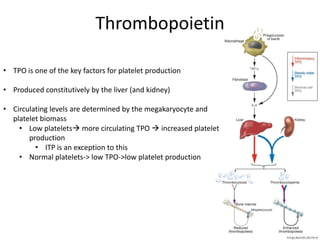
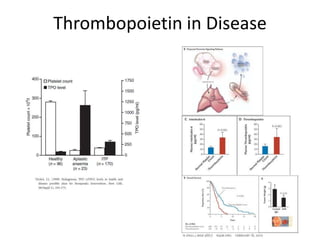
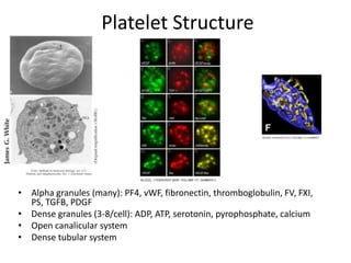
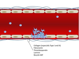
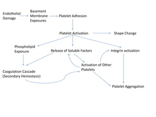
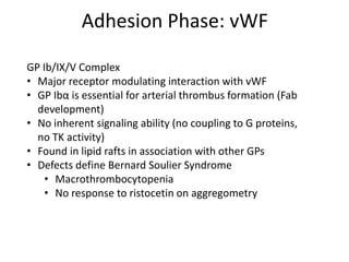
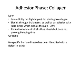
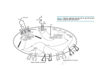
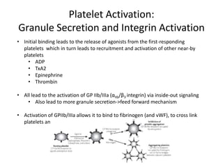
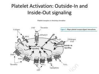
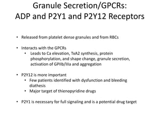
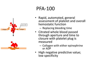
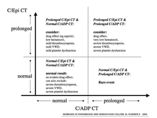
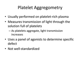
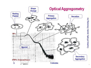
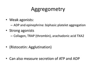
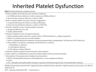
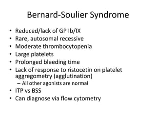
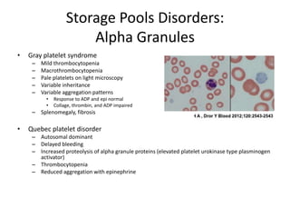
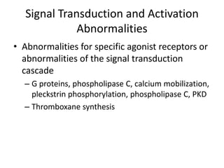
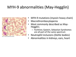
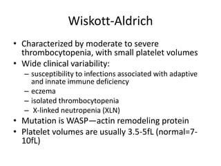
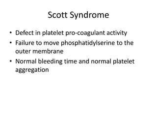
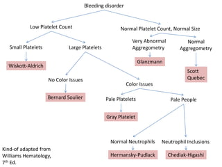
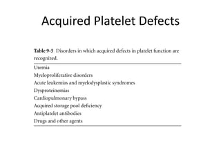
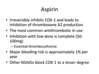
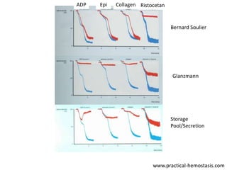
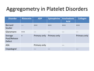
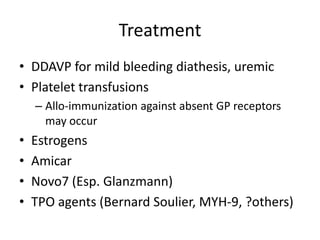
Platelet Function and
Dysfunction
Paul Basciano MD
October 24, 2013
Outline
•
•
•
•
•
•
•
•

Thrombopoiesis
Platelet Structure
Platelet Function
Platelet Activation and Aggregation Biology
Assessment of Platelet Function
Inherited Disorders of Platelet Function
Acquired Disorders of Platelet Function
Treatment of Platelet Dysfunction
PLATELET PRODUCTION,
STRUCTURE, AND FUNCTION
Thrombopoiesis
• Humans produce 1x 10^11 platelets per day
• Can increase 10-20 fold
• Each megakaryocyte can produce 1000
platelets
• The lifespan of a platelet is approximately 10
days
• Platelet production from MK progenitors takes
4-7days
• Most of this time is for the maturation of the
megakaryocyte
• Endomitosis
• Cytoplasmic maturation
Thrombopoietin
• TPO is one of the key factors for platelet production
• Produced constitutively by the liver (and kidney)
• Circulating levels are determined by the megakaryocyte and
platelet biomass
• Low platelets more circulating TPO  increased platelet
production
• ITP is an exception to this
• Normal platelets-> low TPO->low platelet production
Thrombopoietin in Disease
Platelet Structure

• Alpha granules (many): PF4, vWF, fibronectin, thromboglobulin, FV, FXI,
PS, TGFB, PDGF
• Dense granules (3-8/cell): ADP, ATP, serotonin, pyrophosphate, calcium
• Open canalicular system
• Dense tubular system
Platelet Functions
• Primary hemostasis—the “platelet plug”
• Secondary hemostasis—the coagulation
cascade
• Vasculature maintenance

• Reservoir for soluble factors
Collagen (especially Type I and III)
Fibronectin
Thrombospondin
Laminin
Bound-vWF
Endothelial
Damage

Basement
Membrane
Exposures

Platelet Adhesion

Platelet Activation
Phospholipid
Exposure

Coagulation Cascade
(Secondary Hemostasis)

Release of Soluble Factors

Shape Change

Integrin activation

Activation of Other
Platelets

Platelet Aggregation
Adhesion Phase: vWF
GP Ib/IX/V Complex
• Major receptor modulating interaction with vWF
• GP Ibα is essential for arterial thrombus formation (Fab
development)
• No inherent signaling ability (no coupling to G proteins,
no TK activity)
• Found in lipid rafts in association with other GPs
• Defects define Bernard Soulier Syndrome
• Macrothrombocytopenia
• No response to ristocetin on aggregometry
AdhesionPhase: Collagen
GP VI
• Low-affinity but high impact for binding to collagen
• Signals through Src kinases, as well as association with
FcRg dimer which signals through ITAMs
• Ab in development blocks thrombosis but does not
prolong bleeding time
GP Ia/IIa
No specific human disease has been identified with a
defect in either
Platelet Activation:
Granule Secretion and Integrin Activation
• Initial binding leads to the release of agonists from the first-responding
platelets which in turn leads to recruitment and activation of other near-by
platelets
• ADP
• TxA2
• Epinephrine
• Thrombin

• All lead to the activation of GP IIb/IIIa (αIIb/β3 integrin) via inside-out signaling
• Also lead to more granule secretion->feed forward mechanism
• Activation of GPIIb/IIIa allows it to bind to fibrinogen (and vWF), to cross link
platelets and allow the thrombus to form
Platelet Activation: Outside-In and
Inside-Out signaling
Granule Secretion/GPCRs:
ADP and P2Y1 and P2Y12 Receptors
• Released from platelet dense granules and from RBCs
• Interacts with the GPCRs
• Leads to Ca elevation, TxA2 synthesis, protein
phosphorylation, and shape change, granule secretion,
activation of GPIIb/IIIa and aggregation
• P2Y12 is more important
• Few patients identified with dysfunction and bleeding
diathesis
• Major target of thienopyridine drugs
• P2Y1 is necessary for full signaling and is a potential drug target
GPCRs:
Thromboxane A2 and Tpa receptor
• The product of COX and TxA2 synthase enzymes using
arachadonic acid (AA) as a precursor
• Freely diffuses across plasma membrane
• High activity during aggregation when platelets are
closely apposed
• Causes shape change, phospholipid hydrolysis, Ca
mobilization, secretion, and aggregation via the TPa
receptor
• Aspirin inhibits COX-1 irreversibly
GPCRs:
Epinephrine and α2A Receptor
• Weak agonist—cannot induce shape change or activate PLC
• Reduces levels of cAMP within the platelet
GPCRs:
Thrombin and PAR receptors
• Likely the most potent platelet activator

• Acts via PAR-1 and PAR-4 (protease activated receptors)
• The ligand is tethered to the receptor but hidden until it is
cleaved

Nature 407, 258-264(14 September 2000)

• Also Ib/IX/V complex
• Impaired thrombin responsiveness in BSS—Ib/IX possibly localizes
thrombin to the PARs

• PAR-1 is likely the primary mediator, but PAR4 is necessary for full
activation

• No clinical examples of PAR deficiencies have been described
Adhesion

Integrin Activation and
Platelet Aggregation

Secretion
GPCRs activation
Ca++, PKC

Fibrinogen

GP IIb/IIIa (αIIBβIII)
ASSESSMENT OF PLATELET
FUNCTION
Evaluation of Platelet Function
•
•
•
•
•

Peripheral Blood Smear
Bleeding Time
PFA-100
Platelet aggregometry and secretion
Thromboelastography
Peripheral Blood Smear
• Keys to look for:
– Number of platelets
– Size of platelets
– Granulations within platelets
– WBC inclusions
Bleeding Time
• Controversial, and mainly abandoned
• Invasive, difficult to standardize
• Requires dedicated technician to perform test
over a relatively long period of time on one
patient
PFA-100
Epi or ADP

collagen

collagen

• Rapid, automated, general
assessment of platelet and overall
hemostatic function
– Replacing bleeding time

• Citrated whole blood passed
through aperture and time to
closure with platelet plug is
measured
– Collagen with either epinephrine
or ADP

• High negative predictive value;
low specificity
PFA-100
• Influenced by many factors:
– Hematocrit
– Platelet count
– Blood group
– Timing and processing
Platelet Aggregometry
• Usually performed on platelet-rich plasma
• Measures transmission of light through the
solution full of platelets
– As platelets aggregate, light transmission
increases

• Uses a panel of agonists to determine specific
defect
• Not well-standardized
Aggregometry
• Weak agonists:
– ADP and epinephrine: biphasic platelet aggregation

• Strong agonists
– Collagen, TRAP (thrombin), arachadonic acid TXA2

• (Ristocetin: Agglutination)

• Can also measure secretion of ATP and ADP
SPECIFIC PLATELET DISORDERS
Inherited Platelet Dysfunction
Bernard-Soulier Syndrome
•
•
•
•
•
•

Reduced/lack of GP Ib/IX
Rare, autosomal recessive
Moderate thrombocytopenia
Large platelets
Prolonged bleeding time
Lack of response to ristocetin on platelet
aggregometry (agglutination)
– All other agonists are normal

• ITP vs BSS
• Can diagnose via flow cytometry
Glanzmann Thombasthenia
• Qualitative and/or quantitative abnormality of GP
IIb/IIIa
• More severe mucocutaneous bleeding than other
platelet disorders
• Rare, autosomal recessive
• No aggregation response to any agonists except
ristocetin
• Normal secretion to thrombin, but reduced to
weak agonists
• Can diagnose via flow cytometry
Storage Pool Disorders:
Dense Granules
• 3-8 dense granules per platelet; will not see
problems on light microscopy
• Contain ADP, ATP, serotonin, Ca, pyrophosphate
• Moderate bleeding diathesis
• Second wave of aggregation to ADP and Epi is lost
• Reduced collagen response
• Seen in association with Hermansky-Pudlak, ChediakHigashi, and Wiskott Aldrich, TAR, and Griscelli
syndromes
Hermansky-Pudlak
• Rare autosomal disorder
• Largest concentration in
Puerto Rico
• Occulocutaneous albinism,
congenital nystagmus,
decreased visual acuity
• Also granulomatous colitis
and pulmonary fibrosis
Chediak-Higashi
• Occulocutaneous
albinism
• Immune deficiency
• Cytotoxic T and NK cell
dysfunction
• Neurologic symptoms
• Cytoplasmic inclusions
• LYST gene on
chromosome 1
ASH Image Bank
Storage Pools Disorders:
Alpha Granules
•

Gray platelet syndrome
–
–
–
–
–

Mild thrombocytopenia
Macrothrombocytopenia
Pale platelets on light microscopy
Variable inheritance
Variable aggregation patterns
•
•

Response to ADP and epi normal
Collage, thrombin, and ADP impaired

– Splenomegaly, fibrosis

•

Quebec platelet disorder
– Autosomal dominant
– Delayed bleeding
– Increased proteolysis of alpha granule proteins (elevated platelet urokinase type plasminogen
activator)
– Thrombocytopenia
– Reduced aggregation with epinephrine
Signal Transduction and Activation
Abnormalities
• Abnormalities for specific agonist receptors or
abnormalities of the signal transduction
cascade
– G proteins, phospholipase C, calcium mobilization,
pleckstrin phosphorylation, phospholipase C, PKD
– Thromboxane synthesis
MYH-9 abnormalities (May-Hegglin)
• MYH-9 mutations (myosin heavy chain)
• Macrothrombocytopenia
• Most commonly described as MayHegglin;
– Fechtner, Epstein, Sebastian Syndromes
are all part of the same spectrum

• Neutrophil inclusions (Dohle bodies)
• Abnormalities in kidneys, ears, heart
Wiskott-Aldrich
• Characterized by moderate to severe
thrombocytopenia, with small platelet volumes
• Wide clinical variability:
– susceptibility to infections associated with adaptive
and innate immune deficiency
– eczema
– isolated thrombocytopenia
– X-linked neutropenia (XLN)

• Mutation is WASP—actin remodeling protein
• Platelet volumes are usually 3.5-5fL (normal=710fL)
Scott Syndrome
• Defect in platelet pro-coagulant activity
• Failure to move phosphatidylserine to the
outer membrane
• Normal bleeding time and normal platelet
aggregation
Bleeding disorder

Low Platelet Count

Small Platelets

Large Platelets

Normal Platelet Count, Normal Size
Very Abnormal
Aggregometry

Normal
Aggregometry

Glanzmann

Wiskott-Aldrich
No Color Issues
Bernard Soulier

Scott
Quebec
Color Issues
Pale Platelets

Pale People

Gray Platelet

Normal Neutrophils
Kind-of adapted from
Williams Hematology,
7th Ed.

Neutrophil Inclusions

Hermansky-Pudlack

Chediak-Higashi
Acquired Platelet Defects
Aspirin
• Irreversibly inhibits COX-1 and leads to
inhibition of thromboxane A2 production
• The most common antithrombotic in use
• Inhibition with low-dose is complete (50100mg)
– Essential thrombocythemia

• Major bleeding risk is approximately 1% per
year
• Other NSAIDs block COX-1 to a lesser degree
Thienopyridines
• Clopidogrel, ticlopidine, and prasagurel
• Work (mainly? in part?) by blocking the ADP
receptor
• Multiple methods to asses effect:
aggregometry, PFA-100 (ADP), Verify-Now
– All have significant variability in detection of
‘resistance’ and questionable clinical utility
GP IIb/IIIa inhibitors
• Abciximab, eptifibatide, tirofiban
• Complete thrombasthenic state (Glanzman’s
thrombasthenia)
• High incidence of bleeding complications
• May also cause acute thrombocytopenia
–
–
–
–

Rapid onset, 30min to one day
Variable recovery
May be severe
May also induce platelet clumping/
pseudothrombocytopenia
Other medications
• Penicillins (especially high dose)
– Onset 2-3d, lasts 3-10d after discontinuation

• Cephalosporins
• SSRIs
ADP

Epi

Collagen Ristocetan

Bernard Soulier

Glanzmann

Storage
Pool/Secretion

www.practical-hemostasis.com
Aggregometry in Platelet Disorders
Disorder

Ristocetin

ADP

Epinephrine Arachadonic
Acid

Collagen

Bernard
Soulier

---

+++

+++

+++

+++

Glanzmann

+++

---

---

---

---

Storage
Pool/Release
Defect

+

Primary only Primary only

ASA

Primary only

Clopidogrel

---

Primary only

-----

---
Treatment
• DDAVP for mild bleeding diathesis, uremic
• Platelet transfusions
– Allo-immunization against absent GP receptors
may occur

•
•
•
•

Estrogens
Amicar
Novo7 (Esp. Glanzmann)
TPO agents (Bernard Soulier, MYH-9, ?others)

Platelet function and dysfunction

  • 1.
    Platelet Function and Dysfunction PaulBasciano MD October 24, 2013
  • 2.
    Outline • • • • • • • • Thrombopoiesis Platelet Structure Platelet Function PlateletActivation and Aggregation Biology Assessment of Platelet Function Inherited Disorders of Platelet Function Acquired Disorders of Platelet Function Treatment of Platelet Dysfunction
  • 4.
  • 5.
    Thrombopoiesis • Humans produce1x 10^11 platelets per day • Can increase 10-20 fold • Each megakaryocyte can produce 1000 platelets • The lifespan of a platelet is approximately 10 days • Platelet production from MK progenitors takes 4-7days • Most of this time is for the maturation of the megakaryocyte • Endomitosis • Cytoplasmic maturation
  • 6.
    Thrombopoietin • TPO isone of the key factors for platelet production • Produced constitutively by the liver (and kidney) • Circulating levels are determined by the megakaryocyte and platelet biomass • Low platelets more circulating TPO  increased platelet production • ITP is an exception to this • Normal platelets-> low TPO->low platelet production
  • 7.
  • 8.
    Platelet Structure • Alphagranules (many): PF4, vWF, fibronectin, thromboglobulin, FV, FXI, PS, TGFB, PDGF • Dense granules (3-8/cell): ADP, ATP, serotonin, pyrophosphate, calcium • Open canalicular system • Dense tubular system
  • 9.
    Platelet Functions • Primaryhemostasis—the “platelet plug” • Secondary hemostasis—the coagulation cascade • Vasculature maintenance • Reservoir for soluble factors
  • 10.
    Collagen (especially TypeI and III) Fibronectin Thrombospondin Laminin Bound-vWF
  • 11.
    Endothelial Damage Basement Membrane Exposures Platelet Adhesion Platelet Activation Phospholipid Exposure CoagulationCascade (Secondary Hemostasis) Release of Soluble Factors Shape Change Integrin activation Activation of Other Platelets Platelet Aggregation
  • 12.
    Adhesion Phase: vWF GPIb/IX/V Complex • Major receptor modulating interaction with vWF • GP Ibα is essential for arterial thrombus formation (Fab development) • No inherent signaling ability (no coupling to G proteins, no TK activity) • Found in lipid rafts in association with other GPs • Defects define Bernard Soulier Syndrome • Macrothrombocytopenia • No response to ristocetin on aggregometry
  • 13.
    AdhesionPhase: Collagen GP VI •Low-affinity but high impact for binding to collagen • Signals through Src kinases, as well as association with FcRg dimer which signals through ITAMs • Ab in development blocks thrombosis but does not prolong bleeding time GP Ia/IIa No specific human disease has been identified with a defect in either
  • 15.
    Platelet Activation: Granule Secretionand Integrin Activation • Initial binding leads to the release of agonists from the first-responding platelets which in turn leads to recruitment and activation of other near-by platelets • ADP • TxA2 • Epinephrine • Thrombin • All lead to the activation of GP IIb/IIIa (αIIb/β3 integrin) via inside-out signaling • Also lead to more granule secretion->feed forward mechanism • Activation of GPIIb/IIIa allows it to bind to fibrinogen (and vWF), to cross link platelets and allow the thrombus to form
  • 16.
    Platelet Activation: Outside-Inand Inside-Out signaling
  • 17.
    Granule Secretion/GPCRs: ADP andP2Y1 and P2Y12 Receptors • Released from platelet dense granules and from RBCs • Interacts with the GPCRs • Leads to Ca elevation, TxA2 synthesis, protein phosphorylation, and shape change, granule secretion, activation of GPIIb/IIIa and aggregation • P2Y12 is more important • Few patients identified with dysfunction and bleeding diathesis • Major target of thienopyridine drugs • P2Y1 is necessary for full signaling and is a potential drug target
  • 18.
    GPCRs: Thromboxane A2 andTpa receptor • The product of COX and TxA2 synthase enzymes using arachadonic acid (AA) as a precursor • Freely diffuses across plasma membrane • High activity during aggregation when platelets are closely apposed • Causes shape change, phospholipid hydrolysis, Ca mobilization, secretion, and aggregation via the TPa receptor • Aspirin inhibits COX-1 irreversibly
  • 19.
    GPCRs: Epinephrine and α2AReceptor • Weak agonist—cannot induce shape change or activate PLC • Reduces levels of cAMP within the platelet
  • 20.
    GPCRs: Thrombin and PARreceptors • Likely the most potent platelet activator • Acts via PAR-1 and PAR-4 (protease activated receptors) • The ligand is tethered to the receptor but hidden until it is cleaved Nature 407, 258-264(14 September 2000) • Also Ib/IX/V complex • Impaired thrombin responsiveness in BSS—Ib/IX possibly localizes thrombin to the PARs • PAR-1 is likely the primary mediator, but PAR4 is necessary for full activation • No clinical examples of PAR deficiencies have been described
  • 21.
    Adhesion Integrin Activation and PlateletAggregation Secretion GPCRs activation Ca++, PKC Fibrinogen GP IIb/IIIa (αIIBβIII)
  • 22.
  • 23.
    Evaluation of PlateletFunction • • • • • Peripheral Blood Smear Bleeding Time PFA-100 Platelet aggregometry and secretion Thromboelastography
  • 24.
    Peripheral Blood Smear •Keys to look for: – Number of platelets – Size of platelets – Granulations within platelets – WBC inclusions
  • 25.
    Bleeding Time • Controversial,and mainly abandoned • Invasive, difficult to standardize • Requires dedicated technician to perform test over a relatively long period of time on one patient
  • 26.
    PFA-100 Epi or ADP collagen collagen •Rapid, automated, general assessment of platelet and overall hemostatic function – Replacing bleeding time • Citrated whole blood passed through aperture and time to closure with platelet plug is measured – Collagen with either epinephrine or ADP • High negative predictive value; low specificity
  • 27.
    PFA-100 • Influenced bymany factors: – Hematocrit – Platelet count – Blood group – Timing and processing
  • 29.
    Platelet Aggregometry • Usuallyperformed on platelet-rich plasma • Measures transmission of light through the solution full of platelets – As platelets aggregate, light transmission increases • Uses a panel of agonists to determine specific defect • Not well-standardized
  • 31.
    Aggregometry • Weak agonists: –ADP and epinephrine: biphasic platelet aggregation • Strong agonists – Collagen, TRAP (thrombin), arachadonic acid TXA2 • (Ristocetin: Agglutination) • Can also measure secretion of ATP and ADP
  • 32.
  • 33.
  • 34.
    Bernard-Soulier Syndrome • • • • • • Reduced/lack ofGP Ib/IX Rare, autosomal recessive Moderate thrombocytopenia Large platelets Prolonged bleeding time Lack of response to ristocetin on platelet aggregometry (agglutination) – All other agonists are normal • ITP vs BSS • Can diagnose via flow cytometry
  • 35.
    Glanzmann Thombasthenia • Qualitativeand/or quantitative abnormality of GP IIb/IIIa • More severe mucocutaneous bleeding than other platelet disorders • Rare, autosomal recessive • No aggregation response to any agonists except ristocetin • Normal secretion to thrombin, but reduced to weak agonists • Can diagnose via flow cytometry
  • 36.
    Storage Pool Disorders: DenseGranules • 3-8 dense granules per platelet; will not see problems on light microscopy • Contain ADP, ATP, serotonin, Ca, pyrophosphate • Moderate bleeding diathesis • Second wave of aggregation to ADP and Epi is lost • Reduced collagen response • Seen in association with Hermansky-Pudlak, ChediakHigashi, and Wiskott Aldrich, TAR, and Griscelli syndromes
  • 37.
    Hermansky-Pudlak • Rare autosomaldisorder • Largest concentration in Puerto Rico • Occulocutaneous albinism, congenital nystagmus, decreased visual acuity • Also granulomatous colitis and pulmonary fibrosis
  • 38.
    Chediak-Higashi • Occulocutaneous albinism • Immunedeficiency • Cytotoxic T and NK cell dysfunction • Neurologic symptoms • Cytoplasmic inclusions • LYST gene on chromosome 1 ASH Image Bank
  • 39.
    Storage Pools Disorders: AlphaGranules • Gray platelet syndrome – – – – – Mild thrombocytopenia Macrothrombocytopenia Pale platelets on light microscopy Variable inheritance Variable aggregation patterns • • Response to ADP and epi normal Collage, thrombin, and ADP impaired – Splenomegaly, fibrosis • Quebec platelet disorder – Autosomal dominant – Delayed bleeding – Increased proteolysis of alpha granule proteins (elevated platelet urokinase type plasminogen activator) – Thrombocytopenia – Reduced aggregation with epinephrine
  • 40.
    Signal Transduction andActivation Abnormalities • Abnormalities for specific agonist receptors or abnormalities of the signal transduction cascade – G proteins, phospholipase C, calcium mobilization, pleckstrin phosphorylation, phospholipase C, PKD – Thromboxane synthesis
  • 41.
    MYH-9 abnormalities (May-Hegglin) •MYH-9 mutations (myosin heavy chain) • Macrothrombocytopenia • Most commonly described as MayHegglin; – Fechtner, Epstein, Sebastian Syndromes are all part of the same spectrum • Neutrophil inclusions (Dohle bodies) • Abnormalities in kidneys, ears, heart
  • 42.
    Wiskott-Aldrich • Characterized bymoderate to severe thrombocytopenia, with small platelet volumes • Wide clinical variability: – susceptibility to infections associated with adaptive and innate immune deficiency – eczema – isolated thrombocytopenia – X-linked neutropenia (XLN) • Mutation is WASP—actin remodeling protein • Platelet volumes are usually 3.5-5fL (normal=710fL)
  • 43.
    Scott Syndrome • Defectin platelet pro-coagulant activity • Failure to move phosphatidylserine to the outer membrane • Normal bleeding time and normal platelet aggregation
  • 44.
    Bleeding disorder Low PlateletCount Small Platelets Large Platelets Normal Platelet Count, Normal Size Very Abnormal Aggregometry Normal Aggregometry Glanzmann Wiskott-Aldrich No Color Issues Bernard Soulier Scott Quebec Color Issues Pale Platelets Pale People Gray Platelet Normal Neutrophils Kind-of adapted from Williams Hematology, 7th Ed. Neutrophil Inclusions Hermansky-Pudlack Chediak-Higashi
  • 45.
  • 46.
    Aspirin • Irreversibly inhibitsCOX-1 and leads to inhibition of thromboxane A2 production • The most common antithrombotic in use • Inhibition with low-dose is complete (50100mg) – Essential thrombocythemia • Major bleeding risk is approximately 1% per year • Other NSAIDs block COX-1 to a lesser degree
  • 47.
    Thienopyridines • Clopidogrel, ticlopidine,and prasagurel • Work (mainly? in part?) by blocking the ADP receptor • Multiple methods to asses effect: aggregometry, PFA-100 (ADP), Verify-Now – All have significant variability in detection of ‘resistance’ and questionable clinical utility
  • 48.
    GP IIb/IIIa inhibitors •Abciximab, eptifibatide, tirofiban • Complete thrombasthenic state (Glanzman’s thrombasthenia) • High incidence of bleeding complications • May also cause acute thrombocytopenia – – – – Rapid onset, 30min to one day Variable recovery May be severe May also induce platelet clumping/ pseudothrombocytopenia
  • 49.
    Other medications • Penicillins(especially high dose) – Onset 2-3d, lasts 3-10d after discontinuation • Cephalosporins • SSRIs
  • 50.
  • 51.
    Aggregometry in PlateletDisorders Disorder Ristocetin ADP Epinephrine Arachadonic Acid Collagen Bernard Soulier --- +++ +++ +++ +++ Glanzmann +++ --- --- --- --- Storage Pool/Release Defect + Primary only Primary only ASA Primary only Clopidogrel --- Primary only ----- ---
  • 52.
    Treatment • DDAVP formild bleeding diathesis, uremic • Platelet transfusions – Allo-immunization against absent GP receptors may occur • • • • Estrogens Amicar Novo7 (Esp. Glanzmann) TPO agents (Bernard Soulier, MYH-9, ?others)