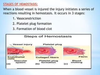
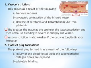
Presented By:- PALVI SINGLA
Hemostasis:
Hemostasis is defined as arrest or stoppage of bleeding.
Coagulation:
The process by which blood changes from a liquid to a gel forming a blood
clot; resulting in hemostasis.
Clot:
Blood clot is defined as the mass of coagulated blood which contains
RBCs; WBCs and platelets entrapped in fibrin meshwork.
Thrombosis:
Thrombosis is the formation of a blood clot, known as a thrombus, within
a blood vessel.
STAGES OF HEMOSTASIS:
When a blood vessel is injured the injury initiates a series of
reactions resulting in hemostasis. It occurs in 3 stages:
1. Vasoconstriction
2. Platelet plug formation
3. Formation of blood clot
1. Vasoconstriction:
This occurs as a result of the following-
a) Nervous reflexes
b) Myogenic contraction of the injured vessel
c) Release of serotonin and Thromboxane A2 from
platelets.
The greater the trauma; the stronger the vasoconstriction and
vice versa; so bleeding is severe in sharply cut vessels.
Vasoconstriction is also weaker if the cut was longitudinal or
irregular.
2. Platelet plug formation:
The platelet plug formed is as a result of the following-
a) injury of the blood vessel wall; the subendothelial
collagen fibres are exposed
b) platelets binding
c) such binding of platelets initiates platelet activation.
The activated platelets swell and develops pseudopodia;
become sticky and release the contents of their granules;
which contain ADP.
d) The released ADP binds to the nearby platelets leading
to their activation.
e) The newly activated platelets stick to originally
activated platelets and also release ADP which in turn
activates more and more platelets.
Thus a vicious circle of platelet activation is elicited leading to
formation of a loose platelet plug .
In addition to ADP; platelet aggregation is also promoted by:
1) A platelet activation factor
2) Thrombin
3) Thromboxane A2
3. Formation of blood clot:
The platelet plug becomes firm when fibrin threads are deposited
and this is essential for stoppage of bleeding from large vessels.
It becomes about one half its original size and a clear non-
coagulable yellow fluid called serum is squeezed out.
As the clot retracts; it pulls the edges of the injured vessels
together which helps hemostasis.
Clot retraction was believed to be produced by a substance
secreted by the platelets called retractozyme.
Thromboxane A2 -
*This is a prostaglandin-related substance that causes
vasoconstriction and promotes platelet aggregation.
*It is formed from arachidonic acid.
*The balance between the actions of thromboxane A2 and
prostacyclin normally favours localised platelet aggregation and
clot formation.
*This prevents excessive extension of the blood clot; which keeps
the blood vessels open and maintains the blood flow.
*Aspirin blocks the cyclooxygenase enzyme; so the production of
both thromboxane A2 and prostacyclin is decreased.
*The formation of prostacyclin is much more rapidly restored than
thromboxane A2 thus platelet aggregation is inhibited and clot
formation is reduced.
*For this reason small doses of aspirin taken for prolonged periods
can prevent intravascular clotting.
Mechanism of formation of
thromboxane A2
Structure of thromboxane A2
MECHANISM OF BLOOD
CLOTTING
 Coagulation of blood occurs through series of reactions due to the activation of
a group of substances.
 Substances necessary for clotting are called clotting factors.
 Total 13 clotting factors are identified.
Easy way to remember the 13 clotting factors
SEQUENCE OF CLOTTING MECHANISM
ENZYME CASCADE THEORY:
Enzyme cascade theory explains how various reactions involved in the conversion
of proenzymes to active enzymes take place in the form of a cascade; occuring in
3 stages:
1. Formation of prothrombin activator
2. Conversion of prothrombin into thrombin
3. Conversion of fibrinogen into fibrin
(A)THE INTRINSIC PATHWAY
This occurs in absence of tissue damage both in vitro by exposing the blood
to electronegatively charged wettable surfaces (e.g. glass) and in vivo
e.g. in cases of intravascular clotting (= thrombosis) which occurs when
blood is exposed to damaged endothelial cells or subendothelial collagen
fibres.
It is a slow process that begins to develop in 1-2 minutes and requires 6
minutes or more to form a clot. It proceeds as follows
(1) Exposure of blood to substances such as glass or collagen fibres leads
to 2 effects
a) Activation of factor XI (catalyzed by HMW kininogen and
kallikrein)
b)Platelet aggregation and release of PL
(2) Factor XIIa activates factor XJ (catalyzed by HMW kininogen).
(3) Factor XIa activates factor IX (the latter is also activated by factor
VIIa formed in the extrinsic system)
(4) Factor IXa forms a complex with factor VIIIa , and in the presence of
PL and Ca2+, it activates factor X.
(5) The same occurs as in steps 3, 4 and 5 in the extrinsic pathway
(B) THE EXTRINSIC PATHWAY
This occurs when there is trauma (damage) to the vascular wall and
the surrounding tissues.
It is a rapid process that may start and terminate in as little as 15
seconds, and it proceeds as follows
1) The damaged tissues release TPL (factor III): This is a protein
phospholipid mixture that activates factor VII
2) Active factor VII (VIIa) in presence of TPL, PL and Ca2+ activates
factor X (an alpha-globulin that is synthesized by the liver).
3) Active factor X (Xa) acts as a prothrombin activator that converts
prothrombin to thrombin (in presence of factor V , Ca 2+ and PL).
4) Thrombin converts the soluble fibrinogen into insoluble fibrin (by
release of 2 pairs of polypeptides from each fibrinogen molecule).
5) The formed clot is soft and loose, and it becomes firm and tight by
factor XIII (which is released from the platelets and becomes
activated by thrombin) in presence of Ca2+ The clot adheres to the
injured part of the vessel and then retracts, thus preventing further
blood loss.
Fibrinolysis
Prevents excessive fibrin deposition
It is the body's defense against vascular occlusion.
Fibrinolysis is essentially a localized, surface-bound
phenomenon, that is catalyzed by fibrin formation i.e. ‘fibrin
initiates its own destruction’.
As well as coagulation there is a balance for dissolution of the
clot.
It also should govern that bleeding doesn't reoccur due to
premature fibrinolysis.
To ensure that fibrin deposition in excess of that required to
prevent blood loss from damaged vessels is either prevented or
rapidly removed.
When fibrin is formed ,plasminogen is bound to the clot (Fibrin
adsorbs the plasminogen from plasma).
t-PA released from the vascular endothelium is also adsorbed on
the fibrin surface and efficiently activates plasminogen to
plasmin.
Diagrammatic representation of fibrinogen showing
pairs of Aα; Bβ and γ chains linked by disulfide
bonds.
Conversion Of Plasminogen To Plasmin
 Thrombosis is the formation of a clotted mas of blood within the
cardiovascular system.
 The clotted mass is called as THROMBUS.
THROMBUS:
 Involves wall of blood vessels; formed elements of blood & blood clotting
system
 Composed of platelets & fibrin
 May be life threatening.
3 Types of Thrombi are:
1. White thrombus-- composed of platelets & fibrin ;relatively poor in
erythrocytes ;forms at site of an injury or abnormal vessel wall;
where blood flow is rapid.
2. Red thrombus–- consists of RBCs & fibrin; morphologically resembles
the clot formed in a test tube ;forms in areas of retarded blood flow
or statsis(e.g. veins) or it may form at a site of injury or in abnormal
vessel in conjunction with an initiating platelet plug.
3. Fibrin deposit– desseminated in very small blood vessels or capillaries.
Platelet Activation
ROLE OF Ca2+ IN BLOOD CLOTTING
Ca 2+ is an essential catalyst in the process of blood clotting.
Except for the initial steps, it is required for all other steps of blood
clotting by both the extrinsic and intrinsic pathways.
Its normal plasma level is 9-11 mg%, and if removed from the blood,
clotting wouldn't occur .
However, in vivo reduction of blood Ca 2+ to levels that stop blood
clotting is incompatible with life.
This is because clotting stops only when the blood Ca 2+ level is severely
decreased (to about 4 mg%), and such level cannot be reached clinically
since death would occur before it is reached due to tetany (severe muscle
spasm) that occurs when the Ca2+ level drops just below 7 mg% .
IMPORTANCE OF VITAMIN K IN BLOOD CLOTTING:
Vitamin K is one of the fat-soluble vitamins, so it requires bile to be
absorbed in the small intestine.
It is a necessary for conversion of glutamic acid to gamma-
carboxyglutamic acid
6 of the proteins involved in blood clotting require this conversion before
they are released into the circulation .
These include clotting factors II,VII,XI and X as well as proteins C and S
Therefore, lack of vitamin K causes bleeding (so it is called
antihemorrhagic vitamin).
This can occur as a result of either
(a) Failure of absorption e.g. due to deficient bile flow
(b) Deficient intestinal bacteria (which normally synthesize vitamin K)
(c) Severe lack of the vitamin in diet.
Molecules involved in Regulation of hemostasis and
thrombosis
Laboratory Tests to measure Coagulation:
*Platelet Count
*Bleeding time
*Activated Partial Thromboplastin Time (aPTT or PTT)
*Prothrombin Time (PT)
*Thrombin Time (TT)
*Concentration of fibrinogen
*Fibrin clot stability
*Measurement of fibrin degradation products.
Conclusion
Blood and its coagulation plays an essential role in the
maintenance of health of a person.
Consequently disorders involving them can have a adverse effect
on it.
It is important to be familiar with these processes and the
disorders that can be caused by it.
One must be familiar with current understanding of various
medical disorders ; their management and the recommendations
provided by the concerned authorities from time to time.
Blood coagulation

Blood coagulation

  • 1.
  • 2.
    Hemostasis: Hemostasis is definedas arrest or stoppage of bleeding. Coagulation: The process by which blood changes from a liquid to a gel forming a blood clot; resulting in hemostasis. Clot: Blood clot is defined as the mass of coagulated blood which contains RBCs; WBCs and platelets entrapped in fibrin meshwork. Thrombosis: Thrombosis is the formation of a blood clot, known as a thrombus, within a blood vessel.
  • 3.
    STAGES OF HEMOSTASIS: Whena blood vessel is injured the injury initiates a series of reactions resulting in hemostasis. It occurs in 3 stages: 1. Vasoconstriction 2. Platelet plug formation 3. Formation of blood clot
  • 4.
    1. Vasoconstriction: This occursas a result of the following- a) Nervous reflexes b) Myogenic contraction of the injured vessel c) Release of serotonin and Thromboxane A2 from platelets. The greater the trauma; the stronger the vasoconstriction and vice versa; so bleeding is severe in sharply cut vessels. Vasoconstriction is also weaker if the cut was longitudinal or irregular. 2. Platelet plug formation: The platelet plug formed is as a result of the following- a) injury of the blood vessel wall; the subendothelial collagen fibres are exposed b) platelets binding
  • 5.
    c) such bindingof platelets initiates platelet activation. The activated platelets swell and develops pseudopodia; become sticky and release the contents of their granules; which contain ADP. d) The released ADP binds to the nearby platelets leading to their activation. e) The newly activated platelets stick to originally activated platelets and also release ADP which in turn activates more and more platelets. Thus a vicious circle of platelet activation is elicited leading to formation of a loose platelet plug . In addition to ADP; platelet aggregation is also promoted by: 1) A platelet activation factor 2) Thrombin 3) Thromboxane A2
  • 6.
    3. Formation ofblood clot: The platelet plug becomes firm when fibrin threads are deposited and this is essential for stoppage of bleeding from large vessels. It becomes about one half its original size and a clear non- coagulable yellow fluid called serum is squeezed out. As the clot retracts; it pulls the edges of the injured vessels together which helps hemostasis. Clot retraction was believed to be produced by a substance secreted by the platelets called retractozyme.
  • 7.
    Thromboxane A2 - *Thisis a prostaglandin-related substance that causes vasoconstriction and promotes platelet aggregation. *It is formed from arachidonic acid. *The balance between the actions of thromboxane A2 and prostacyclin normally favours localised platelet aggregation and clot formation. *This prevents excessive extension of the blood clot; which keeps the blood vessels open and maintains the blood flow. *Aspirin blocks the cyclooxygenase enzyme; so the production of both thromboxane A2 and prostacyclin is decreased. *The formation of prostacyclin is much more rapidly restored than thromboxane A2 thus platelet aggregation is inhibited and clot formation is reduced. *For this reason small doses of aspirin taken for prolonged periods can prevent intravascular clotting.
  • 8.
    Mechanism of formationof thromboxane A2 Structure of thromboxane A2
  • 10.
    MECHANISM OF BLOOD CLOTTING Coagulation of blood occurs through series of reactions due to the activation of a group of substances.  Substances necessary for clotting are called clotting factors.  Total 13 clotting factors are identified.
  • 12.
    Easy way toremember the 13 clotting factors
  • 13.
    SEQUENCE OF CLOTTINGMECHANISM ENZYME CASCADE THEORY: Enzyme cascade theory explains how various reactions involved in the conversion of proenzymes to active enzymes take place in the form of a cascade; occuring in 3 stages: 1. Formation of prothrombin activator 2. Conversion of prothrombin into thrombin 3. Conversion of fibrinogen into fibrin
  • 15.
    (A)THE INTRINSIC PATHWAY Thisoccurs in absence of tissue damage both in vitro by exposing the blood to electronegatively charged wettable surfaces (e.g. glass) and in vivo e.g. in cases of intravascular clotting (= thrombosis) which occurs when blood is exposed to damaged endothelial cells or subendothelial collagen fibres. It is a slow process that begins to develop in 1-2 minutes and requires 6 minutes or more to form a clot. It proceeds as follows (1) Exposure of blood to substances such as glass or collagen fibres leads to 2 effects a) Activation of factor XI (catalyzed by HMW kininogen and kallikrein) b)Platelet aggregation and release of PL (2) Factor XIIa activates factor XJ (catalyzed by HMW kininogen). (3) Factor XIa activates factor IX (the latter is also activated by factor VIIa formed in the extrinsic system) (4) Factor IXa forms a complex with factor VIIIa , and in the presence of PL and Ca2+, it activates factor X. (5) The same occurs as in steps 3, 4 and 5 in the extrinsic pathway
  • 17.
    (B) THE EXTRINSICPATHWAY This occurs when there is trauma (damage) to the vascular wall and the surrounding tissues. It is a rapid process that may start and terminate in as little as 15 seconds, and it proceeds as follows 1) The damaged tissues release TPL (factor III): This is a protein phospholipid mixture that activates factor VII 2) Active factor VII (VIIa) in presence of TPL, PL and Ca2+ activates factor X (an alpha-globulin that is synthesized by the liver). 3) Active factor X (Xa) acts as a prothrombin activator that converts prothrombin to thrombin (in presence of factor V , Ca 2+ and PL). 4) Thrombin converts the soluble fibrinogen into insoluble fibrin (by release of 2 pairs of polypeptides from each fibrinogen molecule). 5) The formed clot is soft and loose, and it becomes firm and tight by factor XIII (which is released from the platelets and becomes activated by thrombin) in presence of Ca2+ The clot adheres to the injured part of the vessel and then retracts, thus preventing further blood loss.
  • 19.
    Fibrinolysis Prevents excessive fibrindeposition It is the body's defense against vascular occlusion. Fibrinolysis is essentially a localized, surface-bound phenomenon, that is catalyzed by fibrin formation i.e. ‘fibrin initiates its own destruction’. As well as coagulation there is a balance for dissolution of the clot. It also should govern that bleeding doesn't reoccur due to premature fibrinolysis. To ensure that fibrin deposition in excess of that required to prevent blood loss from damaged vessels is either prevented or rapidly removed. When fibrin is formed ,plasminogen is bound to the clot (Fibrin adsorbs the plasminogen from plasma). t-PA released from the vascular endothelium is also adsorbed on the fibrin surface and efficiently activates plasminogen to plasmin.
  • 20.
    Diagrammatic representation offibrinogen showing pairs of Aα; Bβ and γ chains linked by disulfide bonds.
  • 21.
  • 22.
     Thrombosis isthe formation of a clotted mas of blood within the cardiovascular system.  The clotted mass is called as THROMBUS. THROMBUS:  Involves wall of blood vessels; formed elements of blood & blood clotting system  Composed of platelets & fibrin  May be life threatening. 3 Types of Thrombi are: 1. White thrombus-- composed of platelets & fibrin ;relatively poor in erythrocytes ;forms at site of an injury or abnormal vessel wall; where blood flow is rapid. 2. Red thrombus–- consists of RBCs & fibrin; morphologically resembles the clot formed in a test tube ;forms in areas of retarded blood flow or statsis(e.g. veins) or it may form at a site of injury or in abnormal vessel in conjunction with an initiating platelet plug. 3. Fibrin deposit– desseminated in very small blood vessels or capillaries.
  • 23.
  • 24.
    ROLE OF Ca2+IN BLOOD CLOTTING Ca 2+ is an essential catalyst in the process of blood clotting. Except for the initial steps, it is required for all other steps of blood clotting by both the extrinsic and intrinsic pathways. Its normal plasma level is 9-11 mg%, and if removed from the blood, clotting wouldn't occur . However, in vivo reduction of blood Ca 2+ to levels that stop blood clotting is incompatible with life. This is because clotting stops only when the blood Ca 2+ level is severely decreased (to about 4 mg%), and such level cannot be reached clinically since death would occur before it is reached due to tetany (severe muscle spasm) that occurs when the Ca2+ level drops just below 7 mg% .
  • 25.
    IMPORTANCE OF VITAMINK IN BLOOD CLOTTING: Vitamin K is one of the fat-soluble vitamins, so it requires bile to be absorbed in the small intestine. It is a necessary for conversion of glutamic acid to gamma- carboxyglutamic acid 6 of the proteins involved in blood clotting require this conversion before they are released into the circulation . These include clotting factors II,VII,XI and X as well as proteins C and S Therefore, lack of vitamin K causes bleeding (so it is called antihemorrhagic vitamin). This can occur as a result of either (a) Failure of absorption e.g. due to deficient bile flow (b) Deficient intestinal bacteria (which normally synthesize vitamin K) (c) Severe lack of the vitamin in diet.
  • 26.
    Molecules involved inRegulation of hemostasis and thrombosis
  • 27.
    Laboratory Tests tomeasure Coagulation: *Platelet Count *Bleeding time *Activated Partial Thromboplastin Time (aPTT or PTT) *Prothrombin Time (PT) *Thrombin Time (TT) *Concentration of fibrinogen *Fibrin clot stability *Measurement of fibrin degradation products.
  • 28.
    Conclusion Blood and itscoagulation plays an essential role in the maintenance of health of a person. Consequently disorders involving them can have a adverse effect on it. It is important to be familiar with these processes and the disorders that can be caused by it. One must be familiar with current understanding of various medical disorders ; their management and the recommendations provided by the concerned authorities from time to time.