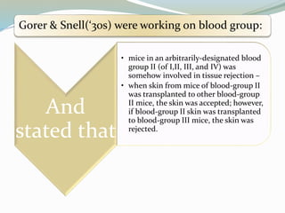
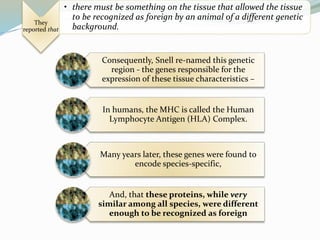
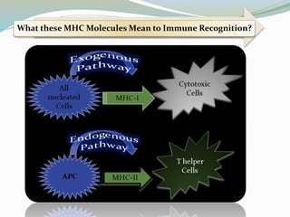
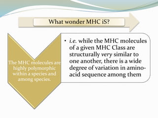
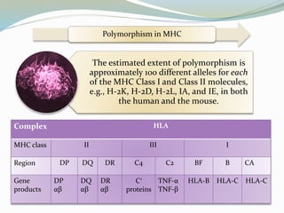
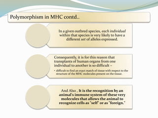
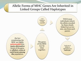
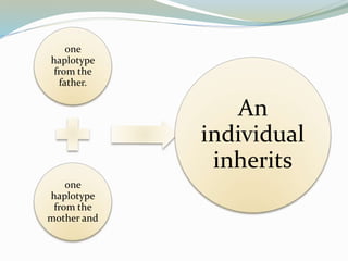
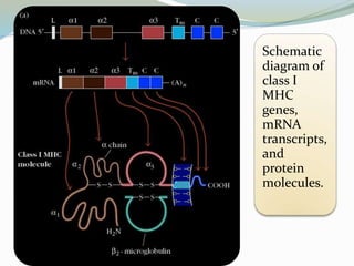
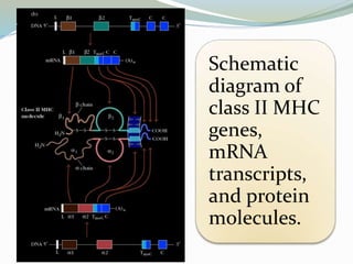
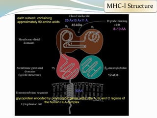
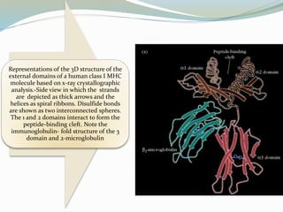
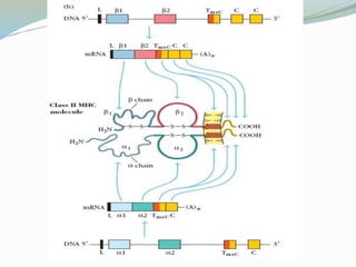
Dr Alok Tripathi
Department of Biotechnology
aquaimmuno@yahoo.co.in
09795894495
Gorer & Snell(‘30s) were working on blood group:
And
stated that
• mice in an arbitrarily-designated blood
group II (of I,II, III, and IV) was
somehow involved in tissue rejection –
• when skin from mice of blood-group II
was transplanted to other blood-group
II mice, the skin was accepted; however,
if blood-group II skin was transplanted
to blood-group III mice, the skin was
rejected.
They
reported that
• there must be something on the tissue that allowed the tissue
to be recognized as foreign by an animal of a different genetic
background.
Consequently, Snell re-named this genetic
region - the genes responsible for the
expression of these tissue characteristics –
In humans, the MHC is called the Human
Lymphocyte Antigen (HLA) Complex.
Many years later, these genes were found to
encode species-specific,
And, that these proteins, while very
similar among all species, were different
enough to be recognized as foreign
What these MHC Molecules Mean to Immune Recognition?
What wonder MHC iS?
The MHC molecules are
highly polymorphic
within a species and
among species.
• i.e. while the MHC molecules
of a given MHC Class are
structurally very similar to
one another, there is a wide
degree of variation in amino-
acid sequence among them
Effector Cells in Adaptive Immunity
Effector
T Cell
Pathogen
Location
Antigen
Presentation
Target Cell
Action
Cellular
Immunity
Tc CD8
cytotoxic
Cytoplasm
Infected cell
MHC I
Infected cell
apoptosis
Th1 CD4
inflammatory
Macrophage
vesicles
Macrophage
MHC II
Macrophage
activation to
kill pathogen
Polymorphism in MHC
The estimated extent of polymorphism is
approximately 100 different alleles for each
of the MHC Class I and Class II molecules,
e.g., H-2K, H-2D, H-2L, IA, and IE, in both
the human and the mouse.
Complex HLA
MHC class II III I
Region DP DQ DR C4 C2 BF B CA
Gene
products
DP
αβ
DQ
αβ
DR
αβ
C′
proteins
TNF-α
TNF-β
HLA-B HLA-C HLA-C
Polymorphism in MHC contd..
In a given outbred species, each individual
within that species is very likely to have a
different set of alleles expressed.
Consequently, it is for this reason that
transplants of human organs from one
individual to another is so difficult –
• difficult to find an exact match of tissue with respect to the
structure of the MHC molecules present on the tissue.
And Also , It is the recognition by an
animal's immune system of these very
molecules that allows the animal to
recognize cells as "self" or as "foreign."
Allelic Forms of MHC Genes Are Inherited in
Linked Groups Called Haplotypes
the loci
constituting the
MHC are highly
polymorphic(i.e.
many alternative
forms of the gene,
or alleles), exist at
each locus among
the population.
The genes
of the MHC
loci lie
close
together;
C/O
frequency
is only
1/200 R/D
THUS most
individuals inherit
the alleles encoded
by these closely
linked loci as two
sets, one from each
parent.
Each set of
alleles is
referred to as a
haplotype.
one
haplotype
from the
father.
one
haplotype
from the
mother and
An
individual
inherits
Schematic
diagram of
class I
MHC
genes,
mRNA
transcripts,
and
protein
molecules.
Schematic
diagram of
class II MHC
genes,
mRNA
transcripts,
and protein
molecules.
Peptide binding by class I and class II MHC molecules
Class I molecules Class II molecules
Peptide-binding domain ἃ1/ἃ2 ἃ1/β2
Nature of peptide-
binding cleft
Closed at both ends Open at both ends
General size of bound
peptides
8–10 amino acids 13–18 amino acids
Peptide motifs involved
in binding to MHC
molecule
Anchor residues at both
ends of peptide; generally
hydrophobic carboxyl-
terminal anchor
Anchor residues
distributed along the
length of the peptide
Nature of bound peptide Extended structure in
which both ends
interact with MHC cleft
but middle
arches up away from
MHC molecule
Extended structure that is
held at a constant
elevation above the floor
of MHC cleft
MHC-I Structure
Representations of the 3D structure of the
external domains of a human class I MHC
molecule based on x-ray crystallographic
analysis.-Side view in which the strands
are depicted as thick arrows and the
helices as spiral ribbons. Disulfide bonds
are shown as two interconnected spheres.
The 1 and 2 domains interact to form the
peptide-binding cleft. Note the
immunoglobulin- fold structure of the 3
domain and 2-microglobulin
Antigen-binding cleft of dimeric class II DR1 molecule in (a) top view and
(b) side view. This molecule crystallized as a dimer of the heterodimer.
The crystallized dimer is shown with one DR1 molecule in red and the
other DR1 molecule in blue. The bound peptides are yellow. The two
peptide-binding clefts in the dimeric molecule face in opposite directions.
MHC: Major Histocompetability Complex

MHC: Major Histocompetability Complex

  • 1.
    Dr Alok Tripathi Departmentof Biotechnology aquaimmuno@yahoo.co.in 09795894495
  • 2.
    Gorer & Snell(‘30s)were working on blood group: And stated that • mice in an arbitrarily-designated blood group II (of I,II, III, and IV) was somehow involved in tissue rejection – • when skin from mice of blood-group II was transplanted to other blood-group II mice, the skin was accepted; however, if blood-group II skin was transplanted to blood-group III mice, the skin was rejected.
  • 3.
    They reported that • theremust be something on the tissue that allowed the tissue to be recognized as foreign by an animal of a different genetic background. Consequently, Snell re-named this genetic region - the genes responsible for the expression of these tissue characteristics – In humans, the MHC is called the Human Lymphocyte Antigen (HLA) Complex. Many years later, these genes were found to encode species-specific, And, that these proteins, while very similar among all species, were different enough to be recognized as foreign
  • 4.
    What these MHCMolecules Mean to Immune Recognition?
  • 5.
    What wonder MHCiS? The MHC molecules are highly polymorphic within a species and among species. • i.e. while the MHC molecules of a given MHC Class are structurally very similar to one another, there is a wide degree of variation in amino- acid sequence among them
  • 6.
    Effector Cells inAdaptive Immunity Effector T Cell Pathogen Location Antigen Presentation Target Cell Action Cellular Immunity Tc CD8 cytotoxic Cytoplasm Infected cell MHC I Infected cell apoptosis Th1 CD4 inflammatory Macrophage vesicles Macrophage MHC II Macrophage activation to kill pathogen
  • 7.
    Polymorphism in MHC Theestimated extent of polymorphism is approximately 100 different alleles for each of the MHC Class I and Class II molecules, e.g., H-2K, H-2D, H-2L, IA, and IE, in both the human and the mouse. Complex HLA MHC class II III I Region DP DQ DR C4 C2 BF B CA Gene products DP αβ DQ αβ DR αβ C′ proteins TNF-α TNF-β HLA-B HLA-C HLA-C
  • 8.
    Polymorphism in MHCcontd.. In a given outbred species, each individual within that species is very likely to have a different set of alleles expressed. Consequently, it is for this reason that transplants of human organs from one individual to another is so difficult – • difficult to find an exact match of tissue with respect to the structure of the MHC molecules present on the tissue. And Also , It is the recognition by an animal's immune system of these very molecules that allows the animal to recognize cells as "self" or as "foreign."
  • 9.
    Allelic Forms ofMHC Genes Are Inherited in Linked Groups Called Haplotypes the loci constituting the MHC are highly polymorphic(i.e. many alternative forms of the gene, or alleles), exist at each locus among the population. The genes of the MHC loci lie close together; C/O frequency is only 1/200 R/D THUS most individuals inherit the alleles encoded by these closely linked loci as two sets, one from each parent. Each set of alleles is referred to as a haplotype.
  • 10.
  • 11.
  • 12.
    Schematic diagram of class IIMHC genes, mRNA transcripts, and protein molecules.
  • 13.
    Peptide binding byclass I and class II MHC molecules Class I molecules Class II molecules Peptide-binding domain ἃ1/ἃ2 ἃ1/β2 Nature of peptide- binding cleft Closed at both ends Open at both ends General size of bound peptides 8–10 amino acids 13–18 amino acids Peptide motifs involved in binding to MHC molecule Anchor residues at both ends of peptide; generally hydrophobic carboxyl- terminal anchor Anchor residues distributed along the length of the peptide Nature of bound peptide Extended structure in which both ends interact with MHC cleft but middle arches up away from MHC molecule Extended structure that is held at a constant elevation above the floor of MHC cleft
  • 14.
  • 15.
    Representations of the3D structure of the external domains of a human class I MHC molecule based on x-ray crystallographic analysis.-Side view in which the strands are depicted as thick arrows and the helices as spiral ribbons. Disulfide bonds are shown as two interconnected spheres. The 1 and 2 domains interact to form the peptide-binding cleft. Note the immunoglobulin- fold structure of the 3 domain and 2-microglobulin
  • 16.
    Antigen-binding cleft ofdimeric class II DR1 molecule in (a) top view and (b) side view. This molecule crystallized as a dimer of the heterodimer. The crystallized dimer is shown with one DR1 molecule in red and the other DR1 molecule in blue. The bound peptides are yellow. The two peptide-binding clefts in the dimeric molecule face in opposite directions.