Downloaded 368 times


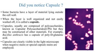

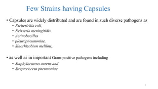
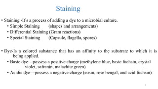
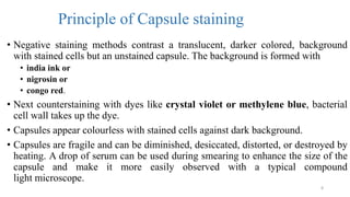
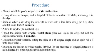
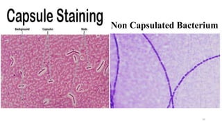
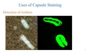


This document discusses capsule staining, which is a technique used to identify the presence of bacterial capsules under a light microscope. It begins by defining bacterial capsules and explaining their functions, which include helping bacteria resist phagocytosis and providing protection. It then discusses the principle of capsule staining, which uses a negative stain to contrast the unstained capsule against stained bacterial cells. The procedure involves smearing a bacterial culture onto a slide with negative stain, staining with a counterstain like crystal violet, and examining under a microscope for unstained capsules surrounding stained cells. Examples of capsule-containing bacteria that can be identified this way include Klebsiella pneumoniae and Bacillus anthracis.