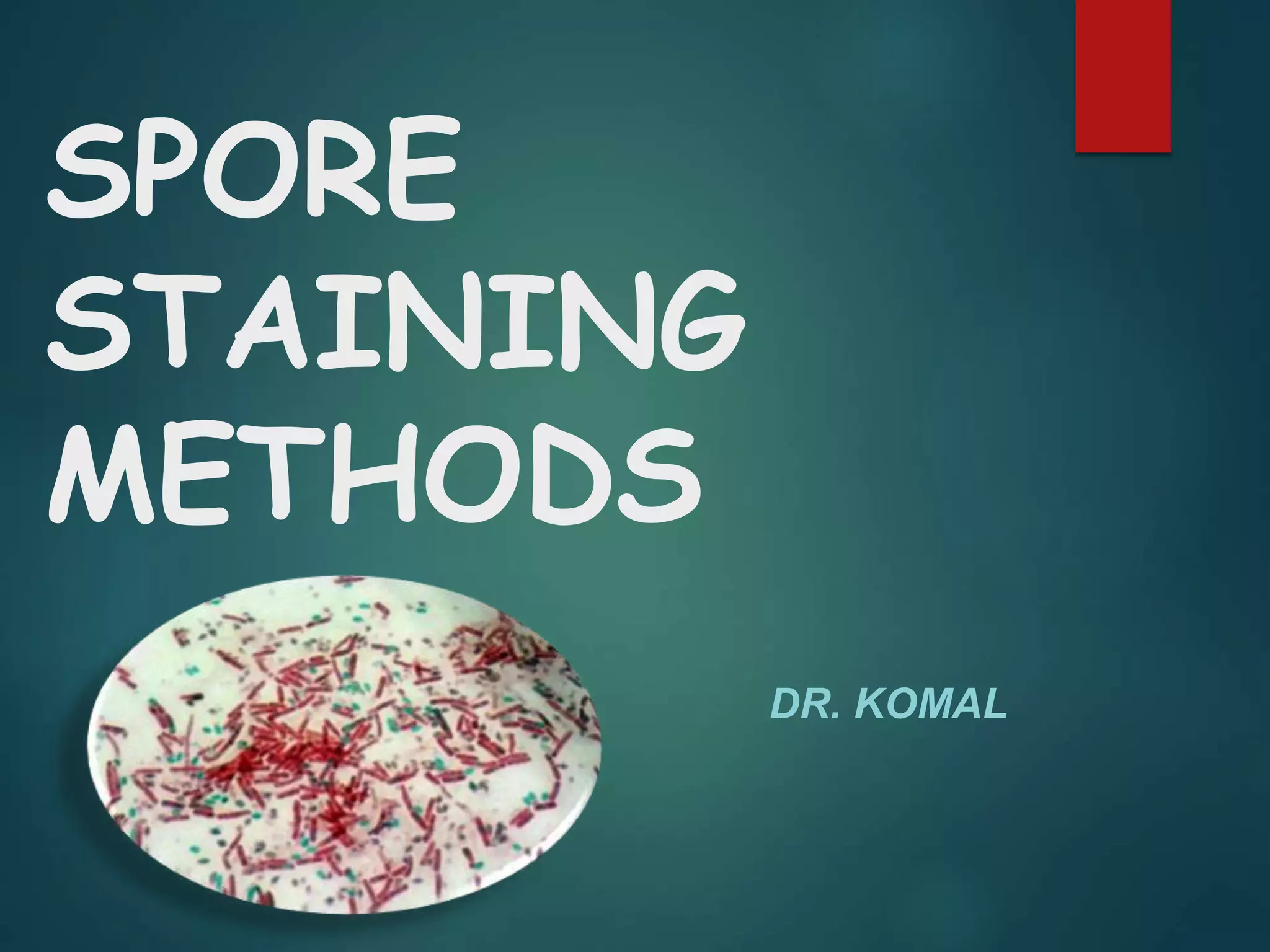
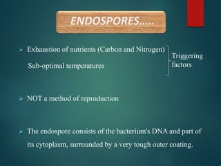
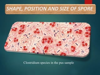
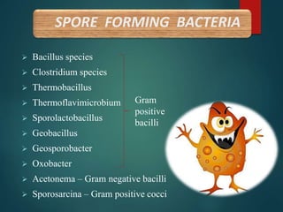
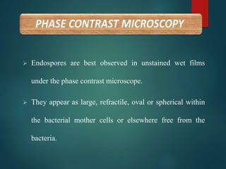
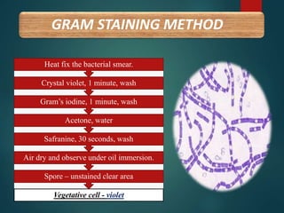
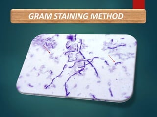
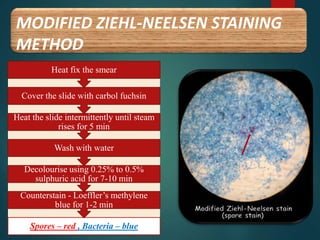
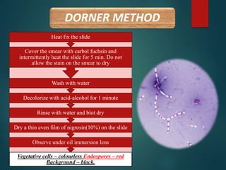
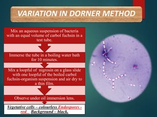
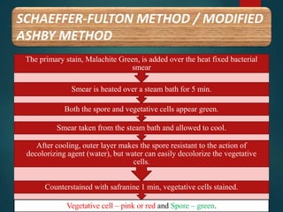
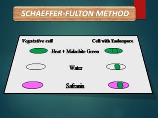
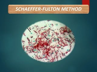
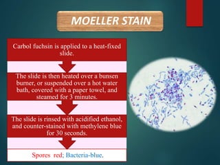
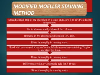
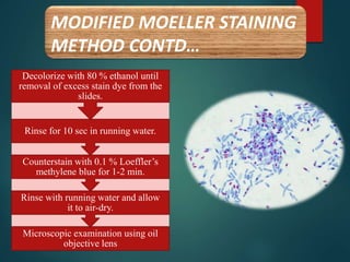
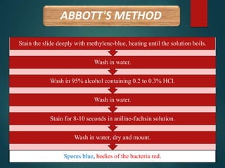
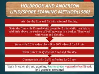
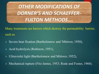
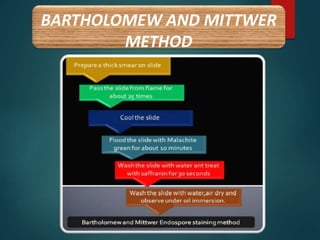
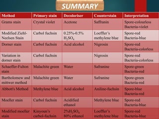
SlideShare a Scribd company logo
SPORE
STAINING
METHODS
DR. KOMAL
LOHI
HISTORY
 Endospores were first described by Cohn (1872) in Bacillus
subtilis and later by Koch(1876) in the pathogen, Bacillus
anthracis.
 Cohn demonstrated the heat resistance of endospores in B.
subtilis, and Koch described the developmental cycle of
spore formation in B. anthracis.
HISTORY CONTD….
 In 1922, Dorner published a method for staining endospores. It
employed a lengthy heating step but resulted in differential
staining of endospores and vegetative cells in the same sample.
 Shaeffer and Fulton modified Dorner’s method in 1933 to
make the process faster.
ENDOSPORES…..
 Endospores are so named because they are formed intracellularly,
although they are eventually released from this mother cell or
sporangium as free spores.
 An endospore is a spherical/oval, thick-walled, highly resistant
structure formed in certain bacterial species that represent a
dormant or resting stage in the growth cycle of an organism. e.g.
Bacillus sp., Clostridium sp.
ENDOSPORES…..
 Exhaustion of nutrients (Carbon and Nitrogen)
Sub-optimal temperatures
 NOT a method of reproduction
 The endospore consists of the bacterium's DNA and part of
its cytoplasm, surrounded by a very tough outer coating.
Triggering
factors
COVERINGS OF SPORE
Spore wall :
• delicate membrane from
which cell wall of future
vegetative bacterium develop
Spore cortex
Spore coat
• tough, multi-layered
Exosporium
RESISTANT TO….
 Ultraviolet radiation
 Desiccation
 High temperature
 Extreme freezing
 Chemical disinfectants
 Starvation
DESTROYED BY….
 Autoclaving
 Tyndallisation
ENDOSPORES…..
Germination of spores-
 Increase in metabolic
activity of spore
 Endospore swells
 Germ cell appears by
rupture or absorption of
spore coat
 Elongates to vegetative
bacterium
 Distend bacillary body –
Clostridium spp.
 Do not distend bacillary body –
B. anthracis
 Central – B. anthracis,
C. bifermentans
 Terminal - C. tetani, C. tertium
 Sub-terminal – C. perfringens
(club-shaped)
 Oval - B. anthracis, C. tertium
(tennis racket)
 Spherical - C. tetani. (drum-stick)
SHAPE, POSITION AND SIZE OF
SPORE RELATIVE TO PARENT CELL
SHAPE, POSITION AND SIZE OF SPORE
Clostridium species in the pus sample
 Bacillus species
 Clostridium species
 Thermobacillus
 Thermoflavimicrobium
 Sporolactobacillus
 Geobacillus
 Geosporobacter
 Oxobacter
 Acetonema – Gram negative bacilli
 Sporosarcina – Gram positive cocci
SPORE FORMING BACTERIA
Gram
positive
bacilli
METHODS OF
DEMONSTRATION OF SPORES
 Endospores are best observed in unstained wet films
under the phase contrast microscope.
 They appear as large, refractile, oval or spherical within
the bacterial mother cells or elsewhere free from the
bacteria.
PHASE CONTRAST MICROSCOPY
B. thuringiensis by Phase contrast microscopy
GRAM STAINING METHOD
Vegetative cell - violet
Spore – unstained clear area
Air dry and observe under oil immersion.
Safranine, 30 seconds, wash
Acetone, water
Gram’s iodine, 1 minute, wash
Crystal violet, 1 minute, wash
Heat fix the bacterial smear.
GRAM STAINING METHOD
MODIFIED ZIEHL-NEELSEN STAINING
METHOD
Spores – red , Bacteria – blue
Counterstain - Loeffler’s methylene
blue for 1-2 min
Decolourise using 0.25% to 0.5%
sulphuric acid for 7-10 min
Wash with water
Heat the slide intermittently until steam
rises for 5 min
Cover the slide with carbol fuchsin
Heat fix the smear
DORNER METHOD
Vegetative cells – colourless Endospores – red
Background – black.
Observe under oil immersion lens
Dry a thin even film of nigrosin(10%) on the slide
Rinse with water and blot dry
Decolorize with acid-alcohol for 1 minute
Wash with water
Cover the smear with carbol fuchsin and
intermittently heat the slide for 5 min. Do not
allow the stain on the smear to dry
Heat fix the slide
VARIATION IN DORNER METHOD
Vegetative cells – colourless Endospores -
red Background – black.
Observe under oil immersion lens.
Mix a loopful of nigrosin on a glass slide
with one loopful of the boiled carbol
fuchsin-organism suspension and air dry to
a thin film.
Immerse the tube in a boiling water bath
for 10 minutes.
Mix an aqueous suspension of bacteria
with an equal volume of carbol fuchsin in a
test tube.
SCHAEFFER-FULTON METHOD / MODIFIED
ASHBY METHOD
Vegetative cell – pink or red and Spore – green.
Counterstained with safranine 1 min, vegetative cells stained.
After cooling, outer layer makes the spore resistant to the action of
decolorizing agent (water), but water can easily decolorize the vegetative
cells.
Smear taken from the steam bath and allowed to cool.
Both the spore and vegetative cells appear green.
Smear is heated over a steam bath for 5 min.
The primary stain, Malachite Green, is added over the heat fixed bacterial
smear
SCHAEFFER-FULTON METHOD
MOELLER STAIN
Spores red; Bacteria-blue.
The slide is rinsed with acidified ethanol,
and counter-stained with methylene blue
for 30 seconds.
The slide is then heated over a bunsen
burner, or suspended over a hot water
bath, covered with a paper towel, and
steamed for 3 minutes.
Carbol fuchsin is applied to a heat-fixed
slide.
MODIFIED MOELLER STAINING
METHOD
Rinse thoroughly in running water
Differentiate with 2 % sulphuric acid for 5-10 sec.
Rinse thoroughly in running water.
Flood with un-steamed Kinyoun’s carbol-fuchsin solution containing Tergitol
7, and stain for 3 min.
Rinse thoroughly in running water.
Immerse in 5% chromic acid solution for 3 min.
Fix in absolute methyl alcohol for 1-3 min.
Spread a small drop of the specimen on a slide, and allow it to air-dry at room
temperature.
MODIFIED MOELLER STAINING
METHOD CONTD…
Microscopic examination using oil
objective lens
Rinse with running water and allow
it to air-dry.
Counterstain with 0.1 % Loeffler’s
methylene blue for 1-2 min.
Rinse for 10 sec in running water.
Decolorize with 80 % ethanol until
removal of excess stain dye from the
slides.
ABBOTT'S METHOD
Spores blue, bodies of the bacteria red.
Wash in water, dry and mount.
Stain for 8-10 seconds in aniline-fuchsin solution.
Wash in water.
Wash in 95% alcohol containing 0.2 to 0.3% HCl.
Wash in water.
Stain the slide deeply with methylene-blue, heating until the solution boils.
HOLBROOK AND ANDERSON
LIPID/SPORE STAINING METHOD(1980)
Wash in water, dry and examine. Spores-green, vegetative bacilli-red,
lipid granules-unstained.
Counterstain with 0.5% safranine for 20 sec.
Wash film with xylene for 5 sec and blot dry.
Stain with 0.3% sudan black B in 70% ethanol for 15 min
Stain the film with 5% malachite green for 2 min while the slide is
held little above the surface of boiling water in a beaker. Then wash
with water and blot dry.
Air dry the film and fix with minimal flaming.
Many treatments are known which destroy the permeability barrier,
such as
 Severe heat fixation (Bartholomew and 'Mittwer, 1950),
 Acid hydrolysis (Robinow, 1951),
 Ultraviolet light (Bartholomew and Mittwer, 1952),
 Mechanical rupture (Fitz-James, 1953; Rode and Foster, 1960).
OTHER MODIFICATIONS OF
DORNER’S AND SCHAEFFER-
FULTON METHODS….
BARTHOLOMEW AND MITTWER
METHOD
SUMMARY
Method Primary stain Decoloriser Counterstain Interpretation
Grams stain Crystal violet Acetone Saffranin Spore-colourless
Bacteria-violet
Modified Ziehl-
Neelsen Stain
Carbol fuchsin 0.25%-0.5%
H2SO4
Loeffler’s
methylene blue
Spore-red
Bacteria-blue
Dorner stain Carbol fuchsin Acid alcohol Nigrosin Spore-red
Bacteria-colorless
Variation in
dorner stain
Carbol fuchsin Nigrosin Spore-red
Bacteria-colorless
Schaeffer-Fulton
stain
Malachite green Water Safranine Spore-green
Bacteria-red
Bartholomew and
mittwer method
Malachite green Water Safranine Spore-green
Bacteria-red
Abbott's Method Methylene blue Acid alcohol Aniline-fuchsin Spore-blue
Bacteria-red
Moeller stain Carbol fuchsin Acidified
ethanol
Methylene blue Spore-red
Bacteria-blue
Modified moeller
stain
Kinyoun’s
carbol-fuchsin
2%H2SO4
80% ethanol
Loeffler’s
methylene blue
Spore-red
Bacteria-blue
REFERENCES
 Colour Atlas & Textbook of Diagnostic Microbiology by Koneman
 Practical Medical Microbiology by Mackie & McCartney
 Monica Cheesbrough
 Textbook of Microbiology by Ananthanarayan
 http://www.microbelibrary.org/component/resource/laboratory-test/3112-
endospore-stain-protocol
 http://chestofbooks.com/health/disease/Pathology/Methods-For-Staining-
Spores.html#
 http://www.generalmicroscience.com/microbial-laboratory-
techniques/endospore-staining-by-bartholomew-and-mittwers-method/
 M. Hayama, K. Oana, T. Kozakai, S. Umeda, J. Fujimoto, H. Ota, Y.
Kawakami Proposal of a simplified technique for staining bacterial spores
without applying heat-successful modification of Moeller’s method. Eur J
Med Res (2007) 12: 356-359.
Spore staining methods

More Related Content

What's hot

Bacillus anthracis
Bacillus anthracisBacillus anthracis
Treponema pallidum
Treponema pallidumTreponema pallidum
Treponema pallidumPrbn Shah
 
Automated system for bacterial identification
Automated system for bacterial identificationAutomated system for bacterial identification
Automated system for bacterial identification
DEEKSHANT KUMAR
 
Flagella Staining.pptx
Flagella Staining.pptxFlagella Staining.pptx
Flagella Staining.pptx
MonishaM73
 
Parasite culture
Parasite cultureParasite culture
Parasite culture
Pradip Hamal
 
laboratory diagnosis of fungal inections
laboratory diagnosis of fungal inectionslaboratory diagnosis of fungal inections
laboratory diagnosis of fungal inections
Aditi Kothari
 
Laboratory diagnosis of fungal infections
Laboratory diagnosis of fungal infectionsLaboratory diagnosis of fungal infections
Laboratory diagnosis of fungal infections
Dr.Dinesh Jain
 
Mycobacterium tuberculosis
Mycobacterium tuberculosisMycobacterium tuberculosis
Mycobacterium tuberculosis
Anushi Jain
 
Quality control in microbiology
Quality control in microbiologyQuality control in microbiology
Quality control in microbiology
Tabeen Bint Mansoor
 
Diagnostic methods of Parasites
Diagnostic methods of ParasitesDiagnostic methods of Parasites
Diagnostic methods of Parasites
SHRUTHI VASAN
 
Antibiotic sensitivity testing (AST)
Antibiotic sensitivity testing (AST) Antibiotic sensitivity testing (AST)
Antibiotic sensitivity testing (AST)
Gul Muhammad
 
Lab diagnosis of fungal infections, Dr Naveen Reddy
Lab diagnosis of fungal infections, Dr Naveen ReddyLab diagnosis of fungal infections, Dr Naveen Reddy
Lab diagnosis of fungal infections, Dr Naveen Reddy
Naveen Parvathareddy
 
Dimorphic fungi
Dimorphic  fungi Dimorphic  fungi
Dimorphic fungi
Prbn Shah
 
16. vibrio cholera
16. vibrio cholera16. vibrio cholera
16. vibrio cholera
Ratheeshkrishnakripa
 
Bacillus species
Bacillus species Bacillus species
Bacillus species
Prasad Gunjal
 
COLLECTION AND TRANSPORTATION OF CLINICAL SAMPLES
COLLECTION AND TRANSPORTATION OF CLINICAL SAMPLESCOLLECTION AND TRANSPORTATION OF CLINICAL SAMPLES
COLLECTION AND TRANSPORTATION OF CLINICAL SAMPLES
NCRIMS, Meerut
 
Bordetella
BordetellaBordetella
Antimicrobial susceptibility testing
Antimicrobial susceptibility testingAntimicrobial susceptibility testing
Antimicrobial susceptibility testing
Saajida Sultaana
 

What's hot (20)

Bacillus anthracis
Bacillus anthracisBacillus anthracis
Bacillus anthracis
 
Bacillus anthracis
Bacillus anthracisBacillus anthracis
Bacillus anthracis
 
Treponema pallidum
Treponema pallidumTreponema pallidum
Treponema pallidum
 
Automated system for bacterial identification
Automated system for bacterial identificationAutomated system for bacterial identification
Automated system for bacterial identification
 
Flagella Staining.pptx
Flagella Staining.pptxFlagella Staining.pptx
Flagella Staining.pptx
 
Parasite culture
Parasite cultureParasite culture
Parasite culture
 
laboratory diagnosis of fungal inections
laboratory diagnosis of fungal inectionslaboratory diagnosis of fungal inections
laboratory diagnosis of fungal inections
 
Laboratory diagnosis of fungal infections
Laboratory diagnosis of fungal infectionsLaboratory diagnosis of fungal infections
Laboratory diagnosis of fungal infections
 
Mycobacterium tuberculosis
Mycobacterium tuberculosisMycobacterium tuberculosis
Mycobacterium tuberculosis
 
Quality control in microbiology
Quality control in microbiologyQuality control in microbiology
Quality control in microbiology
 
Diagnostic methods of Parasites
Diagnostic methods of ParasitesDiagnostic methods of Parasites
Diagnostic methods of Parasites
 
Antibiotic sensitivity testing (AST)
Antibiotic sensitivity testing (AST) Antibiotic sensitivity testing (AST)
Antibiotic sensitivity testing (AST)
 
Lab diagnosis of fungal infections, Dr Naveen Reddy
Lab diagnosis of fungal infections, Dr Naveen ReddyLab diagnosis of fungal infections, Dr Naveen Reddy
Lab diagnosis of fungal infections, Dr Naveen Reddy
 
Dimorphic fungi
Dimorphic  fungi Dimorphic  fungi
Dimorphic fungi
 
16. vibrio cholera
16. vibrio cholera16. vibrio cholera
16. vibrio cholera
 
Streptococcus
StreptococcusStreptococcus
Streptococcus
 
Bacillus species
Bacillus species Bacillus species
Bacillus species
 
COLLECTION AND TRANSPORTATION OF CLINICAL SAMPLES
COLLECTION AND TRANSPORTATION OF CLINICAL SAMPLESCOLLECTION AND TRANSPORTATION OF CLINICAL SAMPLES
COLLECTION AND TRANSPORTATION OF CLINICAL SAMPLES
 
Bordetella
BordetellaBordetella
Bordetella
 
Antimicrobial susceptibility testing
Antimicrobial susceptibility testingAntimicrobial susceptibility testing
Antimicrobial susceptibility testing
 

Similar to Spore staining methods

identificationof bacteria.pptx
identificationof bacteria.pptxidentificationof bacteria.pptx
identificationof bacteria.pptx
obedcudjoe1
 
practical protocol.pdf
practical protocol.pdfpractical protocol.pdf
practical protocol.pdf
SNEHA AGRAWAL GUPTA
 
Identification of bacteria
Identification of bacteriaIdentification of bacteria
Identification of bacteria
Dhanashri Mali
 
Thermus aquaticus gen. n. and sp. n., a nonsporulating extreme thermophile
Thermus aquaticus gen. n. and sp. n., a nonsporulating extreme thermophileThermus aquaticus gen. n. and sp. n., a nonsporulating extreme thermophile
Thermus aquaticus gen. n. and sp. n., a nonsporulating extreme thermophile
José Luis Moreno Garvayo
 
Acid-Fast Bacteria. Spores of Bacteria. Complete methods of staining
Acid-Fast Bacteria. Spores of Bacteria. Complete methods of stainingAcid-Fast Bacteria. Spores of Bacteria. Complete methods of staining
Acid-Fast Bacteria. Spores of Bacteria. Complete methods of staining
Eneutron
 
Staining
StainingStaining
Staining
Madhuri Bobburi
 
Staining Techniques in Microbiology
Staining Techniques in MicrobiologyStaining Techniques in Microbiology
Staining Techniques in Microbiology
Guddeti Prashanth Kumar
 
403149684 stainingtechniquesgp-150203151421-conversion-gate02-pdf
403149684 stainingtechniquesgp-150203151421-conversion-gate02-pdf403149684 stainingtechniquesgp-150203151421-conversion-gate02-pdf
403149684 stainingtechniquesgp-150203151421-conversion-gate02-pdf
ssuserd72fc5
 
GRAM STAINING OF BACTERIA
GRAM STAINING OF BACTERIAGRAM STAINING OF BACTERIA
GRAM STAINING OF BACTERIADr. Anindya Das
 
Exp1 microbial growth
Exp1 microbial growthExp1 microbial growth
Exp1 microbial growth
Rione Drevale
 
Culture methods
Culture methodsCulture methods
bacterial spore.ppt
bacterial spore.pptbacterial spore.ppt
bacterial spore.ppt
Dr. Md Ashraf Ali Namaji
 
5. Staining techniques (Microbiology)
5. Staining techniques (Microbiology)5. Staining techniques (Microbiology)
5. Staining techniques (Microbiology)
Jay Khaniya
 
Cyanobacteria
CyanobacteriaCyanobacteria
Cyanobacteria
Shraddha Chavan
 
Bacillus.ppt
Bacillus.pptBacillus.ppt
Bacillus.ppt
SadamIrshad2
 
Bacteria Project
Bacteria ProjectBacteria Project
Bacteria Project
12hector
 
Bacteria Report
Bacteria Report Bacteria Report
Bacteria Report
12hector
 
Make drawings of both organisms stained in each staining procedure. L.pdf
Make drawings of both organisms stained in each staining procedure. L.pdfMake drawings of both organisms stained in each staining procedure. L.pdf
Make drawings of both organisms stained in each staining procedure. L.pdf
Dhanrajsolanki2091
 
bacterial staining ppt
bacterial staining pptbacterial staining ppt
bacterial staining ppt
AniruddhGupta10
 

Similar to Spore staining methods (20)

identificationof bacteria.pptx
identificationof bacteria.pptxidentificationof bacteria.pptx
identificationof bacteria.pptx
 
practical protocol.pdf
practical protocol.pdfpractical protocol.pdf
practical protocol.pdf
 
Identification of bacteria
Identification of bacteriaIdentification of bacteria
Identification of bacteria
 
Thermus aquaticus gen. n. and sp. n., a nonsporulating extreme thermophile
Thermus aquaticus gen. n. and sp. n., a nonsporulating extreme thermophileThermus aquaticus gen. n. and sp. n., a nonsporulating extreme thermophile
Thermus aquaticus gen. n. and sp. n., a nonsporulating extreme thermophile
 
Acid-Fast Bacteria. Spores of Bacteria. Complete methods of staining
Acid-Fast Bacteria. Spores of Bacteria. Complete methods of stainingAcid-Fast Bacteria. Spores of Bacteria. Complete methods of staining
Acid-Fast Bacteria. Spores of Bacteria. Complete methods of staining
 
Staining
StainingStaining
Staining
 
SPORE ashraf
SPORE ashrafSPORE ashraf
SPORE ashraf
 
Staining Techniques in Microbiology
Staining Techniques in MicrobiologyStaining Techniques in Microbiology
Staining Techniques in Microbiology
 
403149684 stainingtechniquesgp-150203151421-conversion-gate02-pdf
403149684 stainingtechniquesgp-150203151421-conversion-gate02-pdf403149684 stainingtechniquesgp-150203151421-conversion-gate02-pdf
403149684 stainingtechniquesgp-150203151421-conversion-gate02-pdf
 
GRAM STAINING OF BACTERIA
GRAM STAINING OF BACTERIAGRAM STAINING OF BACTERIA
GRAM STAINING OF BACTERIA
 
Exp1 microbial growth
Exp1 microbial growthExp1 microbial growth
Exp1 microbial growth
 
Culture methods
Culture methodsCulture methods
Culture methods
 
bacterial spore.ppt
bacterial spore.pptbacterial spore.ppt
bacterial spore.ppt
 
5. Staining techniques (Microbiology)
5. Staining techniques (Microbiology)5. Staining techniques (Microbiology)
5. Staining techniques (Microbiology)
 
Cyanobacteria
CyanobacteriaCyanobacteria
Cyanobacteria
 
Bacillus.ppt
Bacillus.pptBacillus.ppt
Bacillus.ppt
 
Bacteria Project
Bacteria ProjectBacteria Project
Bacteria Project
 
Bacteria Report
Bacteria Report Bacteria Report
Bacteria Report
 
Make drawings of both organisms stained in each staining procedure. L.pdf
Make drawings of both organisms stained in each staining procedure. L.pdfMake drawings of both organisms stained in each staining procedure. L.pdf
Make drawings of both organisms stained in each staining procedure. L.pdf
 
bacterial staining ppt
bacterial staining pptbacterial staining ppt
bacterial staining ppt
 

Recently uploaded

ANATOMY AND PHYSIOLOGY OF URINARY SYSTEM.pptx
ANATOMY AND PHYSIOLOGY OF URINARY SYSTEM.pptxANATOMY AND PHYSIOLOGY OF URINARY SYSTEM.pptx
ANATOMY AND PHYSIOLOGY OF URINARY SYSTEM.pptx
Swetaba Besh
 
Non-respiratory Functions of the Lungs.pdf
Non-respiratory Functions of the Lungs.pdfNon-respiratory Functions of the Lungs.pdf
Non-respiratory Functions of the Lungs.pdf
MedicoseAcademics
 
Ocular injury ppt Upendra pal optometrist upums saifai etawah
Ocular injury  ppt  Upendra pal  optometrist upums saifai etawahOcular injury  ppt  Upendra pal  optometrist upums saifai etawah
Ocular injury ppt Upendra pal optometrist upums saifai etawah
pal078100
 
Ophthalmology Clinical Tests for OSCE exam
Ophthalmology Clinical Tests for OSCE examOphthalmology Clinical Tests for OSCE exam
Ophthalmology Clinical Tests for OSCE exam
KafrELShiekh University
 
Light House Retreats: Plant Medicine Retreat Europe
Light House Retreats: Plant Medicine Retreat EuropeLight House Retreats: Plant Medicine Retreat Europe
Light House Retreats: Plant Medicine Retreat Europe
Lighthouse Retreat
 
KDIGO 2024 guidelines for diabetologists
KDIGO 2024 guidelines for diabetologistsKDIGO 2024 guidelines for diabetologists
KDIGO 2024 guidelines for diabetologists
د.محمود نجيب
 
263778731218 Abortion Clinic /Pills In Harare ,
263778731218 Abortion Clinic /Pills In Harare ,263778731218 Abortion Clinic /Pills In Harare ,
263778731218 Abortion Clinic /Pills In Harare ,
sisternakatoto
 
Superficial & Deep Fascia of the NECK.pptx
Superficial & Deep Fascia of the NECK.pptxSuperficial & Deep Fascia of the NECK.pptx
Superficial & Deep Fascia of the NECK.pptx
Dr. Rabia Inam Gandapore
 
Cervical & Brachial Plexus By Dr. RIG.pptx
Cervical & Brachial Plexus By Dr. RIG.pptxCervical & Brachial Plexus By Dr. RIG.pptx
Cervical & Brachial Plexus By Dr. RIG.pptx
Dr. Rabia Inam Gandapore
 
ARTIFICIAL INTELLIGENCE IN HEALTHCARE.pdf
ARTIFICIAL INTELLIGENCE IN  HEALTHCARE.pdfARTIFICIAL INTELLIGENCE IN  HEALTHCARE.pdf
ARTIFICIAL INTELLIGENCE IN HEALTHCARE.pdf
Anujkumaranit
 
New Directions in Targeted Therapeutic Approaches for Older Adults With Mantl...
New Directions in Targeted Therapeutic Approaches for Older Adults With Mantl...New Directions in Targeted Therapeutic Approaches for Older Adults With Mantl...
New Directions in Targeted Therapeutic Approaches for Older Adults With Mantl...
i3 Health
 
Tom Selleck Health: A Comprehensive Look at the Iconic Actor’s Wellness Journey
Tom Selleck Health: A Comprehensive Look at the Iconic Actor’s Wellness JourneyTom Selleck Health: A Comprehensive Look at the Iconic Actor’s Wellness Journey
Tom Selleck Health: A Comprehensive Look at the Iconic Actor’s Wellness Journey
greendigital
 
basicmodesofventilation2022-220313203758.pdf
basicmodesofventilation2022-220313203758.pdfbasicmodesofventilation2022-220313203758.pdf
basicmodesofventilation2022-220313203758.pdf
aljamhori teaching hospital
 
TEST BANK for Operations Management, 14th Edition by William J. Stevenson, Ve...
TEST BANK for Operations Management, 14th Edition by William J. Stevenson, Ve...TEST BANK for Operations Management, 14th Edition by William J. Stevenson, Ve...
TEST BANK for Operations Management, 14th Edition by William J. Stevenson, Ve...
kevinkariuki227
 
Physiology of Special Chemical Sensation of Taste
Physiology of Special Chemical Sensation of TastePhysiology of Special Chemical Sensation of Taste
Physiology of Special Chemical Sensation of Taste
MedicoseAcademics
 
Surat @ℂall @Girls ꧁❤8527049040❤꧂@ℂall @Girls Service Vip Top Model Safe
Surat @ℂall @Girls ꧁❤8527049040❤꧂@ℂall @Girls Service Vip Top Model SafeSurat @ℂall @Girls ꧁❤8527049040❤꧂@ℂall @Girls Service Vip Top Model Safe
Surat @ℂall @Girls ꧁❤8527049040❤꧂@ℂall @Girls Service Vip Top Model Safe
Savita Shen $i11
 
Phone Us ❤85270-49040❤ #ℂall #gIRLS In Surat By Surat @ℂall @Girls Hotel With...
Phone Us ❤85270-49040❤ #ℂall #gIRLS In Surat By Surat @ℂall @Girls Hotel With...Phone Us ❤85270-49040❤ #ℂall #gIRLS In Surat By Surat @ℂall @Girls Hotel With...
Phone Us ❤85270-49040❤ #ℂall #gIRLS In Surat By Surat @ℂall @Girls Hotel With...
Savita Shen $i11
 
Pharma Pcd Franchise in Jharkhand - Yodley Lifesciences
Pharma Pcd Franchise in Jharkhand - Yodley LifesciencesPharma Pcd Franchise in Jharkhand - Yodley Lifesciences
Pharma Pcd Franchise in Jharkhand - Yodley Lifesciences
Yodley Lifesciences
 
Lung Cancer: Artificial Intelligence, Synergetics, Complex System Analysis, S...
Lung Cancer: Artificial Intelligence, Synergetics, Complex System Analysis, S...Lung Cancer: Artificial Intelligence, Synergetics, Complex System Analysis, S...
Lung Cancer: Artificial Intelligence, Synergetics, Complex System Analysis, S...
Oleg Kshivets
 
Alcohol_Dr. Jeenal Mistry MD Pharmacology.pdf
Alcohol_Dr. Jeenal Mistry MD Pharmacology.pdfAlcohol_Dr. Jeenal Mistry MD Pharmacology.pdf
Alcohol_Dr. Jeenal Mistry MD Pharmacology.pdf
Dr Jeenal Mistry
 

Recently uploaded (20)

ANATOMY AND PHYSIOLOGY OF URINARY SYSTEM.pptx
ANATOMY AND PHYSIOLOGY OF URINARY SYSTEM.pptxANATOMY AND PHYSIOLOGY OF URINARY SYSTEM.pptx
ANATOMY AND PHYSIOLOGY OF URINARY SYSTEM.pptx
 
Non-respiratory Functions of the Lungs.pdf
Non-respiratory Functions of the Lungs.pdfNon-respiratory Functions of the Lungs.pdf
Non-respiratory Functions of the Lungs.pdf
 
Ocular injury ppt Upendra pal optometrist upums saifai etawah
Ocular injury  ppt  Upendra pal  optometrist upums saifai etawahOcular injury  ppt  Upendra pal  optometrist upums saifai etawah
Ocular injury ppt Upendra pal optometrist upums saifai etawah
 
Ophthalmology Clinical Tests for OSCE exam
Ophthalmology Clinical Tests for OSCE examOphthalmology Clinical Tests for OSCE exam
Ophthalmology Clinical Tests for OSCE exam
 
Light House Retreats: Plant Medicine Retreat Europe
Light House Retreats: Plant Medicine Retreat EuropeLight House Retreats: Plant Medicine Retreat Europe
Light House Retreats: Plant Medicine Retreat Europe
 
KDIGO 2024 guidelines for diabetologists
KDIGO 2024 guidelines for diabetologistsKDIGO 2024 guidelines for diabetologists
KDIGO 2024 guidelines for diabetologists
 
263778731218 Abortion Clinic /Pills In Harare ,
263778731218 Abortion Clinic /Pills In Harare ,263778731218 Abortion Clinic /Pills In Harare ,
263778731218 Abortion Clinic /Pills In Harare ,
 
Superficial & Deep Fascia of the NECK.pptx
Superficial & Deep Fascia of the NECK.pptxSuperficial & Deep Fascia of the NECK.pptx
Superficial & Deep Fascia of the NECK.pptx
 
Cervical & Brachial Plexus By Dr. RIG.pptx
Cervical & Brachial Plexus By Dr. RIG.pptxCervical & Brachial Plexus By Dr. RIG.pptx
Cervical & Brachial Plexus By Dr. RIG.pptx
 
ARTIFICIAL INTELLIGENCE IN HEALTHCARE.pdf
ARTIFICIAL INTELLIGENCE IN  HEALTHCARE.pdfARTIFICIAL INTELLIGENCE IN  HEALTHCARE.pdf
ARTIFICIAL INTELLIGENCE IN HEALTHCARE.pdf
 
New Directions in Targeted Therapeutic Approaches for Older Adults With Mantl...
New Directions in Targeted Therapeutic Approaches for Older Adults With Mantl...New Directions in Targeted Therapeutic Approaches for Older Adults With Mantl...
New Directions in Targeted Therapeutic Approaches for Older Adults With Mantl...
 
Tom Selleck Health: A Comprehensive Look at the Iconic Actor’s Wellness Journey
Tom Selleck Health: A Comprehensive Look at the Iconic Actor’s Wellness JourneyTom Selleck Health: A Comprehensive Look at the Iconic Actor’s Wellness Journey
Tom Selleck Health: A Comprehensive Look at the Iconic Actor’s Wellness Journey
 
basicmodesofventilation2022-220313203758.pdf
basicmodesofventilation2022-220313203758.pdfbasicmodesofventilation2022-220313203758.pdf
basicmodesofventilation2022-220313203758.pdf
 
TEST BANK for Operations Management, 14th Edition by William J. Stevenson, Ve...
TEST BANK for Operations Management, 14th Edition by William J. Stevenson, Ve...TEST BANK for Operations Management, 14th Edition by William J. Stevenson, Ve...
TEST BANK for Operations Management, 14th Edition by William J. Stevenson, Ve...
 
Physiology of Special Chemical Sensation of Taste
Physiology of Special Chemical Sensation of TastePhysiology of Special Chemical Sensation of Taste
Physiology of Special Chemical Sensation of Taste
 
Surat @ℂall @Girls ꧁❤8527049040❤꧂@ℂall @Girls Service Vip Top Model Safe
Surat @ℂall @Girls ꧁❤8527049040❤꧂@ℂall @Girls Service Vip Top Model SafeSurat @ℂall @Girls ꧁❤8527049040❤꧂@ℂall @Girls Service Vip Top Model Safe
Surat @ℂall @Girls ꧁❤8527049040❤꧂@ℂall @Girls Service Vip Top Model Safe
 
Phone Us ❤85270-49040❤ #ℂall #gIRLS In Surat By Surat @ℂall @Girls Hotel With...
Phone Us ❤85270-49040❤ #ℂall #gIRLS In Surat By Surat @ℂall @Girls Hotel With...Phone Us ❤85270-49040❤ #ℂall #gIRLS In Surat By Surat @ℂall @Girls Hotel With...
Phone Us ❤85270-49040❤ #ℂall #gIRLS In Surat By Surat @ℂall @Girls Hotel With...
 
Pharma Pcd Franchise in Jharkhand - Yodley Lifesciences
Pharma Pcd Franchise in Jharkhand - Yodley LifesciencesPharma Pcd Franchise in Jharkhand - Yodley Lifesciences
Pharma Pcd Franchise in Jharkhand - Yodley Lifesciences
 
Lung Cancer: Artificial Intelligence, Synergetics, Complex System Analysis, S...
Lung Cancer: Artificial Intelligence, Synergetics, Complex System Analysis, S...Lung Cancer: Artificial Intelligence, Synergetics, Complex System Analysis, S...
Lung Cancer: Artificial Intelligence, Synergetics, Complex System Analysis, S...
 
Alcohol_Dr. Jeenal Mistry MD Pharmacology.pdf
Alcohol_Dr. Jeenal Mistry MD Pharmacology.pdfAlcohol_Dr. Jeenal Mistry MD Pharmacology.pdf
Alcohol_Dr. Jeenal Mistry MD Pharmacology.pdf
 

Spore staining methods

  • 2. HISTORY  Endospores were first described by Cohn (1872) in Bacillus subtilis and later by Koch(1876) in the pathogen, Bacillus anthracis.  Cohn demonstrated the heat resistance of endospores in B. subtilis, and Koch described the developmental cycle of spore formation in B. anthracis.
  • 3. HISTORY CONTD….  In 1922, Dorner published a method for staining endospores. It employed a lengthy heating step but resulted in differential staining of endospores and vegetative cells in the same sample.  Shaeffer and Fulton modified Dorner’s method in 1933 to make the process faster.
  • 4. ENDOSPORES…..  Endospores are so named because they are formed intracellularly, although they are eventually released from this mother cell or sporangium as free spores.  An endospore is a spherical/oval, thick-walled, highly resistant structure formed in certain bacterial species that represent a dormant or resting stage in the growth cycle of an organism. e.g. Bacillus sp., Clostridium sp.
  • 5. ENDOSPORES…..  Exhaustion of nutrients (Carbon and Nitrogen) Sub-optimal temperatures  NOT a method of reproduction  The endospore consists of the bacterium's DNA and part of its cytoplasm, surrounded by a very tough outer coating. Triggering factors
  • 6. COVERINGS OF SPORE Spore wall : • delicate membrane from which cell wall of future vegetative bacterium develop Spore cortex Spore coat • tough, multi-layered Exosporium
  • 7. RESISTANT TO….  Ultraviolet radiation  Desiccation  High temperature  Extreme freezing  Chemical disinfectants  Starvation DESTROYED BY….  Autoclaving  Tyndallisation ENDOSPORES…..
  • 8. Germination of spores-  Increase in metabolic activity of spore  Endospore swells  Germ cell appears by rupture or absorption of spore coat  Elongates to vegetative bacterium
  • 9.  Distend bacillary body – Clostridium spp.  Do not distend bacillary body – B. anthracis  Central – B. anthracis, C. bifermentans  Terminal - C. tetani, C. tertium  Sub-terminal – C. perfringens (club-shaped)  Oval - B. anthracis, C. tertium (tennis racket)  Spherical - C. tetani. (drum-stick) SHAPE, POSITION AND SIZE OF SPORE RELATIVE TO PARENT CELL
  • 10. SHAPE, POSITION AND SIZE OF SPORE Clostridium species in the pus sample
  • 11.  Bacillus species  Clostridium species  Thermobacillus  Thermoflavimicrobium  Sporolactobacillus  Geobacillus  Geosporobacter  Oxobacter  Acetonema – Gram negative bacilli  Sporosarcina – Gram positive cocci SPORE FORMING BACTERIA Gram positive bacilli
  • 13.  Endospores are best observed in unstained wet films under the phase contrast microscope.  They appear as large, refractile, oval or spherical within the bacterial mother cells or elsewhere free from the bacteria.
  • 14. PHASE CONTRAST MICROSCOPY B. thuringiensis by Phase contrast microscopy
  • 15. GRAM STAINING METHOD Vegetative cell - violet Spore – unstained clear area Air dry and observe under oil immersion. Safranine, 30 seconds, wash Acetone, water Gram’s iodine, 1 minute, wash Crystal violet, 1 minute, wash Heat fix the bacterial smear.
  • 17. MODIFIED ZIEHL-NEELSEN STAINING METHOD Spores – red , Bacteria – blue Counterstain - Loeffler’s methylene blue for 1-2 min Decolourise using 0.25% to 0.5% sulphuric acid for 7-10 min Wash with water Heat the slide intermittently until steam rises for 5 min Cover the slide with carbol fuchsin Heat fix the smear
  • 18. DORNER METHOD Vegetative cells – colourless Endospores – red Background – black. Observe under oil immersion lens Dry a thin even film of nigrosin(10%) on the slide Rinse with water and blot dry Decolorize with acid-alcohol for 1 minute Wash with water Cover the smear with carbol fuchsin and intermittently heat the slide for 5 min. Do not allow the stain on the smear to dry Heat fix the slide
  • 19. VARIATION IN DORNER METHOD Vegetative cells – colourless Endospores - red Background – black. Observe under oil immersion lens. Mix a loopful of nigrosin on a glass slide with one loopful of the boiled carbol fuchsin-organism suspension and air dry to a thin film. Immerse the tube in a boiling water bath for 10 minutes. Mix an aqueous suspension of bacteria with an equal volume of carbol fuchsin in a test tube.
  • 20. SCHAEFFER-FULTON METHOD / MODIFIED ASHBY METHOD Vegetative cell – pink or red and Spore – green. Counterstained with safranine 1 min, vegetative cells stained. After cooling, outer layer makes the spore resistant to the action of decolorizing agent (water), but water can easily decolorize the vegetative cells. Smear taken from the steam bath and allowed to cool. Both the spore and vegetative cells appear green. Smear is heated over a steam bath for 5 min. The primary stain, Malachite Green, is added over the heat fixed bacterial smear
  • 23. MOELLER STAIN Spores red; Bacteria-blue. The slide is rinsed with acidified ethanol, and counter-stained with methylene blue for 30 seconds. The slide is then heated over a bunsen burner, or suspended over a hot water bath, covered with a paper towel, and steamed for 3 minutes. Carbol fuchsin is applied to a heat-fixed slide.
  • 24. MODIFIED MOELLER STAINING METHOD Rinse thoroughly in running water Differentiate with 2 % sulphuric acid for 5-10 sec. Rinse thoroughly in running water. Flood with un-steamed Kinyoun’s carbol-fuchsin solution containing Tergitol 7, and stain for 3 min. Rinse thoroughly in running water. Immerse in 5% chromic acid solution for 3 min. Fix in absolute methyl alcohol for 1-3 min. Spread a small drop of the specimen on a slide, and allow it to air-dry at room temperature.
  • 25. MODIFIED MOELLER STAINING METHOD CONTD… Microscopic examination using oil objective lens Rinse with running water and allow it to air-dry. Counterstain with 0.1 % Loeffler’s methylene blue for 1-2 min. Rinse for 10 sec in running water. Decolorize with 80 % ethanol until removal of excess stain dye from the slides.
  • 26. ABBOTT'S METHOD Spores blue, bodies of the bacteria red. Wash in water, dry and mount. Stain for 8-10 seconds in aniline-fuchsin solution. Wash in water. Wash in 95% alcohol containing 0.2 to 0.3% HCl. Wash in water. Stain the slide deeply with methylene-blue, heating until the solution boils.
  • 27. HOLBROOK AND ANDERSON LIPID/SPORE STAINING METHOD(1980) Wash in water, dry and examine. Spores-green, vegetative bacilli-red, lipid granules-unstained. Counterstain with 0.5% safranine for 20 sec. Wash film with xylene for 5 sec and blot dry. Stain with 0.3% sudan black B in 70% ethanol for 15 min Stain the film with 5% malachite green for 2 min while the slide is held little above the surface of boiling water in a beaker. Then wash with water and blot dry. Air dry the film and fix with minimal flaming.
  • 28. Many treatments are known which destroy the permeability barrier, such as  Severe heat fixation (Bartholomew and 'Mittwer, 1950),  Acid hydrolysis (Robinow, 1951),  Ultraviolet light (Bartholomew and Mittwer, 1952),  Mechanical rupture (Fitz-James, 1953; Rode and Foster, 1960). OTHER MODIFICATIONS OF DORNER’S AND SCHAEFFER- FULTON METHODS….
  • 30. SUMMARY Method Primary stain Decoloriser Counterstain Interpretation Grams stain Crystal violet Acetone Saffranin Spore-colourless Bacteria-violet Modified Ziehl- Neelsen Stain Carbol fuchsin 0.25%-0.5% H2SO4 Loeffler’s methylene blue Spore-red Bacteria-blue Dorner stain Carbol fuchsin Acid alcohol Nigrosin Spore-red Bacteria-colorless Variation in dorner stain Carbol fuchsin Nigrosin Spore-red Bacteria-colorless Schaeffer-Fulton stain Malachite green Water Safranine Spore-green Bacteria-red Bartholomew and mittwer method Malachite green Water Safranine Spore-green Bacteria-red Abbott's Method Methylene blue Acid alcohol Aniline-fuchsin Spore-blue Bacteria-red Moeller stain Carbol fuchsin Acidified ethanol Methylene blue Spore-red Bacteria-blue Modified moeller stain Kinyoun’s carbol-fuchsin 2%H2SO4 80% ethanol Loeffler’s methylene blue Spore-red Bacteria-blue
  • 31. REFERENCES  Colour Atlas & Textbook of Diagnostic Microbiology by Koneman  Practical Medical Microbiology by Mackie & McCartney  Monica Cheesbrough  Textbook of Microbiology by Ananthanarayan  http://www.microbelibrary.org/component/resource/laboratory-test/3112- endospore-stain-protocol  http://chestofbooks.com/health/disease/Pathology/Methods-For-Staining- Spores.html#  http://www.generalmicroscience.com/microbial-laboratory- techniques/endospore-staining-by-bartholomew-and-mittwers-method/  M. Hayama, K. Oana, T. Kozakai, S. Umeda, J. Fujimoto, H. Ota, Y. Kawakami Proposal of a simplified technique for staining bacterial spores without applying heat-successful modification of Moeller’s method. Eur J Med Res (2007) 12: 356-359.

Editor's Notes

  1. Bacteria produce a single endospore internally. The spore is sometimes surrounded by a thin covering known as the exosporium, which overlies the spore coat. The spore coat, which acts like a sieve that excludes large toxic molecules like lysozyme, is resistant to many toxic molecules and may also contain enzymes that are involved in germination. The cortex lies beneath the spore coat and consists of peptidoglycan. The core wall lies beneath the cortex and surrounds the protoplast or core of the endospore. The core contains the spore chromosomal DNA which is encased in chromatin-like proteins known as SASPs (small acid-soluble spore proteins), that protect the spore DNA from UV radiation and heat. The core also contains normal cell structures, such as ribosomes and other enzymes, but is not metabolically active. Up to 20% of the dry weight of the endospore consists of calcium dipicolinate within the core, which is thought to stabilize the DNA. Dipicolinic acid could be responsible for the heat resistance of the spore, and calcium may aid in resistance to heat and oxidizing agents. However, mutants resistant to heat but lacking dipicolinic acid have been isolated, suggesting other mechanisms contributing to heat resistance are also at work.[5] Visualising endospores under the light microscope can be difficult due to the impermeability of the endospore wall to dyes and stains. While the rest of a bacterial cell may stain, the endospore is left colourless. To combat this, a special stain technique called a Moeller stain is used. That allows the endospore to show up as red, while the rest of the cell stains blue. Another staining technique for endospores is the Schaeffer-Fulton stain, which stains endospores green and bacterial bodies red. The arrangement of spore layers is as follows: Exosporium Spore coat Spore cortex Core wall
  2. Because of their tough protein coats made of keratin, spores are highly resistant to normal staining procedures.
  3. C. Perfringens also called as C.welchii. Other subterminals- C.sporogenes, c. botulinum, c.novyi type C. other terminals- C. innocuum, C. putrifiucm, C. melanominatum. C.bifermentans and C. sordelli don’t typically bulge. Central – B.cereus, B.megaterium
  4. It is equivalent to combining Burdon’s stain for lipid with Ashby’s stain for spores.