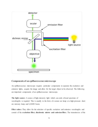
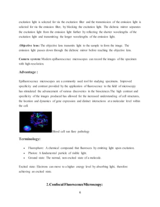
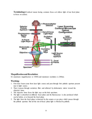
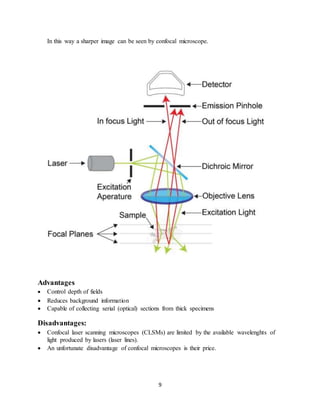
1
FLORESCENCE MICROSCOPY
FRAZ AHMAD MAZARI
Department of Bio Chemistry
Bahauddin Zakariya University Multan
MICROSCOPE:
An optical instrument used for viewing very small object such as mineral sample animal or plant
cell typically magnified several hundred times.
MICROSCOPY:
Use of microscope is called microscopy.
INTRODUCTION:
Fluorescence microscopy:
Fluorescence microscopy is basically a method of studying material which can be made to
fluoresce,either in its natural form or when treated with chemicals capable of fluorescing with
the help of fluorescence microscope.
2
Fluorescence microscope:
A fluorescence microscope is an optical microscope that usess fluorescence and
phosphorescence instead of,or in addition to scattering ,reflection and attenution or absorption to
study properties of organic and inorganic substances.
History:
Fluorescence microscopy was discovered by August Kohler in 1904 .
INSTRUMENTATION:
Typicall fluorescence microscope consist of following components.
1.Fluorescentdyes (Fluorophore):
 Most cellular components are colorless and cannot be clearly distinguished under a
microscope. The basic premise of fluorescence microscopy is to stain the components
with dyes.
 A fluorophore is a fluorescent chemical compound that can re-emit light upon light
excitation.
 Fluorophores typically contain several combined aromatic groups, or plane or cyclic
molecules with several π bonds.
 Many fluorescent stains have been designed for a range of biological molecules.
 Some of these are small molecules which are intrinsically fluorescent and bind a
biological molecule of interest. Major examples of these are nucleic acid stains like DAPI
and Hoechst, phalloidin which is used to stain actin fibres in mammalian cells.
2.Light source:
 Four main types of light source are used, including xenon arc lamps or mercury-vapor
lamps with an excitation filter, lasers, and high- power LEDs.
 Lasers are mostly used for complex fluorescence microscopy techniques, while xenon
lamps, and mercury lamps, and LEDs with a dichroic excitation filter are commonly used
for wide field epifluorescence microscopes.
3.The excitationfilter:
 The excitation filter is an bandpass filter that passes only the wavelengths absorbed by
the fluorophore, thus minimizing excitation of other sources of fluorescence.
4.The dichroic mirror:
 A dichroic filter or thin- film filter, is a very accurate color filter used to selectively pass
light of a small range of colors while reflecting other colors.
5.The emissionfilter.
 The emitter is typically a bandpass filter that passes only the wavelengths emitted by the
fluorophore and blocks all undesired light outside this band – especially the excitation
light.
3
 By blocking unwanted excitation energy (including UV and IR) or sample and system
autofluorescence, optical filters ensure the darkest background.
(Fluorescence microscope)
Princple:
 Fluorescent dyes, also known as fluorophores are molecules that absorb excitation light at
a given wavelength (generally UV), and after a short delay emit light at a longer
wavelength.The delay between absorption and emission is negligible, generally on the
order of nanoseconds.
 The emission light can then be filtered from the excitation light to reveal the location of
the fluorophores.
 Fluorescence microscopy uses a much higher intensity light to illuminate the sample.
This light excites fluorescence species in the sample, which then emit light of a longer
wavelength.
 The image produced is based on the second light source or the emission wavelength of
the fluorescent species — rathe rthan from the light originally used to illuminate, and
excite, the sample.
TYPES OF FLUORESCENCEMICROSCOPY:
1.Epifluorescencemicroscopy
History:
In 1843, George Gabriel Stokes described fluorescence as being characterized by the
wavelength of an emitted light that is longer than the wavelength of the excitinglight.
This discovery led to the invention of the epifluorescence microscope, now a ubiquitous
instrument in biological and medical laboratories.
Basic:
4
 The specimen are dyed with fluorescence dyes .
 These fluorescence dyes have fluorophores which absorb the incident light which have
greater energy and smaller wave- length.
 Fluorophores atoms gets excited to higher energy levels
 Some energy is wasted or released in the form of heat.
 Than atoms as being unstable prone to get stable liberate or emit rays of different
wavelength which have lower energy and larger wavelength.
Principle:
Fluorescence microscopy is a particular form of light microscopy in which, instead of utilizing
visible light to illuminate specimens, a higher intensity light source excites a fluorescent
molecule called a fluorophore. The fluorophore absorbs photons leading to electrons moving to a
higher energy state. When the electrons return to the ground state by losing energy, the
fluorophore emits light of a longer wavelength.The emitted light is then separated from the
original excitation light via a filter and this produces a magnified image of the specimen being
studied. The use of fluorescence allows for the object of interest to be specifically targeted. As
the excitation light is filtered out, the emitted fluorescence allows only the fluorescent objects of
interest to remain visible.
5
Components of an epifluorescencemicroscope
An epifluorescence microscope requires particular components to separate the excitation and
emission lights, acquire the image and allow for the target object to be observed. The following
are important components of an epifluorescence microscope.
The light source: A source of high-intensity light which can emit a broad spectrum of
wavelengths is required. This is usually in the form of a xenon arc lamp or a high-pressure short
arc mercury lamp and LASER beam.
Filter cubes: They allow for the selection of specific excitation and emission wavelengths and
consist of the excitation filter, diachronic mirror and emission filter. The transmission of the
6
excitation light is selected for via the excitation filter and the transmission of the emission light is
selected for via the emission filter, by blocking the excitation light. The dichroic mirror separates
the excitation light from the emission light further by reflecting the shorter wavelengths of the
excitation light and transmitting the longer wavelengths of the emission light.
.Objective lens: The objective lens transmits light to the sample to form the image. The
emission light passes down through the dichroic mirror before reaching the objective lens.
Camera system: Modern epifluorescence microscopes can record the images of the specimen
with high-resolution.
Advantage :
Epifluorescence microscopes are a commonly used tool for studying specimens. Improved
specificity and contrast provided by the application of fluorescence to the field of microscopy
has stimulated the advancement of various discoveries in the biosciences.The high contrast and
specificity of the images produced has allowed for the increased understanding of cell structures,
the location and dynamics of gene expression and distinct interactions at a molecular level within
the cell.
Blood cell sun flare pathology
Terminology:
 Fluorophore: A chemical compound that fluoresces by emitting light upon excitation.
 Photon: A fundamental particle of visible light.
 Ground state: The normal, non-excited state of a molecule.
Excited state: Electrons can move to a higher energy level by absorbing light, therefore
achieving an excited state.
2.Confocal FluorscenceMicroscopy:
7
It is an optical imaging technique for increasing optical resolution and contrast of a micrograph
by means of using a spatial pinhole to block out of light in image formation.
History:
The basic concept of confocal microscopy was originally developed by Marvin Minsky in the
mid 1950s.
Instrumentation:
All the instrumentation is same but it contains additional two parts:
 Source: laser light
 Filter:pinholeasspatial filter
Principle:
Coherent light emitted by the laser system (excitation source) passes through a pinhole aperture
that is situated in a conjugate plane (confocal) with a scanning point on the specimen and a
second pinhole aperture positioned in the front of the detector (a photomultiplier tube). As the
laser is reflected by a dichromatic mirror and scanned across the specimen in a defined focal
plane, secondary fluorescence emitted from the points on the specimen (in the same focal plane)
pass back through the dichromatic mirror and are focused as a confocal point at the detector
pinhole aperture.
 Similar to the widefield microscope the confocal microscope uses fluorescence optics.
Instead of illuminating the whole sample at once, laser light is focused onto a defined spot at
a specific depth within the sample . This leads to the emission of fluorescent light at exactly
this point.
8
Terminology:Confocal means having common focus as it allows light of one focal plane
to focus on camera.
Magnificationand Resolution:
It’s maximum magnification is 1500X and maximum resolution is 200nm.
Working:
1. First laser beam come from laser light source and pass through first pinhole aperture present
near to light source.
2. Then it passes through excitation filter and reflected by dichromatic mirror toward the
objective lense.
3. The objective lense focus the light rays on the hole specimen.
4. The light is absorbed on different focal plane and the fluorescence is also produced which
can be before, at or beyond the focal plane.
5. The light from the focal plane is detected by the camera or eye piece which passes through
the pinhole aperture. But all the out-of-focus plane light is blocked by pinhole.
9
In this way a sharper image can be seen by confocal microscope.
Advantages
 Control depth of fields
 Reduces background information
 Capable of collecting serial (optical) sections from thick specimens
Disadvantages:
 Confocal laser scanning microscopes (CLSMs) are limited by the available wavelenghts of
light produced by lasers (laser lines).
 An unfortunate disadvantage of confocal microscopes is their price.
10
Two photon excitation Microscopy(Multiphoton)
History:
Two-photon microscopy was pioneered and patented by Winfried Denk and James Strickler in
the lab of Watt W. Webb at Cornell University in 1990.
Working Principle:
 Two-photon excitation can be a superior alternative to confocal microscopy due to its deeper
tissue penetration, efficient light detection, and reduced photobleaching.
 The principle of two-photon excitation is based on the idea that two photons of comparably
lower photon energy than needed for one photon excitation, can also excite a in one quantum
event.
 Each photon carries approximately half the energy necessary to excite the molecule.
 An excitation results in the subsequent emission of a fluorescence photon, typically at a
higher energy than either of the two excitatory photons. The probability of the near-
simultaneous absorption of two photons is extremely low.
 Therefore, a high flux of excitation photons is typically required, usually from a femtosecond
laser.
 The purpose of employing the two-photon effect is that the axial spread of the point spread
function is substantially lower than for single-photon excitation.
Advantages:
 Two-photon microscopy finds its applications in numerous fields including: physiology,
neurobiology, embryology and tissue engineering.
 Transparent tissues (such as skin cells) have been visualized with clear detail due to this
technique.
11
 Two-photon microscopy have high speed imaging capabilities.
 Two-photon microscopy penetrating power is high, it can penetrate up to 1mm in to
tissues.
 Laser used can excite approximately in a range of 700-1000nm.
Disadvantages:
 Muh more expensive
 the absorption is much less compare toone photon excitation
 This leads very often to high intensities which can destroy the cell
4. Totalinternal reflectionfluorescence microscopy
History:
Total internal reflection fluorescence (TIRF) is a special
technique in fluorescence microscopy developed by Daniel Axelrod at the
University of Michigan, Ann Arbor in the early 1980s. TIRFmicroscopy delivers
images with an outstandingly high axial resolution below 100 nm. This allows the
observation of membrane-associated processes.
Explanation
It allows imaging of fluorescent molecules located close
to the glass/water (or glass/specimen) interface. This is achieved by employing an
evanescent wave for excitation of the fluorophores instead of direct illumination
via light delivered by an arc lamp, LEDs or lasers. The evanescent field occurs if
incident light is totally reflected at the interface of two transparent media with
different refractive indices. In biological applications the incident light is usually
laser light and the interface the glass of the coverslip and a film of aqueous
solution between coverslip and adherent cells.
As the energy of an evanescent field decreases exponentially with distance to
the interface, only fluorophores in a certain proximity to the coverslip are excited.
12
This allows the creation of images with outstanding signal-to-noise ratio, as
fluorophores in the rest of the cell are hardly excited.
Additionally, TIRF microscopy delivers images with an outstandingly high axial
resolution below 100 nm.
This allows the observation of membrane-associated
processes like cell adhesion, hormone binding, molecule transport and
 Whenever
light encounters the interface of two transparent media with different refractive
indices, it will be partially diffracted and partially reflected. At a certain angle of
incidence, the so called critical angle, the light will be completely reflected and a
phenomenon called total internal reflection occurs On occurrence of total internal reflection, a
portion of the energy of the incident
light will be converted to an electromagnetic field and pass through the interface to
form an evanescent wave originating at the interface. The emerging evanescent
wave has the same frequency as the incident light and its amplitude decays
13
exponentially with depth of penetration On occurrence of total internal reflection, a portion of
the energy of the incident
Advantages
 The main advantage of total internal reflection fluorescence microscopy is its outstanding
signal to noise ratio due to its lofwer penetration depth of the evanescent field
 There fore out of field the fluorescence is dramatically minimized and almost no
background fluorescence occurs.
14

Florescense microscopy

  • 1.
    1 FLORESCENCE MICROSCOPY FRAZ AHMADMAZARI Department of Bio Chemistry Bahauddin Zakariya University Multan MICROSCOPE: An optical instrument used for viewing very small object such as mineral sample animal or plant cell typically magnified several hundred times. MICROSCOPY: Use of microscope is called microscopy. INTRODUCTION: Fluorescence microscopy: Fluorescence microscopy is basically a method of studying material which can be made to fluoresce,either in its natural form or when treated with chemicals capable of fluorescing with the help of fluorescence microscope.
  • 2.
    2 Fluorescence microscope: A fluorescencemicroscope is an optical microscope that usess fluorescence and phosphorescence instead of,or in addition to scattering ,reflection and attenution or absorption to study properties of organic and inorganic substances. History: Fluorescence microscopy was discovered by August Kohler in 1904 . INSTRUMENTATION: Typicall fluorescence microscope consist of following components. 1.Fluorescentdyes (Fluorophore):  Most cellular components are colorless and cannot be clearly distinguished under a microscope. The basic premise of fluorescence microscopy is to stain the components with dyes.  A fluorophore is a fluorescent chemical compound that can re-emit light upon light excitation.  Fluorophores typically contain several combined aromatic groups, or plane or cyclic molecules with several π bonds.  Many fluorescent stains have been designed for a range of biological molecules.  Some of these are small molecules which are intrinsically fluorescent and bind a biological molecule of interest. Major examples of these are nucleic acid stains like DAPI and Hoechst, phalloidin which is used to stain actin fibres in mammalian cells. 2.Light source:  Four main types of light source are used, including xenon arc lamps or mercury-vapor lamps with an excitation filter, lasers, and high- power LEDs.  Lasers are mostly used for complex fluorescence microscopy techniques, while xenon lamps, and mercury lamps, and LEDs with a dichroic excitation filter are commonly used for wide field epifluorescence microscopes. 3.The excitationfilter:  The excitation filter is an bandpass filter that passes only the wavelengths absorbed by the fluorophore, thus minimizing excitation of other sources of fluorescence. 4.The dichroic mirror:  A dichroic filter or thin- film filter, is a very accurate color filter used to selectively pass light of a small range of colors while reflecting other colors. 5.The emissionfilter.  The emitter is typically a bandpass filter that passes only the wavelengths emitted by the fluorophore and blocks all undesired light outside this band – especially the excitation light.
  • 3.
    3  By blockingunwanted excitation energy (including UV and IR) or sample and system autofluorescence, optical filters ensure the darkest background. (Fluorescence microscope) Princple:  Fluorescent dyes, also known as fluorophores are molecules that absorb excitation light at a given wavelength (generally UV), and after a short delay emit light at a longer wavelength.The delay between absorption and emission is negligible, generally on the order of nanoseconds.  The emission light can then be filtered from the excitation light to reveal the location of the fluorophores.  Fluorescence microscopy uses a much higher intensity light to illuminate the sample. This light excites fluorescence species in the sample, which then emit light of a longer wavelength.  The image produced is based on the second light source or the emission wavelength of the fluorescent species — rathe rthan from the light originally used to illuminate, and excite, the sample. TYPES OF FLUORESCENCEMICROSCOPY: 1.Epifluorescencemicroscopy History: In 1843, George Gabriel Stokes described fluorescence as being characterized by the wavelength of an emitted light that is longer than the wavelength of the excitinglight. This discovery led to the invention of the epifluorescence microscope, now a ubiquitous instrument in biological and medical laboratories. Basic:
  • 4.
    4  The specimenare dyed with fluorescence dyes .  These fluorescence dyes have fluorophores which absorb the incident light which have greater energy and smaller wave- length.  Fluorophores atoms gets excited to higher energy levels  Some energy is wasted or released in the form of heat.  Than atoms as being unstable prone to get stable liberate or emit rays of different wavelength which have lower energy and larger wavelength. Principle: Fluorescence microscopy is a particular form of light microscopy in which, instead of utilizing visible light to illuminate specimens, a higher intensity light source excites a fluorescent molecule called a fluorophore. The fluorophore absorbs photons leading to electrons moving to a higher energy state. When the electrons return to the ground state by losing energy, the fluorophore emits light of a longer wavelength.The emitted light is then separated from the original excitation light via a filter and this produces a magnified image of the specimen being studied. The use of fluorescence allows for the object of interest to be specifically targeted. As the excitation light is filtered out, the emitted fluorescence allows only the fluorescent objects of interest to remain visible.
  • 5.
    5 Components of anepifluorescencemicroscope An epifluorescence microscope requires particular components to separate the excitation and emission lights, acquire the image and allow for the target object to be observed. The following are important components of an epifluorescence microscope. The light source: A source of high-intensity light which can emit a broad spectrum of wavelengths is required. This is usually in the form of a xenon arc lamp or a high-pressure short arc mercury lamp and LASER beam. Filter cubes: They allow for the selection of specific excitation and emission wavelengths and consist of the excitation filter, diachronic mirror and emission filter. The transmission of the
  • 6.
    6 excitation light isselected for via the excitation filter and the transmission of the emission light is selected for via the emission filter, by blocking the excitation light. The dichroic mirror separates the excitation light from the emission light further by reflecting the shorter wavelengths of the excitation light and transmitting the longer wavelengths of the emission light. .Objective lens: The objective lens transmits light to the sample to form the image. The emission light passes down through the dichroic mirror before reaching the objective lens. Camera system: Modern epifluorescence microscopes can record the images of the specimen with high-resolution. Advantage : Epifluorescence microscopes are a commonly used tool for studying specimens. Improved specificity and contrast provided by the application of fluorescence to the field of microscopy has stimulated the advancement of various discoveries in the biosciences.The high contrast and specificity of the images produced has allowed for the increased understanding of cell structures, the location and dynamics of gene expression and distinct interactions at a molecular level within the cell. Blood cell sun flare pathology Terminology:  Fluorophore: A chemical compound that fluoresces by emitting light upon excitation.  Photon: A fundamental particle of visible light.  Ground state: The normal, non-excited state of a molecule. Excited state: Electrons can move to a higher energy level by absorbing light, therefore achieving an excited state. 2.Confocal FluorscenceMicroscopy:
  • 7.
    7 It is anoptical imaging technique for increasing optical resolution and contrast of a micrograph by means of using a spatial pinhole to block out of light in image formation. History: The basic concept of confocal microscopy was originally developed by Marvin Minsky in the mid 1950s. Instrumentation: All the instrumentation is same but it contains additional two parts:  Source: laser light  Filter:pinholeasspatial filter Principle: Coherent light emitted by the laser system (excitation source) passes through a pinhole aperture that is situated in a conjugate plane (confocal) with a scanning point on the specimen and a second pinhole aperture positioned in the front of the detector (a photomultiplier tube). As the laser is reflected by a dichromatic mirror and scanned across the specimen in a defined focal plane, secondary fluorescence emitted from the points on the specimen (in the same focal plane) pass back through the dichromatic mirror and are focused as a confocal point at the detector pinhole aperture.  Similar to the widefield microscope the confocal microscope uses fluorescence optics. Instead of illuminating the whole sample at once, laser light is focused onto a defined spot at a specific depth within the sample . This leads to the emission of fluorescent light at exactly this point.
  • 8.
    8 Terminology:Confocal means havingcommon focus as it allows light of one focal plane to focus on camera. Magnificationand Resolution: It’s maximum magnification is 1500X and maximum resolution is 200nm. Working: 1. First laser beam come from laser light source and pass through first pinhole aperture present near to light source. 2. Then it passes through excitation filter and reflected by dichromatic mirror toward the objective lense. 3. The objective lense focus the light rays on the hole specimen. 4. The light is absorbed on different focal plane and the fluorescence is also produced which can be before, at or beyond the focal plane. 5. The light from the focal plane is detected by the camera or eye piece which passes through the pinhole aperture. But all the out-of-focus plane light is blocked by pinhole.
  • 9.
    9 In this waya sharper image can be seen by confocal microscope. Advantages  Control depth of fields  Reduces background information  Capable of collecting serial (optical) sections from thick specimens Disadvantages:  Confocal laser scanning microscopes (CLSMs) are limited by the available wavelenghts of light produced by lasers (laser lines).  An unfortunate disadvantage of confocal microscopes is their price.
  • 10.
    10 Two photon excitationMicroscopy(Multiphoton) History: Two-photon microscopy was pioneered and patented by Winfried Denk and James Strickler in the lab of Watt W. Webb at Cornell University in 1990. Working Principle:  Two-photon excitation can be a superior alternative to confocal microscopy due to its deeper tissue penetration, efficient light detection, and reduced photobleaching.  The principle of two-photon excitation is based on the idea that two photons of comparably lower photon energy than needed for one photon excitation, can also excite a in one quantum event.  Each photon carries approximately half the energy necessary to excite the molecule.  An excitation results in the subsequent emission of a fluorescence photon, typically at a higher energy than either of the two excitatory photons. The probability of the near- simultaneous absorption of two photons is extremely low.  Therefore, a high flux of excitation photons is typically required, usually from a femtosecond laser.  The purpose of employing the two-photon effect is that the axial spread of the point spread function is substantially lower than for single-photon excitation. Advantages:  Two-photon microscopy finds its applications in numerous fields including: physiology, neurobiology, embryology and tissue engineering.  Transparent tissues (such as skin cells) have been visualized with clear detail due to this technique.
  • 11.
    11  Two-photon microscopyhave high speed imaging capabilities.  Two-photon microscopy penetrating power is high, it can penetrate up to 1mm in to tissues.  Laser used can excite approximately in a range of 700-1000nm. Disadvantages:  Muh more expensive  the absorption is much less compare toone photon excitation  This leads very often to high intensities which can destroy the cell 4. Totalinternal reflectionfluorescence microscopy History: Total internal reflection fluorescence (TIRF) is a special technique in fluorescence microscopy developed by Daniel Axelrod at the University of Michigan, Ann Arbor in the early 1980s. TIRFmicroscopy delivers images with an outstandingly high axial resolution below 100 nm. This allows the observation of membrane-associated processes. Explanation It allows imaging of fluorescent molecules located close to the glass/water (or glass/specimen) interface. This is achieved by employing an evanescent wave for excitation of the fluorophores instead of direct illumination via light delivered by an arc lamp, LEDs or lasers. The evanescent field occurs if incident light is totally reflected at the interface of two transparent media with different refractive indices. In biological applications the incident light is usually laser light and the interface the glass of the coverslip and a film of aqueous solution between coverslip and adherent cells. As the energy of an evanescent field decreases exponentially with distance to the interface, only fluorophores in a certain proximity to the coverslip are excited.
  • 12.
    12 This allows thecreation of images with outstanding signal-to-noise ratio, as fluorophores in the rest of the cell are hardly excited. Additionally, TIRF microscopy delivers images with an outstandingly high axial resolution below 100 nm. This allows the observation of membrane-associated processes like cell adhesion, hormone binding, molecule transport and  Whenever light encounters the interface of two transparent media with different refractive indices, it will be partially diffracted and partially reflected. At a certain angle of incidence, the so called critical angle, the light will be completely reflected and a phenomenon called total internal reflection occurs On occurrence of total internal reflection, a portion of the energy of the incident light will be converted to an electromagnetic field and pass through the interface to form an evanescent wave originating at the interface. The emerging evanescent wave has the same frequency as the incident light and its amplitude decays
  • 13.
    13 exponentially with depthof penetration On occurrence of total internal reflection, a portion of the energy of the incident Advantages  The main advantage of total internal reflection fluorescence microscopy is its outstanding signal to noise ratio due to its lofwer penetration depth of the evanescent field  There fore out of field the fluorescence is dramatically minimized and almost no background fluorescence occurs.
  • 14.