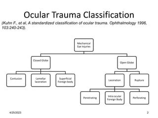
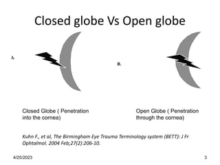
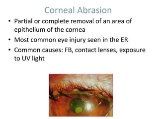
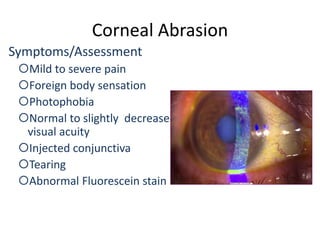
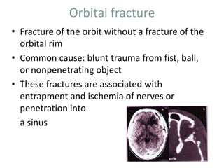
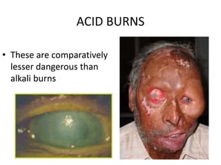
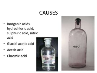
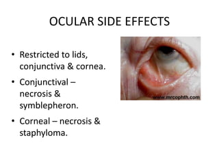
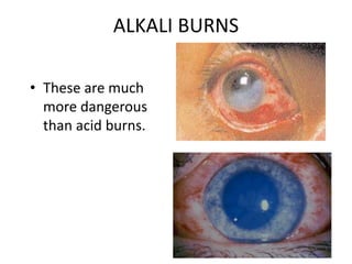
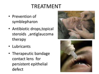
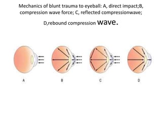
This document discusses various types of ocular trauma including classification, symptoms, assessment, diagnostics, and treatment. It covers closed and open globe injuries, corneal abrasions, extraocular foreign bodies, orbital fractures, hyphema, eyelid lacerations, globe ruptures, and chemical burns. Common injuries from blunt trauma include commotio retinae, retinal hemorrhages, choroidal ruptures, and vitreous hemorrhage or detachment. Assessment examines mechanism of injury and complications involving iris, lens, vitreous, choroid, and retina. Treatment priorities are irrigation, antibiotics, patching, referral to ophthalmologist, and surgery if needed to repair damage and prevent complications.
eye emergency occurs any time we have foreign objects or chemical in our eyes. this slide contain definition, classification, types of injury, identification, management, medical management, nursing management. care of eye in the condition.
In this PPT it included defination, etiology, classification, pathophysiology, diagnostic procedure and variuos medical and surgical management of glaucoma. A students from diploma or master can easily understand this presentation.
Cranial Nerve is a very important topics in field of ophthalmology and optometry. a stuent with knowledge with cranial nerves can easily understand muscle palsy and diagnosis of neuroohthalmology
2024.06.01 Introducing a competency framework for languag learning materials ...Sandy Millin
http://sandymillin.wordpress.com/iateflwebinar2024
Published classroom materials form the basis of syllabuses, drive teacher professional development, and have a potentially huge influence on learners, teachers and education systems. All teachers also create their own materials, whether a few sentences on a blackboard, a highly-structured fully-realised online course, or anything in between. Despite this, the knowledge and skills needed to create effective language learning materials are rarely part of teacher training, and are mostly learnt by trial and error.
Knowledge and skills frameworks, generally called competency frameworks, for ELT teachers, trainers and managers have existed for a few years now. However, until I created one for my MA dissertation, there wasn’t one drawing together what we need to know and do to be able to effectively produce language learning materials.
This webinar will introduce you to my framework, highlighting the key competencies I identified from my research. It will also show how anybody involved in language teaching (any language, not just English!), teacher training, managing schools or developing language learning materials can benefit from using the framework.
June 3, 2024 Anti-Semitism Letter Sent to MIT President Kornbluth and MIT Cor...Levi Shapiro
Letter from the Congress of the United States regarding Anti-Semitism sent June 3rd to MIT President Sally Kornbluth, MIT Corp Chair, Mark Gorenberg
Dear Dr. Kornbluth and Mr. Gorenberg,
The US House of Representatives is deeply concerned by ongoing and pervasive acts of antisemitic
harassment and intimidation at the Massachusetts Institute of Technology (MIT). Failing to act decisively to ensure a safe learning environment for all students would be a grave dereliction of your responsibilities as President of MIT and Chair of the MIT Corporation.
This Congress will not stand idly by and allow an environment hostile to Jewish students to persist. The House believes that your institution is in violation of Title VI of the Civil Rights Act, and the inability or
unwillingness to rectify this violation through action requires accountability.
Postsecondary education is a unique opportunity for students to learn and have their ideas and beliefs challenged. However, universities receiving hundreds of millions of federal funds annually have denied
students that opportunity and have been hijacked to become venues for the promotion of terrorism, antisemitic harassment and intimidation, unlawful encampments, and in some cases, assaults and riots.
The House of Representatives will not countenance the use of federal funds to indoctrinate students into hateful, antisemitic, anti-American supporters of terrorism. Investigations into campus antisemitism by the Committee on Education and the Workforce and the Committee on Ways and Means have been expanded into a Congress-wide probe across all relevant jurisdictions to address this national crisis. The undersigned Committees will conduct oversight into the use of federal funds at MIT and its learning environment under authorities granted to each Committee.
• The Committee on Education and the Workforce has been investigating your institution since December 7, 2023. The Committee has broad jurisdiction over postsecondary education, including its compliance with Title VI of the Civil Rights Act, campus safety concerns over disruptions to the learning environment, and the awarding of federal student aid under the Higher Education Act.
• The Committee on Oversight and Accountability is investigating the sources of funding and other support flowing to groups espousing pro-Hamas propaganda and engaged in antisemitic harassment and intimidation of students. The Committee on Oversight and Accountability is the principal oversight committee of the US House of Representatives and has broad authority to investigate “any matter” at “any time” under House Rule X.
• The Committee on Ways and Means has been investigating several universities since November 15, 2023, when the Committee held a hearing entitled From Ivory Towers to Dark Corners: Investigating the Nexus Between Antisemitism, Tax-Exempt Universities, and Terror Financing. The Committee followed the hearing with letters to those institutions on January 10, 202
Acetabularia Information For Class 9 .docxvaibhavrinwa19
Acetabularia acetabulum is a single-celled green alga that in its vegetative state is morphologically differentiated into a basal rhizoid and an axially elongated stalk, which bears whorls of branching hairs. The single diploid nucleus resides in the rhizoid.
Safalta Digital marketing institute in Noida, provide complete applications that encompass a huge range of virtual advertising and marketing additives, which includes search engine optimization, virtual communication advertising, pay-per-click on marketing, content material advertising, internet analytics, and greater. These university courses are designed for students who possess a comprehensive understanding of virtual marketing strategies and attributes.Safalta Digital Marketing Institute in Noida is a first choice for young individuals or students who are looking to start their careers in the field of digital advertising. The institute gives specialized courses designed and certification.
for beginners, providing thorough training in areas such as SEO, digital communication marketing, and PPC training in Noida. After finishing the program, students receive the certifications recognised by top different universitie, setting a strong foundation for a successful career in digital marketing.
How to Make a Field invisible in Odoo 17Celine George
It is possible to hide or invisible some fields in odoo. Commonly using “invisible” attribute in the field definition to invisible the fields. This slide will show how to make a field invisible in odoo 17.
Introduction to AI for Nonprofits with Tapp NetworkTechSoup
Dive into the world of AI! Experts Jon Hill and Tareq Monaur will guide you through AI's role in enhancing nonprofit websites and basic marketing strategies, making it easy to understand and apply.
Unit 8 - Information and Communication Technology (Paper I).pdfThiyagu K
This slides describes the basic concepts of ICT, basics of Email, Emerging Technology and Digital Initiatives in Education. This presentations aligns with the UGC Paper I syllabus.
Synthetic Fiber Construction in lab .pptxPavel ( NSTU)
Synthetic fiber production is a fascinating and complex field that blends chemistry, engineering, and environmental science. By understanding these aspects, students can gain a comprehensive view of synthetic fiber production, its impact on society and the environment, and the potential for future innovations. Synthetic fibers play a crucial role in modern society, impacting various aspects of daily life, industry, and the environment. ynthetic fibers are integral to modern life, offering a range of benefits from cost-effectiveness and versatility to innovative applications and performance characteristics. While they pose environmental challenges, ongoing research and development aim to create more sustainable and eco-friendly alternatives. Understanding the importance of synthetic fibers helps in appreciating their role in the economy, industry, and daily life, while also emphasizing the need for sustainable practices and innovation.
Embracing GenAI - A Strategic ImperativePeter Windle
Artificial Intelligence (AI) technologies such as Generative AI, Image Generators and Large Language Models have had a dramatic impact on teaching, learning and assessment over the past 18 months. The most immediate threat AI posed was to Academic Integrity with Higher Education Institutes (HEIs) focusing their efforts on combating the use of GenAI in assessment. Guidelines were developed for staff and students, policies put in place too. Innovative educators have forged paths in the use of Generative AI for teaching, learning and assessments leading to pockets of transformation springing up across HEIs, often with little or no top-down guidance, support or direction.
This Gasta posits a strategic approach to integrating AI into HEIs to prepare staff, students and the curriculum for an evolving world and workplace. We will highlight the advantages of working with these technologies beyond the realm of teaching, learning and assessment by considering prompt engineering skills, industry impact, curriculum changes, and the need for staff upskilling. In contrast, not engaging strategically with Generative AI poses risks, including falling behind peers, missed opportunities and failing to ensure our graduates remain employable. The rapid evolution of AI technologies necessitates a proactive and strategic approach if we are to remain relevant.
2. Ocular Trauma Classification
Mechanical
Eye injuries
Closed Globe
Contusion Lamellar
laceration
Superficial
Foreign body
Open Globe
Laceration
Penetrating
Intra ocular
Foreign Body
Perforating
Rupture
4/25/2023
(Kuhn F., et al, A standardized classification of ocular trauma. Ophthalmology 1996,
103:240-243).
2
3. Closed globe Vs Open globe
Closed Globe ( Penetration
into the cornea)
Open Globe ( Penetration
through the cornea)
Kuhn F., et al, The Birmingham Eye Trauma Terminology system (BETT): J Fr
Ophtalmol. 2004 Feb;27(2):206-10.
4/25/2023 3
4. TYPE OF INJURY
• Refers to the mechanism of injury.
A. Rupture
B. Penetrating
C. Intraocular foreign body
D. Perforating
E. Mixed
4/25/2023 4
5. Complications
• Common anterior segment complications were
– iris prolapse (43%)
– cataract (36% )
– hyphema (17%).
– glaucoma
• Common posterior segment complications were
– Vitreous haemorrhage (31%),
– Berlin’s oedema (15%),
– retinal detachment (9%) and
– RAPD (7%).
– Signs of infection at presentation 11%
– IOFB (9%) –
• 4% in anterior segment
• 5% in posterior segment
4/25/2023 5
6. Corneal Abrasion
• Partial or complete removal of an area of
epithelium of the cornea
• Most common eye injury seen in the ER
• Common causes: FB, contact lenses, exposure
to UV light
7. Corneal Abrasion
Symptoms/Assessment
Mild to severe pain
Foreign body sensation
Photophobia
Normal to slightly decreased
visual acuity
Injected conjunctiva
Tearing
Abnormal Fluorescein stain
8. Corneal Abrasion
Treatment
– Topical analgesic
– Topical ophthalmic
antibiotic
– Tight patch to affected
eye for 12-24 hours
Education
– Follow-up care
– Proper patching
techniques
– Instillation of meds
– S/S of infection
– Use extra precaution
with activities requiring
depth perception
9. Extraocular Foreign Body
• Can enter as a result from hammering,
grinding, working under cars, or working
above the head
• “Something going into my eye”
• Metal, sawdust, dust particles
• Metal can form a rust ring on the cornea
10. Extraocular Foreign Body
Symptoms/Assessment
– Pain
– Foreign body sensation
– Tearing
– Redness
– Normal to slightly abnormal
visual acuity
– Fluorscein stain abnormal
– FB visualized
Diagnostics
– Magnifying lens
– Fluorescein stain
– Slit-lamp
11. Extraocular Foreign Body
Treatment
–Topical anesthetic
• Topical anesthetic inhibit
wound healing and are
toxic to corneal
epithelium
–Gentle irrigation with NS
– FB removal with moist
cotton swab, needle, eye
spud if irrigation
– Patch both eyes to
reduce unsuccessful
consensual movement
– Possible admission
12. Orbital fracture
• Fracture of the orbit without a fracture of the
orbital rim
• Common cause: blunt trauma from fist, ball,
or nonpenetrating object
• These fractures are associated with
entrapment and ischemia of nerves or
penetration into
a sinus
13. Orbital fracture
Symptoms/Assessment
Hx of blunt trauma
Diplopia
Facial anesthesia
Pain
Sunken appearance of the
eye
Limited vertical eye movement
EOM abnormal
Crepitus
Periorbital edema,
hematoma, ecchymosis
Subconjunctival
hemorrhage
Look for other injuries
14. Orbital fracture
Diagnostics
– Visual acuity
– Fundoscopy
– CT scan
– X-rays
• Orbits
• Facial
• Waters’
Treatment/Education
– Ophthalmological consult
– Analgesics
– Antibiotics
– Ice pack
– Refrain from blowing nose
– Follow-up care
– Possible admission or
surgery
15. Hyphema
• Blood in the anterior chamber from the iris
bleeding
• Usually result of blunt trauma
• Significant risk of secondary bleeding in 3-5
days with outcomes poor
17. Hyphema
Treatment/Education
– Have patient sit upright or
bedrest with HOB 30°
– Patch or shield both eyes
– Diuretics to decrease
intraocular pressure
– Refrain from taking aspirin
– Refer to ophthalmologist
– Admission
18. Eyelid Laceration
Symptoms/Assessment
– MOI
– Visual disturbance
– Laceration
– Protrusion of fat
– Upper lid does not raise
– Assess for ocular injuries
– Bleeding
Treatment/Education
– Stop bleeding: Avoid
direct pressure on the
eye
– Surgical repair
– Topical analgesic
– Td
– Wound care
– S/S of infection
– Follow-up
20. Globe Rupture
Symptoms/Assessment
– MOI
• Blunt
• Penetrating
– Sudden visual impairment or
loss
– Pain
– Asymmetry of globe
– Extrusion of aqueous or
vitreous humor
– Direct visualization of FB
– Irregularities in pupillary
borders
– Diagnostics
• CT scan
• MRI
• Orbit films
• Slit-lamp exam
21. Globe Rupture
Treatment
– Ophthalmological referral
– Do not open eye
– Keep patient in Semi-
Fowlers position
– Patch/shield affected both
eyes
– IV analgesics
– IV antibiotics
– Calm, supportive
environment
– Admission/Surgery
– If impaled object: Secure
it.
Do Not
Remove IT!
22. CHEMICAL BURNS
• Caused due to external contact by acids or
alkalis.
• Only ocular injury, requiring immediate
treatment.
• It is an ophthalmic emergency.
23. MODE OF INJURY
• These usually occur with external contact with
chemical under following circumstance
• Domestic accidents ex: with ammonia,solvents
detergents & cosmetics
• Chemical laboratory accidents with alkali ,acids
• Chemical warfare injuries
• Self inflicted chemical injuries seen in
malingerers & psychopaths
26. MECHANISM
• Forms a precipitate of tissue proteins which
coagulate thus causing tissue damage & scar
formation.
• The coagulation forms a barrier that prevents
deep penetration.
30. MECHANISM
• The hydroxyl group causes saponification of fats
& proteins & also causes release of proteolytic
enzymes like collagenase, elastase.
• Thus severe melting of the tissues occur.
• The pH of tissues correlates with tissue damage.
• Irreversible damage is seen with pH of 11.5
31. • Alkalis dissociate the fatty acids of cell membrane &
therefore destroy the tissue.
• In hydroscopic- they extract water from cells causing
necrosis.
• Combines with lipids of cells to form soluble
compounds thus causing softening of tissue.
• This all increases in increased penetration of the
alkali in the tissue.
• Alkali burn therefore spreads widely & the action
continues for some days
32. MANAGEMENT
• To minimize the damage
• Avoidance of complication
• Control of acute inflammation
• Support regeneration of epithelium &
reparative process
33. GRADES OF CHEMICAL
BURNS
good
a) Corneal
haze
b) Iris details
visible
a) Congestion
b) Chemosis
c) Ischemic(< 1/3
)
II
good
epithelial
damage only
a) Chemosis
b) No ischaemia
I
VISUAL
PROGNOSIS
CORNEA
CONJUNCTIVA
GRAD
E
34. doubtful
a)Total
epithelial loss
b)Stromal haze
c)Iris details not
visible
a) Ischemia (1/3-
½)
III
poor
a) Total corneal
opacity
b) No view of
iris & pupil
a) Ischaemia > ½
IV
VISUAL
PROGNOSIS
CORNEA
CONJUNCTIVA
GRADE
35. TREATMENT
• Immediate & thorough
wash with available
clean water or saline
• chemical neutralization
• Mechanical removal of
contaminant
• Removal of
contaminated & necrotic
tissue
37. SURGICAL REHABILITATION
• Conjunctival grafting
• Penetrating or preparatory lamellar
keratoplasty may be done
• Optical penetrating keratoplasty may be
done after one year of complete healing
38. ROLE OF OPTOMETRIST
• Irrigation
• History taking
• Visual assessment
• Therapeutic contact lens
• Referring to ophthalmologist
• Prosthetic contact lens
39. Mechanics of blunt trauma to eyeball: A, direct impact;B,
compression wave force; C, reflected compressionwave;
D,rebound compression wave.
40. Mechanics of blunt trauma to
eyeball
Blunt trauma of eyeball produces damage by
different forces as described below:
1. Direct impact on the globe. It produces
maximum damage at the point where the blow is
received Fig A
2. Compression wave force. It is transmitted
through the fluid contents in all the directions and
strikes the angle of anterior chamber, pushes the
iris lens diaphragm posteriorly, and also strikes the
retina and choroid. This may cause considerable
damage.
41. Sometimes the compression wave may be so explosive, that
maximum damage may be produced at a point distant from the
actual place of impact. This is called contre-coup damage.(Fig-B)
3. Reflected compression wave force. After striking the outer
coats the compression waves are reflected towards the posterior
pole and may cause foveal damage (Fig-C).
4. Rebound compression wave force. After striking the posterior
wall of the globe, the compression waves rebound back
anteriorly. This force damages the retina and choroid by forward
pull and lens-iris diaphragm by forward thrust from the back .(Fig
D)
42. 5. Indirect force. Ocular damage may also be
caused by the indirect forces from the bony
walls and elastic contents of the orbit, when
globe suddenly strikes against these structures.
43. Iris, pupil and ciliary body
1. Traumatic miosis. It occurs initially due to
irritation of ciliary nerves. It may be associated
with spasm of accommodation.
2. Traumatic mydriasis (Iridoplegia). It is usually
permanent and may be associated with
traumatic cycloplegia.
3. Rupture of the pupillary margin is a
commonoccurrence in closed-globe injury.
44. 4. Radiating tears in the iris stroma, sometimes
reaching up to ciliary body, may occur
occasionally.
5. Iridodialysis i.e., detachment of iris from its
root at the ciliary body occurs frequently. It
results in a D-shaped pupil and a black biconvex
area seen at the periphery.
6. Antiflexion of the iris. It refers to rotation of
the detached portion of iris, in which its
posterior surface faces anteriorly
45. 7. Retroflexion of the iris. This term is used when
whole of the iris is doubled back into the ciliary
region and becomes invisible.
8. Traumatic aniridia or iridremia. In this
condition, the completely torn iris (from ciliary
body) sinks to the bottom of anterior chamber
in the form of a minute ball.
46. 9. Angle recession refers to the tear between
longitudinal and circular muscle fibres of the
ciliary body. It is characterized by deepening of
the anterior chamber and widening of the ciliary
body band on gonioscopy. Later on it is
complicated by glaucoma.
47. 10. Inflammatory changes. These include
traumatic iridocyclitis, haemophthalmitis, post-
traumatic iris atrophy and pigmentary changes.
Treatment. It consists of atropine, antibiotics
and steroids. In the presence of ruptures of
pupillary margins and subluxation of lens,
atropine is contraindicated
48. Lens It may show following
changes:
1. Vossius ring. It is a circular ring of brown
pigment seen on the anterior capsule. It occurs
due to striking of the contracted pupillary
margin against the crystalline lens. It is always
smaller than the size of the pupil.
2. Concussion cataract. It occurs mainly due to
imbibition of aqueous and partly due to direct
mechanical effects of the injury on lens fibres.
49. Traumatic absorption of the lens. It may occur
sometimes in young children resulting in
aphakia.
4. Subluxation of the lens. It may occur due to
partial tear of zonules. The subluxated lens is
slightly displaced but still present in the
pupillary area. On dilatation of the pupil its edge
may be seen.
50. Depending upon the site of zonular tear
subluxation may be vertical (upward or
downward), or lateral (nasal or temporal).
5. Dislocation of the lens. It occurs when rupture
of the zonules is complete. It may be intraocular
(commonly) or extraocular (sometimes).
51. Intraocular dislocation may be anterior into the
anterior chamber, or posterior into the vitreous.
Extraocular dislocation may be in the
subconjunctival space (phakocele) or it may fall
outside the eye.
52. Vitreous
1. Liquefaction and appearance of clouds of fine
pigmentary opacities (a most common change).
2. Detachment of the vitreous either anterior or
posterior.
3. Vitreous haemorrhage. It is of common
occurrence .
4. Vitreous herniation in the anterior chamber may
occur with subluxation or dislocation of the lens.
53. Choroid
• 1. Rupture of the choroid. The rupture of
choroid is concentric to the optic disc and
situated temporal to it. Rupture may be single
or multiple. On fundus examination, the
choroidal rupture looks like a whitish crescent
(due to underlying sclera) with fine
pigmentation at its margins. Retinal vessels
pass over it.
54. 2. Choroidal haemorrhage may occur under the
retina (subretinal) or may even enter the
vitreous if retina is also torn.
3. Choroidal detachment is also known occur
following blunt trauma. Traumatic choroiditis
may be seen on fundus examination as patches
of pigmentation and discoloration after the eye
becomes silent.
55. Retina
1. Commotio retinae (Berlin’s oedema). It is of
common occurrence following a blow on the
eye. It manifests as milky white cloudiness
involving a considerable area of the posterior
pole with a ‘cherry-red spot’ in the foveal region.
It may disappear after some days or may be
followed by pigmentary changes.
56. 2. Retinal haemorrhages. These are quite common
following concussion trauma. Multiple haemorrhages
including flame-shaped and preretinal (subhyaloid) D-
shaped haemorrhage may be associated with traumatic
retinopathy.
3. Retinal tears. These may follow a contusion,
particularly in the peripheral region, especially ineyes
already suffering from myopia or senile
degenerations.
57. 4. Traumatic proliferative retinopathy (Retinitis
proliferans). It may occur secondary to vitreous
haemorrhage, forming tractional bands.
5. Retinal detachment. It may follow retinal tears or
vitreo-retinal tractional bands.
6. Concussion changes at macula. Traumatic
macular oedema is usually followed by pigmentary
degeneration. Sometimes, a macular cyst is formed,
which on rupture may be converted into a lamellar or
full thickness macular hole.
58. Intraocular pressure changes in closed-globe
injury
1. Traumatic glaucoma. It may occur due to
multiple factors.
2. Traumatic hypotony. It may follow damage to
the ciliary body and may even result in phthisis
bulbi.
59. Traumatic changes in the refraction
1. Myopia may follow ciliary spasm or rupture of
zonules or anterior shift of the lens.
2. Hypermetropia and loss of accommodation
may result from damage to the ciliary body
60. INTRAOCULAR FOREIGN BODIES
Penetrating injuries with foreign bodies are not
infrequent. Seriousness of such injuries is
compounded by the retention of intraocular
foreign bodies (IOFB).
• Common foreign bodies responsible for such
injuries include: chips of iron and steel (90%)
particles of glass, stone, lead pellets, copper
percussion caps, aluminium, plastic and wood.
61. Mechanical effects of IOFB
Mechanical effects depend upon the size,
velocity and type of the foreign body. Foreign
bodies greater than 2 mm in size cause
extensive damage. The lesions caused also
depend upon the route of entry and the site up
to which a foreign body has travelled. In general
these include: Corneal or/and scleral
perforation, hyphaema, iris hole; Rupture of the
lens and traumatic cataract;
62. • Vitreous haemorrhage and/or degeneration;
• Choroidal perforation, haemorrhage and
inflammation;
• Retinal hole, haemorrhages, oedema and
detachment
63. Locations of IOFB
Having entered the eye through the cornea or
sclera a foreign body may be retained at
any of the following sites
1. Anterior chamber. In the anterior chamber,
the IOFB usually sinks at the bottom. A tiny
foreign body may be concealed in the angle of
anterior chamber, and visualised only on
gonioscopy.
64. 2. Iris. Here the foreign body is usually entangledin the
stroma.
3. Posterior chamber. Rarely a foreign body may
sink behind the iris after entering through pupil or after
making a hole in the iris.
4. Lens. Foreign body may be present on the anterior
surface or inside the lens. Either an opaque track may be
seen in the lens or the lens may become completely
cataractous.
5. Vitreous cavity. A foreign body may reach here through
various routes .
65. Common sites for retention of an
intraocular foreign body: 1, anterior
chamber; 2, iris; 3, lens; 4, vitreous;
5, retina; 6, choroid; 7, sclera; 8,
orbital cavity
diagrammatic depiction of routes of access
of a foreign body in the vitreous,
through: A, cornea, pupil, lens; B, cornea,
iris, lens; C, cornea, iris, zonules; D, sclera,
choroid, retina.
66. Reactions of the foreign body,
Inorganic foreign body
Depending upon its chemical nature following 4
types of reactions are noted in the ocular tissues
1. No reaction is produced by the inert
substances which include glass, plastic,
porcelain, gold silver and platinum.
2. Local irritative reaction leading to
encapsulation of the foreign body occurs with
lead and aluminium particles.
67. 3. Suppurative reaction is excited by pure
copper, zinc, nickel and mercury particles.
4. Specific reactions are produced by iron
(Siderosis bulbi) and copper alloys (Chalcosis):
68. • Siderosis bulbi It refers to the degenerative
changes produced by an iron foreign body.
Sidesosis bulbi usually occurs after 2 months
to 2 years of the injury. However, earliest
changes have been reported after 9 days of
trauma.
69. Mechanism
The iron particle undergoes electrolytic
dissociation by the current of rest and its ions
are disseminated throughout the eye. These
ions combine with the intracellular proteins and
produce degenerative changes. In this process,
the epithelial structures of the eye are most
affected.
70. Clinical manifestations
1. The anterior epithelium and capsule of the lens are
involved first of all. Here, the rusty deposits are
arranged radially in a ring. Eventually, the lens becomes
cataractous.
2. Iris It is first stained greenish and later on turns
reddish brown.
3. Retina develops pigmentary degeneration which
resembles retinitis pigmentosa.
4. Secondary open angle type of glaucoma occurs due
to degenerative changes in the trabecular meshwork.
71. Chalcosis
It refers to the specific changes produced by the
alloy of copper in the eye.
Mechanism. Copper ions from the alloy are
dissociated electrolytically and deposited under
the membranous structures of the eye. Unlike
iron ions these do not enter into a chemical
combination with the proteins of the cells and
thus produce no degenerative changes.
72. Clinical manifestations
1. Kayser-Fleischer ring. It is a golden brown ring which
occurs due to deposition of copper under peripheral
parts of the Descemet’s membrane of the cornea.
2. Sunflower cataract. It is produced by deposition of
copper under the posterior capsule of the lens. It is
brilliant golden green in colour and arrangedlike the
petals of a sun flower.
3. Retina. It may show deposition of golden plaques at
the posterior pole which reflect the light with a metallic
sheen
73. Reaction of organic foreign bodies
• The organic foreign bodies such as wood and other
vegetative materials produce a proliferative reaction
characterised by the formation of giant cells.
• Caterpillar hair produces ophthalmia nodosum,
which is characterised by a severe granulomatous
iridocyclitis with nodule formation.