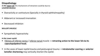
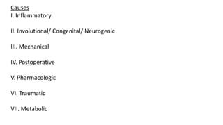
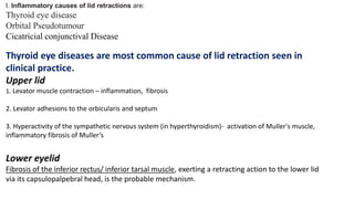
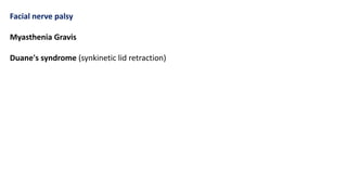
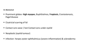
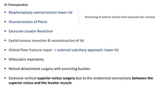
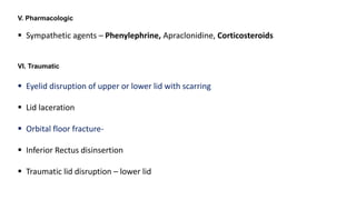
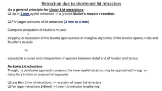
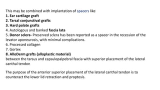
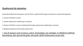
SYMPOSIUM ON LID RETRACTION
Presenter- Dr. Tanvi Gupta
Moderator- Dr. Jayanta Kumar Das
Abnormally high/ low lid position in primary position of upper/ lower lid respectively that
exposes the superior/ inferior sclera
Upper lid retaction
Lower Lid Retraction
Etiopathology
In the Upper Lid, the mechanisms of retraction could be due to:
LEVATOR MUSCLE
 Overactivity or contracture (Specially in thyroid ophthalmopathy)
 Abberant or increased innervation
 Decreased inhibition
MULLER’S MUSCLE
 Sympathetic hyperactivity
In the Lower eyelid
 Fibrosis of inferior rectus / inferior tarsal muscle -> retracting action to the lower lid via its
capsulopalpebral head.
 In the cases of lower eyelid trauma and postsurgical trauma -> intralamellar scarring or anterior
lamellar shortening may vertically shorten the eyelid.
Causes
I. Inflammatory
II. Involutional/ Congenital/ Neurogenic
III. Mechanical
IV. Postoperative
V. Pharmacologic
VI. Traumatic
VII. Metabolic
I. Inflammatory causes of lid retractions are:
Thyroid eye disease
Orbital Pseudotumour
Cicatricial conjunctival Disease
Thyroid eye diseases are most common cause of lid retraction seen in
clinical practice.
Upper lid
1. Levator muscle contraction – inflammation, fibrosis
2. Levator adhesions to the orbicularis and septum
3. Hyperactivity of the sympathetic nervous system (in hyperthyroidism)- activation of Muller's muscle,
inflammatory fibrosis of Muller’s
Lower eyelid
Fibrosis of the inferior rectus/ inferior tarsal muscle, exerting a retracting action to the lower lid
via its capsulopalpebral head, is the probable mechanism.
Common finding in thyroid-related eyelid retraction is lateral flare.
In this condition, the eyelid retraction is more severe laterally than medially, resulting
in an abnormal upper eyelid contour that appears to flare upward along the lateral half
of the eyelid margin.
II. Involutional/ Congenital/ Neurogenic
Congenital
typically result from neurological causes.
 Primary congenital eyelid retraction is a diagnosis of exclusion.
 Lid retraction may be due to a combination of both anterior and posterior lamellar
shortening that results from developmental abnormalities.
 May involve the upper, lower, or both eyelids.
 Congenital hyperthyroidism, aberrant innervation of the third cranial nerve, Marcus
Gunn (jaw-winking) syndrome, seventh cranial nerve palsy, and orbital tumor must be
ruled out.
Aberrant regeneration of Third Nerve
Acquired - Slow growing intra-cavernous aneurysm or meningioma can press upon the 3rd
nerve.
Congenital - Marcus gunn jaw winking phenomenon (commenest form of trigemino oculo
synkinesis)
Midbrain lesion (pretectal syndrome) –lid retraction mostly results from damage to
supranuclear posterior commissure.
Supranuclear lesion (disinhibition of levator muscle)
Nuclear lesion (+/- lid syndrome) - ipsilateral ptosis, contra lateral eyelid retraction
Sympathetic overactivity
Claude Bernard syndrome- cyclic spasm of pupil and lid retraction (associated with facial
hyperhidrosis and headache)
Anxious & psychotic pts
Facial nerve palsy
Myasthenia Gravis
Duane's syndrome (synkinetic lid retraction)
III. Mechanical
 Prominent globes- high myopes, Buphthalmos, Proptosis, Craniostenosis,
Paget'disease
 Cicatricial scarring of lid
 Contact Lens wear / lost Contact Lens under eyelid
 Neoplastic (eyelid tumour)
 Infection- herpes zoster ophthalmicus (severe inflammation) & scleroderma
IV. Postoperative
 Blepharoplasty overcorrection-lower lid
 Overcorrection of Ptosis
 Excessive Levator Resection
 Eyelid tumour resection & reconstruction of lid
 Orbital floor fracture repair -> external subciliary approach- lower lid
 Orbicularis myectomy
 Retinal detachment surgery with encircling buckles
 Excessive vertical superior rectus surgery due to the anatomical connections between the
superior rectus and the levator muscle
Shortening of anterior lamella from excessive skin removal
 Postenucleation syndrome.
V. Pharmacologic
 Sympathetic agents – Phenylephrine, Apraclonidine, Corticosteroids
VI. Traumatic
 Eyelid disruption of upper or lower lid with scarring
 Lid laceration
 Orbital floor fracture-
 Inferior Rectus disinsertion
 Traumatic lid disruption – lower lid
VII. Metabolic syndromes
Cirrhosis
Cushing syndrome
Hypokalemic periodic paralysis
Uremia)
Christopher I. Zoumalan, MD; Richard D. Lisman, MD. Evaluation and Management of Unilateral Ptosis and Avoiding
Contralateral Ptosis. December 14, 2009.
Transient
Normal infants-80% of normal infants (eye popping reflex)
Preterm infants - due to immature myelinisation of extrageniculate visual pathway
Maternal hyperthyroidism
Dorsal mesencephalic lesions (Collier's Lid retraction sign)
Multiple sclerosis (third nerve fascicle involved)
Bilateral episodic lid retraction in petitmal /myoclonic seizures
Oculogyric crisis
Voluntary/ non organic finding
Myasthenia gravis
Post tetanic facilitation of levator muscles.
HISTORY
 Ocular discomfort,
 foreign body sensation,
 photophobia,
 epiphora,
 other symptoms of exposure keratopathy- due to decreased excursion and lagophthalmos
 Alteration in appearance and disfigurement.
In thyroid-related orbitopathy, upper eyelid retraction present in primary gaze is called
Dalrymple sign.
SYMPTOMS
Palpebral fissure and levator function - measured with a millimeter ruler.
Great care should be taken to maintain the ruler perpendicular to the line of gaze, to
ensure that the patient maintains exact primary gaze, and with the examiner sitting at eye
level relative to the patient.
Patients is instructed to relax their eyebrows and eyelids during clinical examination and
measurement procedures.
The amount of eyelid retraction is assessed quantitatively in each eye individually as
fixation is maintained.
Vertical phoria and tropia are coincidentally assessed.
EXAMINATION
MANAGEMENT
INVESTIGATIONS
The presence of proptosis, lid lag, lid edema or ophthalmoplegia with the retraction
should prompt evaluation for thyroid eye disease (TED)
Laboratory testing
Thyroid Profile
Thyroid Stimulating Hormone (TSH)
Free T3
Free T4
Some patients with thyroid ophthalmopathy have no serologic evidence for thyroid
disease
Thyroid autoantibodies may be useful in testing for euthyroid - Grave’s
ophthalmopathy, Hashimoto thyroiditis
anti-TPO antibodies,
anti thyroglobulin(TG) antibodies
Orbital ultrasound or orbital computed tomography may confirm thyroid eye disease.
Autoantibodies against various eye muscles membrane antigens are detected in
96% patients of lid retractions, but may be present in 20% of normal patients.
‘64-K Da’ membrane antigens is most specific for eyelid retraction
TREATMENT
Treatment of underlying etiology
Protection of cornea Botulinum Toxin
Surgical treatment options
The treatment of lid retraction should be aimed at the underlying etiology.
Corneal exposure is the most serious complication of lid retraction.
Prior to correction of lid retraction surgically , the immediate treatment
goal is to protect the cornea.
1. Preservative-free tear supplements,
2. Lubricating ointment or gels
3. Punctal plugs
4. Moist chamber shields
5. Taping the eyelid during sleep
Temporarily treat eyelid retraction secondary to thyroid-related orbitopathy, because
of its effect on striated fibers in the levator muscle.
The effects of a single injection can last for 3 to 4 months.
Botulinum A toxin injection can be used to reduce the lid retraction in symptomatic
cases.
SURGICAL TREATMENT
Retraction can be with-
Shortage of skin
Shortage of Conjunctiva
No shortage of skin or conjunctiva
1. Shortage of skin- Ectropion, retraction of lid-> Skin Graft, Z plasty
2. Cicatricial changes in Conjunctiva- mostly with entropion- lengthening of
posterior lamella
3. No shortage of skin or conjunctiva
Retraction due to shortened lid retractors
(i) Muller’s muscle excision (upto 2 mm)
(ii) Muller’s + Levator aponeurosis recession/ myotomy (upto 3 mm)
(iii) Muller’s + Levator aponeurosis + Spacers (4 mm or more)
As a general principle for Upper Lid retractions:
 Up to 3 mm eyelid retraction -> a graded Muller's muscle resection.
 For larger amounts of lid retraction (3 mm to 4 mm)
Complete extirpation of Muller's muscle
+
stripping or recession of the levator aponeurosis or marginal myotomy of the levator aponeurosis and
Mueller's muscle
+/-
adjustable sutures and interposition of spacers between distal end of levator and tarsus
For Lower Lid retractions
Though, no conclusive approach is present, the lower eyelid retractors may be approached through an
infraciliary incision or conjunctival approach.
 Less than 2mm of retractions, -> recession of Lower Lid retractors
 For larger retractions (>2mm) -> Lower Lid retractor lengthening
Retraction due to shortened lid retractors
This may be combined with implantation of spacers like
1. Ear cartilage graft
2. Tarsal conjunctival grafts
3. Hard palate grafts
4. Autologous and banked fascia lata
5. Donor sclera- Preserved sclera has been reported as a spacer in the recession of the
levator aponeurosis, with minimal complications.
6. Processed collagen
7. Gortex
8. AlloDerm grafts (alloplastic material)
between the tarsus and capsulopalpebral fascia with superior placement of the lateral
canthal tendon
The purpose of the anterior superior placement of the lateral canthal tendon is to
counteract the lower lid retraction and proptosis.
Other supplemented options may be:
Mid face lift
Augmentation of bony support below the eye
Dysthyroid lid retraction
Classically divided into five groups, each of which is performed through an external or conjunctival approach:
1. Excision of Muller's muscle
2. Levator aponeurosis weakening or recession
3. Excision of Muller's muscle combined with levator aponeurosis weakening or recession
4. Marginal myotomy of the levator aponeurosis
5. Use of spacers such as tarsus, sclera, hard palate, ear cartilage, or AlloDerm (LifeCell,
Branchburg, NJ), dermal fat graft, skin graft, SOOF (Subocularis ocular fat).
For retractions with other etiologies, the general surgical principles followed are:
 In traumatic scarring -> excision of scar, and closure of healthy tissue
 Tissue transfer - Z plasty or VY plasty
 In ant lamellar shortening- > release the scar and place full thickness skin graft
 For facial nerve palsy - Mullers muscle excision and lateral tarsorrhaphy
In greater degree of Lid Retraction- lid loading with gold weight, palpebral spring,
temporalis muscle transfer, or levator recession.
A graded recession of Levator-Muller muscle complex, more temporal & central than nasal;
leaving the orbital septum intact, severing all the fibrotic bands between Muller’s muscle
and conjunctiva- gives satisfactory outcome.
REFERENCES
1. DELHI OPHTHALMOLOGICAL SOCIETY –LID RETRACTION-> Lohia DOMS , Sandhya
Makhija MNAMS, FRS, DO, P.A. Lamba MS
2. Collin’s Colour Atlas of Ophthalmic Plastic Surgery
3.THE GRADED LEVATOR HINGE PROCEDURE FOR THE CORRECTION OF UPPER EYELID
RETRACTION (AN AMERICAN OPHTHALMOLOGICAL SOCIETY THESIS). BY Daniel P.
Schaefer MD. Trans Am Ophthalmol Soc 2007;105:481-512
4. Zoumalan, MD; and Richard D. Lisman, MD. Evaluation and Management of Unilateral
Ptosis and Avoiding Contralateral Ptosis. Christopher I. The American Society for
Aesthetic Plastic Surgery
5. Google images
THANK YOU

Lid retraction

  • 1.
    SYMPOSIUM ON LIDRETRACTION Presenter- Dr. Tanvi Gupta Moderator- Dr. Jayanta Kumar Das
  • 2.
    Abnormally high/ lowlid position in primary position of upper/ lower lid respectively that exposes the superior/ inferior sclera Upper lid retaction Lower Lid Retraction
  • 3.
    Etiopathology In the UpperLid, the mechanisms of retraction could be due to: LEVATOR MUSCLE  Overactivity or contracture (Specially in thyroid ophthalmopathy)  Abberant or increased innervation  Decreased inhibition MULLER’S MUSCLE  Sympathetic hyperactivity In the Lower eyelid  Fibrosis of inferior rectus / inferior tarsal muscle -> retracting action to the lower lid via its capsulopalpebral head.  In the cases of lower eyelid trauma and postsurgical trauma -> intralamellar scarring or anterior lamellar shortening may vertically shorten the eyelid.
  • 4.
    Causes I. Inflammatory II. Involutional/Congenital/ Neurogenic III. Mechanical IV. Postoperative V. Pharmacologic VI. Traumatic VII. Metabolic
  • 5.
    I. Inflammatory causesof lid retractions are: Thyroid eye disease Orbital Pseudotumour Cicatricial conjunctival Disease Thyroid eye diseases are most common cause of lid retraction seen in clinical practice. Upper lid 1. Levator muscle contraction – inflammation, fibrosis 2. Levator adhesions to the orbicularis and septum 3. Hyperactivity of the sympathetic nervous system (in hyperthyroidism)- activation of Muller's muscle, inflammatory fibrosis of Muller’s Lower eyelid Fibrosis of the inferior rectus/ inferior tarsal muscle, exerting a retracting action to the lower lid via its capsulopalpebral head, is the probable mechanism.
  • 6.
    Common finding inthyroid-related eyelid retraction is lateral flare. In this condition, the eyelid retraction is more severe laterally than medially, resulting in an abnormal upper eyelid contour that appears to flare upward along the lateral half of the eyelid margin.
  • 7.
    II. Involutional/ Congenital/Neurogenic Congenital typically result from neurological causes.  Primary congenital eyelid retraction is a diagnosis of exclusion.  Lid retraction may be due to a combination of both anterior and posterior lamellar shortening that results from developmental abnormalities.  May involve the upper, lower, or both eyelids.  Congenital hyperthyroidism, aberrant innervation of the third cranial nerve, Marcus Gunn (jaw-winking) syndrome, seventh cranial nerve palsy, and orbital tumor must be ruled out.
  • 8.
    Aberrant regeneration ofThird Nerve Acquired - Slow growing intra-cavernous aneurysm or meningioma can press upon the 3rd nerve. Congenital - Marcus gunn jaw winking phenomenon (commenest form of trigemino oculo synkinesis) Midbrain lesion (pretectal syndrome) –lid retraction mostly results from damage to supranuclear posterior commissure. Supranuclear lesion (disinhibition of levator muscle) Nuclear lesion (+/- lid syndrome) - ipsilateral ptosis, contra lateral eyelid retraction Sympathetic overactivity Claude Bernard syndrome- cyclic spasm of pupil and lid retraction (associated with facial hyperhidrosis and headache) Anxious & psychotic pts
  • 9.
    Facial nerve palsy MyastheniaGravis Duane's syndrome (synkinetic lid retraction)
  • 10.
    III. Mechanical  Prominentglobes- high myopes, Buphthalmos, Proptosis, Craniostenosis, Paget'disease  Cicatricial scarring of lid  Contact Lens wear / lost Contact Lens under eyelid  Neoplastic (eyelid tumour)  Infection- herpes zoster ophthalmicus (severe inflammation) & scleroderma
  • 11.
    IV. Postoperative  Blepharoplastyovercorrection-lower lid  Overcorrection of Ptosis  Excessive Levator Resection  Eyelid tumour resection & reconstruction of lid  Orbital floor fracture repair -> external subciliary approach- lower lid  Orbicularis myectomy  Retinal detachment surgery with encircling buckles  Excessive vertical superior rectus surgery due to the anatomical connections between the superior rectus and the levator muscle Shortening of anterior lamella from excessive skin removal
  • 12.
  • 13.
    V. Pharmacologic  Sympatheticagents – Phenylephrine, Apraclonidine, Corticosteroids VI. Traumatic  Eyelid disruption of upper or lower lid with scarring  Lid laceration  Orbital floor fracture-  Inferior Rectus disinsertion  Traumatic lid disruption – lower lid
  • 14.
    VII. Metabolic syndromes Cirrhosis Cushingsyndrome Hypokalemic periodic paralysis Uremia)
  • 15.
    Christopher I. Zoumalan,MD; Richard D. Lisman, MD. Evaluation and Management of Unilateral Ptosis and Avoiding Contralateral Ptosis. December 14, 2009.
  • 16.
    Transient Normal infants-80% ofnormal infants (eye popping reflex) Preterm infants - due to immature myelinisation of extrageniculate visual pathway Maternal hyperthyroidism Dorsal mesencephalic lesions (Collier's Lid retraction sign) Multiple sclerosis (third nerve fascicle involved) Bilateral episodic lid retraction in petitmal /myoclonic seizures Oculogyric crisis
  • 17.
    Voluntary/ non organicfinding Myasthenia gravis Post tetanic facilitation of levator muscles.
  • 18.
  • 19.
     Ocular discomfort, foreign body sensation,  photophobia,  epiphora,  other symptoms of exposure keratopathy- due to decreased excursion and lagophthalmos  Alteration in appearance and disfigurement. In thyroid-related orbitopathy, upper eyelid retraction present in primary gaze is called Dalrymple sign. SYMPTOMS
  • 20.
    Palpebral fissure andlevator function - measured with a millimeter ruler. Great care should be taken to maintain the ruler perpendicular to the line of gaze, to ensure that the patient maintains exact primary gaze, and with the examiner sitting at eye level relative to the patient. Patients is instructed to relax their eyebrows and eyelids during clinical examination and measurement procedures. The amount of eyelid retraction is assessed quantitatively in each eye individually as fixation is maintained. Vertical phoria and tropia are coincidentally assessed. EXAMINATION
  • 21.
  • 22.
  • 23.
    The presence ofproptosis, lid lag, lid edema or ophthalmoplegia with the retraction should prompt evaluation for thyroid eye disease (TED) Laboratory testing Thyroid Profile Thyroid Stimulating Hormone (TSH) Free T3 Free T4 Some patients with thyroid ophthalmopathy have no serologic evidence for thyroid disease Thyroid autoantibodies may be useful in testing for euthyroid - Grave’s ophthalmopathy, Hashimoto thyroiditis anti-TPO antibodies, anti thyroglobulin(TG) antibodies Orbital ultrasound or orbital computed tomography may confirm thyroid eye disease.
  • 24.
    Autoantibodies against variouseye muscles membrane antigens are detected in 96% patients of lid retractions, but may be present in 20% of normal patients. ‘64-K Da’ membrane antigens is most specific for eyelid retraction
  • 25.
    TREATMENT Treatment of underlyingetiology Protection of cornea Botulinum Toxin Surgical treatment options
  • 26.
    The treatment oflid retraction should be aimed at the underlying etiology.
  • 27.
    Corneal exposure isthe most serious complication of lid retraction. Prior to correction of lid retraction surgically , the immediate treatment goal is to protect the cornea. 1. Preservative-free tear supplements, 2. Lubricating ointment or gels 3. Punctal plugs 4. Moist chamber shields 5. Taping the eyelid during sleep
  • 28.
    Temporarily treat eyelidretraction secondary to thyroid-related orbitopathy, because of its effect on striated fibers in the levator muscle. The effects of a single injection can last for 3 to 4 months. Botulinum A toxin injection can be used to reduce the lid retraction in symptomatic cases.
  • 29.
  • 30.
    Retraction can bewith- Shortage of skin Shortage of Conjunctiva No shortage of skin or conjunctiva 1. Shortage of skin- Ectropion, retraction of lid-> Skin Graft, Z plasty 2. Cicatricial changes in Conjunctiva- mostly with entropion- lengthening of posterior lamella 3. No shortage of skin or conjunctiva Retraction due to shortened lid retractors (i) Muller’s muscle excision (upto 2 mm) (ii) Muller’s + Levator aponeurosis recession/ myotomy (upto 3 mm) (iii) Muller’s + Levator aponeurosis + Spacers (4 mm or more)
  • 31.
    As a generalprinciple for Upper Lid retractions:  Up to 3 mm eyelid retraction -> a graded Muller's muscle resection.  For larger amounts of lid retraction (3 mm to 4 mm) Complete extirpation of Muller's muscle + stripping or recession of the levator aponeurosis or marginal myotomy of the levator aponeurosis and Mueller's muscle +/- adjustable sutures and interposition of spacers between distal end of levator and tarsus For Lower Lid retractions Though, no conclusive approach is present, the lower eyelid retractors may be approached through an infraciliary incision or conjunctival approach.  Less than 2mm of retractions, -> recession of Lower Lid retractors  For larger retractions (>2mm) -> Lower Lid retractor lengthening Retraction due to shortened lid retractors
  • 32.
    This may becombined with implantation of spacers like 1. Ear cartilage graft 2. Tarsal conjunctival grafts 3. Hard palate grafts 4. Autologous and banked fascia lata 5. Donor sclera- Preserved sclera has been reported as a spacer in the recession of the levator aponeurosis, with minimal complications. 6. Processed collagen 7. Gortex 8. AlloDerm grafts (alloplastic material) between the tarsus and capsulopalpebral fascia with superior placement of the lateral canthal tendon The purpose of the anterior superior placement of the lateral canthal tendon is to counteract the lower lid retraction and proptosis.
  • 33.
    Other supplemented optionsmay be: Mid face lift Augmentation of bony support below the eye
  • 34.
    Dysthyroid lid retraction Classicallydivided into five groups, each of which is performed through an external or conjunctival approach: 1. Excision of Muller's muscle 2. Levator aponeurosis weakening or recession 3. Excision of Muller's muscle combined with levator aponeurosis weakening or recession 4. Marginal myotomy of the levator aponeurosis 5. Use of spacers such as tarsus, sclera, hard palate, ear cartilage, or AlloDerm (LifeCell, Branchburg, NJ), dermal fat graft, skin graft, SOOF (Subocularis ocular fat).
  • 35.
    For retractions withother etiologies, the general surgical principles followed are:  In traumatic scarring -> excision of scar, and closure of healthy tissue  Tissue transfer - Z plasty or VY plasty
  • 37.
     In antlamellar shortening- > release the scar and place full thickness skin graft  For facial nerve palsy - Mullers muscle excision and lateral tarsorrhaphy In greater degree of Lid Retraction- lid loading with gold weight, palpebral spring, temporalis muscle transfer, or levator recession. A graded recession of Levator-Muller muscle complex, more temporal & central than nasal; leaving the orbital septum intact, severing all the fibrotic bands between Muller’s muscle and conjunctiva- gives satisfactory outcome.
  • 38.
    REFERENCES 1. DELHI OPHTHALMOLOGICALSOCIETY –LID RETRACTION-> Lohia DOMS , Sandhya Makhija MNAMS, FRS, DO, P.A. Lamba MS 2. Collin’s Colour Atlas of Ophthalmic Plastic Surgery 3.THE GRADED LEVATOR HINGE PROCEDURE FOR THE CORRECTION OF UPPER EYELID RETRACTION (AN AMERICAN OPHTHALMOLOGICAL SOCIETY THESIS). BY Daniel P. Schaefer MD. Trans Am Ophthalmol Soc 2007;105:481-512 4. Zoumalan, MD; and Richard D. Lisman, MD. Evaluation and Management of Unilateral Ptosis and Avoiding Contralateral Ptosis. Christopher I. The American Society for Aesthetic Plastic Surgery 5. Google images
  • 39.

Editor's Notes

  • #6 PROPTOSIS These include inflammatory fibrosis of Müller’s muscle, abnormal sympathetic tone in Müller’s muscle, proptosis, contracture of the inferior rectus muscle with superior rectus hyperactivity (fixation duress), and overmedication with thyroid replacement
  • #14 With inferior rectus restriction (thyroid, trauma), any effort to maintain vertical eye alignment results in overaction of the superior rectus muscle, and because the levator muscle is linked to the superior rectus muscle action, increased innervation of the levator muscle will also occur secondarily.  If upper eyelid retraction increases on attempted upgaze and resolves on downgaze, tight inferior rectus muscles are probably contributing to the upper eyelid retraction and should be surgically recessed first, before surgical correction of the eyelid retraction is entertained.
  • #16 Unmasking Pseudoretraction- Hering’s test Manual elevation of ptotic eye- > measurement of MRD this permits the patient to continue fixation with the eye but removes the need for excessive innervation to both eyelids in an attempt to reduce the ptosis. As a result, the contralateral lid returns to its normal position after several seconds. Once the manually elevated lid is then released, the afferent input is once again increased to both eyelids and results in an increase in the lid position of the contralateral eye.
  • #20 Lagophthalmos- inability to close eyelids completely abnormally high upper eyelid in downgaze is called the von Graefe sign or lid lag
  • #24 TPO - thyroperoxidase