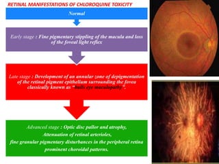
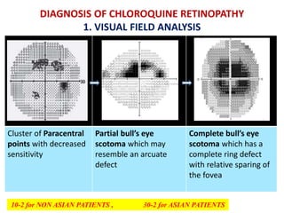
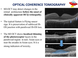
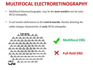
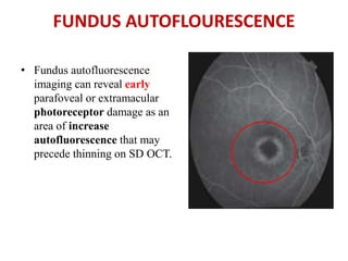
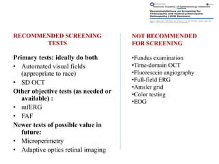
CHLOROQUINE RETINOPATHY
INTRODUCTION
• Chloroquine and hydroxychloroquine are the most
commonly antimalarials. They also used for the treatment
of many autoimmune and connective tissue diseases .
• Retinal toxicity from chloroquine (CQ) and its analogue
hydroxychloroquine (HCQ) has been recognized for many
years1
• Thus, it is important for ophthalmologists and other
physicians to understand the prevalence and risk factors for
retinopathy
PATHOPHYSIOLOGY1
• The mechanism of CQ and HCQ toxicity is not well
understood
• Binding to melanin in the RPE may serve to concentrate the
drugs and contribute to toxic effects.
• The primary damage is seen in photoreceptors and outer
nuclear layer and there is also disruption of the RPE.
• No anatomic features of the retina and RPE are known to
correlate with the parafoveal or extramacular patterns of
damage.
STATISTICAL RISK OF TOXICITY1
• Earlier literature shows that, the prevalence of CQ or HCQ
retinopathy seen only in few patients on long-term therapy and
only recognized in severe toxicity stage. (bull’s-eye changes).
• But now the prevalence of hydroxychloroquine retinopathy
ranges upto 7.5%.1,3
• However, some studies estimate that the percentage of toxic
maculopathy reaches up to 13.1%.5
• Daily dose was the most critical determinant of risk
• Particularly, Very thin patients are at increased risk when dose
is calculated by ideal weight
Advanced stage : Optic disc pallor and atrophy,
Attenuation of retinal arterioles,
fine granular pigmentary disturbances in the peripheral retina
prominent choroidal patterns.
Late stage : Development of an annular zone of depigmentation
of the retinal pigment epithelium surrounding the fovea
classically known as “bulls eye maculopathy”.
Early stage : Fine pigmentary stippling of the macula and loss
of the foveal light reflex
Normal
RETINAL MANIFESTATIONS OF CHLOROQUINE TOXICITY
DIAGNOSIS OF CHLOROQUINE RETINOPATHY
1. VISUAL FIELD ANALYSIS
Cluster of Paracentral
points with decreased
sensitivity
Partial bull’s eye
scotoma which may
resemble an arcuate
defect
Complete bull’s eye
scotoma which has a
complete ring defect
with relative sparing of
the fovea
10-2 for NON ASIAN PATIENTS , 30-2 for ASIAN PATIENTS
OPTICAL COHERENCE TOMOGRAPHY
• SD-OCT may detect changes in the
retinal architecture before the onset of
clinically apparent HCQ retinopathy.
• The typical feature is Flying saucer
sign. It is preservation of subfoveal IS-
OS junction with parafoveal IS-OS loss.
• The SD OCT shows localized thinning
of the photoreceptor layers in the
parafoveal region in non- Asian eyes or
near the arcades in Asian eyes. It is a
strong indicators of toxicity.
MULTIFOCAL ELECTRORETINOGRAPHY
• Multifocal Electroretinography may be the most sensitive test for early
HCQ retinopathy.
• It can localize deficiencies to the central macula, thereby detecting the
subtle changes characteristic of early HCQ retinopathy.
Multifocal ERG
Full field ERG
FUNDUS AUTOFLOURESCENCE
• Fundus autofluorescence
imaging can reveal early
parafoveal or extramacular
photoreceptor damage as an
area of increase
autofluorescence that may
precede thinning on SD OCT.
SCREENING FREQUENCY
Baseline Screening
• Fundus examination within first year of use
• Add visual fields and SD OCT if maculopathy is present
Annual Screening
• Begin after 5 yrs of use
• Sooner in the presence of major risk factors
MAJOR RISK FACTOR FOR TOXIC RETINOPATHY
2016 Guidelines
MINOR RISK FACTOR – AGE , LIVER DISEASE , GENETIC FACTOR
RECOMMENDED SCREENING
TESTS
Primary tests: ideally do both
• Automated visual fields
(appropriate to race)
• SD OCT
Other objective tests (as needed or
available) :
• mfERG
• FAF
Newer tests of possible value in
future:
• Microperimetry
• Adaptive optics retinal imaging
NOT RECOMMENDED
FOR SCREENING
•Fundus examination
•Time-domain OCT
•Fluorescein angiography
•Full-field ERG
•Amsler grid
•Color testing
•EOG
• This presentation is to emphasis the importance of screening and
newer AAO Guideliness regarding chloroquine retinopathy
• Although “Bulls eye maculopathy” is a classical feature , it is seen in
later stage. i.e. the time at which bulls eye maculopathy is seen, the
progression of disease is irreversible even after stoppage of drug.
• Similarly , initial photoreceptor damage in the classic parafoveal
distribution is common only in European population. But, most patients
of Asian population will show initial damage in a more peripheral
extramacular distribution i.e. near the vascular arcades
• Hence the screening tests are recommended which will detect
Chloroquine toxicity long before RPE damage is visible by imaging or
fundus examination.
REFERENCE
1. Marmor MF, Kellner U, Lai TY, Melles RB, Mieler WF; American Academy of
Ophthalmology. Ophthalmology. 2016 Jun;123(6):1386-94. doi:
10.1016/j.ophtha.2016.01.058. Epub 2016
2. Marmor MF, Kellner U, Lai TY, et al. Revised recommendations
on screening for chloroquine and hydroxychloroquine
retinopathy. Ophthalmology 2011;118:415–22.
3. Melles RB, Marmor MF. JAMA Ophthalmol. 2014 Dec;132(12):1453-60. doi:
10.1001/jamaophthalmol.2014.3459. Erratum in: JAMA Ophthalmol. 2014
Dec;132(12):1493.
4. Michaelides M, Stover NB, Francis PJ, Weleber RG. Arch Ophthalmol. 2011
Jan;129(1):30-9. doi: 10.1001/archophthalmol.2010.321.
5. Palma Sánchez D, Rubio Velazquez E, Soro Marín S, Reyes García R. Retinal
toxicity due to antimalarials: frequency and risk factors. Reumatol Clin.
THANK YOU

Chloroquine retinopathy

  • 1.
  • 2.
    INTRODUCTION • Chloroquine andhydroxychloroquine are the most commonly antimalarials. They also used for the treatment of many autoimmune and connective tissue diseases . • Retinal toxicity from chloroquine (CQ) and its analogue hydroxychloroquine (HCQ) has been recognized for many years1 • Thus, it is important for ophthalmologists and other physicians to understand the prevalence and risk factors for retinopathy
  • 3.
    PATHOPHYSIOLOGY1 • The mechanismof CQ and HCQ toxicity is not well understood • Binding to melanin in the RPE may serve to concentrate the drugs and contribute to toxic effects. • The primary damage is seen in photoreceptors and outer nuclear layer and there is also disruption of the RPE. • No anatomic features of the retina and RPE are known to correlate with the parafoveal or extramacular patterns of damage.
  • 4.
    STATISTICAL RISK OFTOXICITY1 • Earlier literature shows that, the prevalence of CQ or HCQ retinopathy seen only in few patients on long-term therapy and only recognized in severe toxicity stage. (bull’s-eye changes). • But now the prevalence of hydroxychloroquine retinopathy ranges upto 7.5%.1,3 • However, some studies estimate that the percentage of toxic maculopathy reaches up to 13.1%.5 • Daily dose was the most critical determinant of risk • Particularly, Very thin patients are at increased risk when dose is calculated by ideal weight
  • 5.
    Advanced stage :Optic disc pallor and atrophy, Attenuation of retinal arterioles, fine granular pigmentary disturbances in the peripheral retina prominent choroidal patterns. Late stage : Development of an annular zone of depigmentation of the retinal pigment epithelium surrounding the fovea classically known as “bulls eye maculopathy”. Early stage : Fine pigmentary stippling of the macula and loss of the foveal light reflex Normal RETINAL MANIFESTATIONS OF CHLOROQUINE TOXICITY
  • 6.
    DIAGNOSIS OF CHLOROQUINERETINOPATHY 1. VISUAL FIELD ANALYSIS Cluster of Paracentral points with decreased sensitivity Partial bull’s eye scotoma which may resemble an arcuate defect Complete bull’s eye scotoma which has a complete ring defect with relative sparing of the fovea 10-2 for NON ASIAN PATIENTS , 30-2 for ASIAN PATIENTS
  • 7.
    OPTICAL COHERENCE TOMOGRAPHY •SD-OCT may detect changes in the retinal architecture before the onset of clinically apparent HCQ retinopathy. • The typical feature is Flying saucer sign. It is preservation of subfoveal IS- OS junction with parafoveal IS-OS loss. • The SD OCT shows localized thinning of the photoreceptor layers in the parafoveal region in non- Asian eyes or near the arcades in Asian eyes. It is a strong indicators of toxicity.
  • 8.
    MULTIFOCAL ELECTRORETINOGRAPHY • MultifocalElectroretinography may be the most sensitive test for early HCQ retinopathy. • It can localize deficiencies to the central macula, thereby detecting the subtle changes characteristic of early HCQ retinopathy. Multifocal ERG Full field ERG
  • 9.
    FUNDUS AUTOFLOURESCENCE • Fundusautofluorescence imaging can reveal early parafoveal or extramacular photoreceptor damage as an area of increase autofluorescence that may precede thinning on SD OCT.
  • 11.
    SCREENING FREQUENCY Baseline Screening •Fundus examination within first year of use • Add visual fields and SD OCT if maculopathy is present Annual Screening • Begin after 5 yrs of use • Sooner in the presence of major risk factors
  • 12.
    MAJOR RISK FACTORFOR TOXIC RETINOPATHY 2016 Guidelines MINOR RISK FACTOR – AGE , LIVER DISEASE , GENETIC FACTOR
  • 14.
    RECOMMENDED SCREENING TESTS Primary tests:ideally do both • Automated visual fields (appropriate to race) • SD OCT Other objective tests (as needed or available) : • mfERG • FAF Newer tests of possible value in future: • Microperimetry • Adaptive optics retinal imaging NOT RECOMMENDED FOR SCREENING •Fundus examination •Time-domain OCT •Fluorescein angiography •Full-field ERG •Amsler grid •Color testing •EOG
  • 15.
    • This presentationis to emphasis the importance of screening and newer AAO Guideliness regarding chloroquine retinopathy • Although “Bulls eye maculopathy” is a classical feature , it is seen in later stage. i.e. the time at which bulls eye maculopathy is seen, the progression of disease is irreversible even after stoppage of drug. • Similarly , initial photoreceptor damage in the classic parafoveal distribution is common only in European population. But, most patients of Asian population will show initial damage in a more peripheral extramacular distribution i.e. near the vascular arcades • Hence the screening tests are recommended which will detect Chloroquine toxicity long before RPE damage is visible by imaging or fundus examination.
  • 16.
    REFERENCE 1. Marmor MF,Kellner U, Lai TY, Melles RB, Mieler WF; American Academy of Ophthalmology. Ophthalmology. 2016 Jun;123(6):1386-94. doi: 10.1016/j.ophtha.2016.01.058. Epub 2016 2. Marmor MF, Kellner U, Lai TY, et al. Revised recommendations on screening for chloroquine and hydroxychloroquine retinopathy. Ophthalmology 2011;118:415–22. 3. Melles RB, Marmor MF. JAMA Ophthalmol. 2014 Dec;132(12):1453-60. doi: 10.1001/jamaophthalmol.2014.3459. Erratum in: JAMA Ophthalmol. 2014 Dec;132(12):1493. 4. Michaelides M, Stover NB, Francis PJ, Weleber RG. Arch Ophthalmol. 2011 Jan;129(1):30-9. doi: 10.1001/archophthalmol.2010.321. 5. Palma Sánchez D, Rubio Velazquez E, Soro Marín S, Reyes García R. Retinal toxicity due to antimalarials: frequency and risk factors. Reumatol Clin.
  • 17.

Editor's Notes

  • #7 Whereas the 10-2 field pattern is excellent for testing the Non- Asian patients, 30-2 fields must be done for Asian patients who often present with a more pericentral defect rather than parafoveal (
  • #12 interference with the interpretation of screening tests