
Recommended
More Related Content
What's hot
What's hot (20)
Similar to Foreign body & trauma to the eye
Similar to Foreign body & trauma to the eye (20)
Recently uploaded
Recently uploaded (20)
Foreign body & trauma to the eye
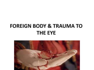
- 1. FOREIGN BODY & TRAUMA TO THE EYE
- 2. A foreign body in the eye is something that enters the eye from outside the body. Foreign body in the Eye
- 3. Eye Trauma Refers to any injury to the eye. The injury may be due to mechanical trauma (blunt or penetrating), chemical agents, or radiation (ultraviolet or ionizing).
- 4. The Birmingham Eye Trauma (BETTS)- An international society of ocular trauma classification of eye injuries
- 5. Closed globe injury No full- thickness wound of eye wall Open globe injury Full- thickness wound of the eye wall. Contusion Injury due to direct force by a blunt object or fist Lamellar laceration Partial- thickness wound of the eye wall caused by a sharp object
- 6. Rupture Injury of the globe with tear’s in the ocular layers usually the sclera, caused by a blunt object. Laceration or perforation Cutting or penetration of tissue may occur in eye lid, conjunctiva, cornea, sclera or globe Penetrating injury It is due to glass, metal, wood, knife, stick or other large objects. Intra ocular foreign bodies Foreign body on the surface of the cornea can cause eye injury, which include particles of glass, wood and metals
- 7. Orbital fractures Fracture and dislocation of walls of orbit due to trauma. Burns by Chemicals Burns caused by alkali or acids. Burns by Thermal Burns to the eye due to fire or intense heat Burns by Ultra violet rays Ultraviolet rays from sunlight, welding machine, germicidal lamp, lasers, etc.
- 8. ETIOLOGY •Ocular injuries are due to automobile accidents, assaults, falls, sports and games or work related accidents. •Blunt injury may due to hit by a fist or ball or any other blunt objects •Foreign body due to flying metal parts, sand, dust or wood chips. • Laceration or perforation is due to scratch by finger nails, dog bite, by knife, pen , pencil or eye cosmetics. •Rupture may be due to bullet or sharp penetrating objects • Burns are due to strong cleansing agents or acids • thermal burns are due to fire, high pressure steam, sunlight, snow blindness or from welding equipments.
- 9. Globe rupture occurs when there is a defect in the cornea, sclera, or both structures. When a blunt force is applied to the eye, the intraocular pressure can increase enough to rupture the sclera. (normal IOP 12-22 mmHg) Hematoma occurs when there is an accumulation of blood. Blood collects behind the eye, there is increased intraocular pressure, which can subsequently cause stretching of the optic nerve and decreased ocular perfusion can lead to permanent blindness. Acid causes coagulation in the cornea and prevents further penetration Alkaline substance penetrates the corneal epithelium and cause corneal necrosis and perforation PATHOPHYSIOLOGY
- 10. SIGNS AND SYMPTOMS •Pain • Photophobia •Redness •Swelling •Tearing •Blood in the anterior chamber •Echymosis •Absence of eye movements •Visible foreign body •Prolapsed globe •Abnormal or decreased vision •Abnormal intra ocular pressure •Burned skin with blisters •Rhinorhea •Contusion •Diplopia •Headache
- 11. Management •History collection is important because of future legal problems • Eye injury is usually associated with head injury, so stabilize the general medical condition • Ensure Airway, Breathing and Circulation •Determine mechanism of injury •Assess for chemical exposure •Immediate eye irrigation •Cover the eye •Elevate head of bed 45 degree •Transport to hospital
- 12. Chemical Burns • Tap water/ saline irrigation • instill local anesthetic drops (lidocaine) • Remove small visible coagulated matter with moistened cotton tips •Irrigation continue until reach to hospital and the conjunctival pH normalizes(7.3-7.6) •Litmus paper may be used to detect conjunctival pH. •Instill antibiotics and cover the eye with sterile dressing / eye pads •Surgical restoration of ocular surface through grafting
- 13. Burns by heat •Wash with plain water or saline •Transport to hospital •Instill antibiotic drops •Apply sterile dressing •Analgesics •Skin grafting
- 14. Mechanical trauma *CT scan or MRI •Suturing •Antibiotics •Analgesics •Cycloplegic eye drops (Atropine) •Cold compress •Sterile dressing/ eye pads
- 15. Foreign Bodies •Irrigate with plain water/ normal saline •Instill anesthetic drops •Stain with fluorescent stain (orange dye) •Antibiotic ointment •Remove FB with cotton tip applicator •Metal FB can be removed with magnet •Surgical removal •Cover the eye with eye protector
- 16. Penetrating injury/ laceration/ruptured globe *Do not try to remove object *Place protection shield *Prevent eye movement *X-Ray, CT Scan *Surgical repair *Enucleation ( complete removal of eyeball) *Antibiotics *Analgesics *Steroids
- 17. Nursing Aseesment *History of present injuries *Assess signs and symptoms *Assess level of discomfort /pain *Assess vital signs *Neurological assessment *Assess visual acuity
- 18. Nursing Diagnosis *Acute pain related to the injury/inflammation or surgical procedures *Fear and anxiety related to visual impairment and loss of autonomy *Disturbed sensory perception related to ocular trauma, inflammation or infection *Knowledge deficit related to the limited ability to participate in diversional and social activities secondary to impaired vision
- 19. Nursing Intervention •History collection *Reassurance *Continuous eye irrigation •Pre and post operative care •Medications as per order •Monitor vital signs •Eye patching or shielding •Maintain safe environment •Provide psychological support •Community education to prevent or reduce incidence of ocular trauma
- 20. Health Education •Medication administration •Use of patch or shield •Teach about the surgical management •Teach signs and symptoms of infection •Importance of follow up care •Attempt to prevent future trauma