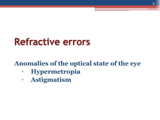
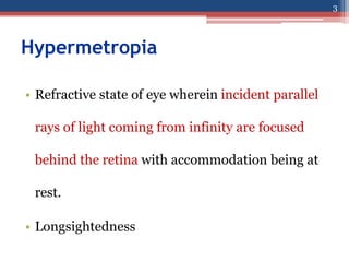
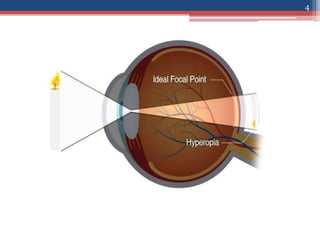
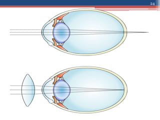
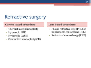
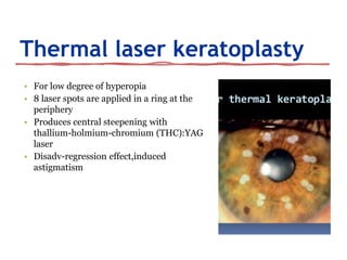
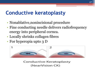
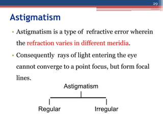
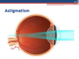
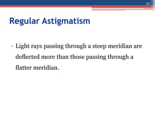
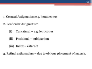
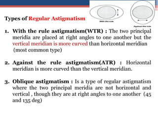
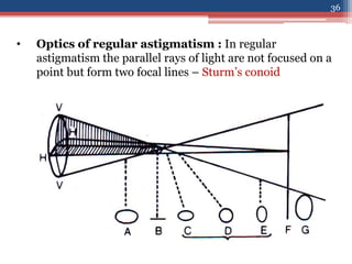
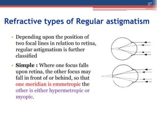
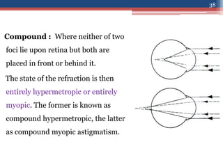
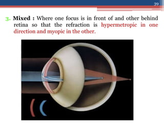
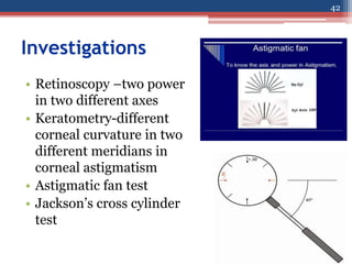
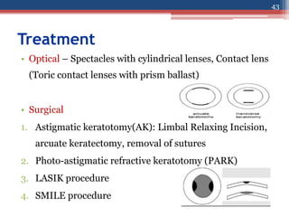
Anomalies of the optical state of the eye
• Hypermetropia
• Astigmatism
2
Hypermetropia
• Refractive state of eye wherein incident parallel
rays of light coming from infinity are focused
behind the retina with accommodation being at
rest.
• Longsightedness
3
4
• Near images can be blurred unless there is
sufficient accommodation, as in a child.
• They have blurred images for distant objects also
• Most children are born about +3 D hyperopic,
but this usually resolves by age 12 years.
5
Types
|
| | | | |
Axial Curvature Index Positional Absence of lens
• Axial is the commonest form.
• In this condition the total refractive power of eye is
normal but there is axial shortening of eye ball.
6
Axial
• Each millimeter of shortening represents approximately
3D of refractive change and thus a hypermetropia of over
6D is uncommon.
• Physiological: Infant, child.
• Pathological: Orbital tumour,micro-
ophthalmos,nanophthalmos or inflammatory mass may
indent the posterior pole of the eye and flatten it
7
• Curvature Hypermetropia : When the radius of
curvature of any of the refracting surfaces is increased,
• congenitally (cornea plana) or as a result of trauma
• Increase of 1 mm produces a hypermetropia of 6 D.
8
• Index Hypermetropia : Usually manifests
itself as a decrease in the effective refractivity of
the lens and is responsible for the
hypermetropia which occurs physiologically in
old age d/t cortical sclerosis and pathologically
9
• Positional Hypermetropia : Posterior placed lens
also produced hypermetropia whether it occurs as a
congenital anomaly or as a result of trauma and disease.
• Aphakia : Surgical, posterior dislocation of lens
10
Clinical Types:
• Simple Hypermetropia : Commonest form.
• It results from normal biological variations in the
development of eye e.g., axial and curvatural.
• Pathological Hypermetropia : Either congenital or
acquired conditions of eyeball which are outside the normal
biological variation of development
• Example: index , positional (Aphakia).
• Functional Hypermetropia : Results from paralysis of
accommodation as seen in patients with third nerve palsy
and internal ophthalmoplegia.
11
Components of hypermetropia
Total hypermetropia = Latent+manifest
(facultative + absolute)
Accommodation in Hypermetropia
• Contraction of ciliary muscle in the act of
accommodation increases the refractive power
of the lens so that it corrects a certain amount of
hypermetropia.
12
Latent hypermetropia
• Normally there is an appreciable amount corrected by
contraction involved in physiological tone of ciliary muscle.
• Consequently the full degree of hypermetropia is revealed
only when this muscle is paralysed by the use of a drug such
as atropine.
• This is called latent hypermetropia, normally 1D.
13
Manifest Hypermetropia consists of:
• Facultative Hypermetropia: Corrected by effort of
accomodation
• Absolute Hypermetropia: Cannot be overcome by
effort of accomodation
• As tone of ciliary muscle decreases with age, some latent
hypermetropia becomes manifest
• As range of accomodation reduces with age, more
facultative hypermetropia becomes absolute, all of it
after age 60.
14
Symptoms
• Asymptomatic
• Vary with degree of hypermetropia and accomodative effort
• Blurred vision: near>distant
• Accomodative asthenopia
• Convergent squint due to continuous effort of accomodation,
excess of convergence leads to dissociation of muscle balance
• Early onset of presbyopia
15
Signs
• Small eyeball
• Smaller cornea
• Shallow anterior chamber predisposes to angle closure
glaucoma since size of lens is normal
• Apparent divergent squint
• A-scan-short AP length of the eyeball
16
Fundus
• Retina : Has peculiar sheen : a reflex effect so
called “shot silk retina” on ophthalmoscopy.
• Optic disc : small,Characteristic appearance
which may resemble optic neuritis
(Pseudopapillitis).
17
18
19
Complications
• Recurrent stye,blepharitis,chalazion
• Accomodative convergent squint
• Amblyopia
• Primary narrow angle glaucoma-care must be
taken while instilling mydriatics.
20
Treatment
• In young children below the age of 6-7 years:
some degree of hypermetropia is physiological
and a correction need be given only if the error is
high or if strabismus is present.
• In those between 6 and 16 years:
smaller error may require correction.
21
• Refractive correction is required
• in middle aged patients
• in high hypermetropia
• and if patient is symptomatic
• Optical:
▫ Glasses
▫ Contact lens
22
• Convex lenses prescribed after full cycloplegic
refraction, particularly in children
• Child with convergent squint may need “full
atropine correction”
• Contact lens power is a little more than spectacle
power
23
24
Refractive surgery
Cornea based procedure Lens based procedure
• Thermal laser keratoplasty
• Hyperopic PRK
• Hyperopic LASIK
• Conductive keratoplasty(CK)
• Phakic refractive lens (PRL) or
implantable contact lens (ICL)
• Refractive lens exchange(RLE)
25
Thermal laser keratoplasty
• For low degree of hyperopia
• 8 laser spots are applied in a ring at the
periphery
• Produces central steepening with
thallium-holmium-chromium (THC):YAG
laser
• Disadv-regression effect,induced
astigmatism
26
Conductive keratoplasty
• Nonablative,nonincisional procedure
• Fine conducting needle delivers radiofrequency
energy into peripheral cornea.
• Locally shrinks collagen fibres
• For hyperopia upto 3 D
27
28
Astigmatism
|
| |
Regular Irregular
Astigmatism
• Astigmatism is a type of refractive error wherein
the refraction varies in different meridia.
• Consequently rays of light entering the eye
cannot converge to a point focus, but form focal
lines.
29
Astigmatism
30
Regular Astigmatism
• Light rays passing through a steep meridian are
deflected more than those passing through a
flatter meridian.
31
1. Corneal Astigmatism e.g. keratoconus
2. Lenticular Astigmatism
(i) Curvatural – e.g. lenticonus
(ii) Positional – subluxation
(iii) Index – cataract
3. Retinal astigmatism – due to oblique placement of macula.
32
Types of Regular Astigmatism
1. With the rule astigmatism(WTR) : The two principal
meridia are placed at right angles to one another but the
vertical meridian is more curved than horizontal meridian
(most common type)
2. Against the rule astigmatism(ATR) : Horizontal
meridian is more curved than the vertical meridian.
3. Oblique astigmatism : Is a type of regular astigmatism
where the two principal meridia are not horizontal and
vertical , though they are at right angles to one another (45
and 135 deg)
33
34
Oblique astigmatism :
(i) Symmetrical : Cylindrical lens required at same axis
in both eyes.
(ii) Complementary : Cylindrical lens required at 30o in
one eye and at 150o in the other eye.
4. Bi-oblique astigmatism : In this type of regular
astigmatism the two principal meridia are not at
right angles to each other, one eye at 30o and other
at 100o.
35
• Optics of regular astigmatism : In regular
astigmatism the parallel rays of light are not focused on a
point but form two focal lines – Sturm’s conoid
36
Refractive types of Regular astigmatism
• Depending upon the position of
two focal lines in relation to retina,
regular astigmatism is further
classified
• Simple : Where one focus falls
upon retina, the other focus may
fall in front of or behind, so that
one meridian is emmetropic the
other is either hypermetropic or
myopic.
37
Compound : Where neither of two
foci lie upon retina but both are
placed in front or behind it.
The state of the refraction is then
entirely hypermetropic or entirely
myopic. The former is known as
compound hypermetropic, the latter
as compound myopic astigmatism.
38
3. Mixed : Where one focus is in front of and other behind
retina so that the refraction is hypermetropic in one
direction and myopic in the other.
39
Symptoms
1. Asthenopic symptoms
2. Blurred vision
3. Elongation of objects
4. Keeping the reading material close to the eye
40
Signs
• Half closure of the lid
• Head tilt
• Oval or tilted optic disc
• Different power in two meridians on retinoscopy
or autorefractometry
41
Investigations
• Retinoscopy –two power
in two different axes
• Keratometry-different
corneal curvature in two
different meridians in
corneal astigmatism
• Astigmatic fan test
• Jackson’s cross cylinder
test
42
Treatment
43
• Optical – Spectacles with cylindrical lenses, Contact lens
(Toric contact lenses with prism ballast)
• Surgical
1. Astigmatic keratotomy(AK): Limbal Relaxing Incision,
arcuate keratectomy, removal of sutures
2. Photo-astigmatic refractive keratotomy (PARK)
3. LASIK procedure
4. SMILE procedure
44
• Irregular Astigmatism : Refraction in different
meridia are irregular.
Etiological types:
1. Curvatural irregular astigmatism: irregular healing of
cornea after trauma and inflammation (particularly
ulceration & keratoconus)
2. Index irregular astigmatism : incipient cataract
45
Symptoms
• Defective vision
• Distortion of objects
• Polyopia (seeing multiple objects)
46
Signs
• Retinoscopy-irregular
pupillary reflex
• Slit-lamp exam-corneal
irregularity or
keratoconus
• Placido’sdisc test-
distorted circles
• Photokeratoscopy and
corneal topography-
irregular corneal
curvature
47
Treatment
• Optical – Spectacles with cylindrical lenses, Contact lens
(Toric contact lenses with prism ballast)
• Surgical
1. Penetrating keratoplasty or DALK(deep anterior
lamellar keratoplasty)-indication corneal scarring
2. Phototherapeutic keratectomy(PTK)-excimer laser
48
Thank you
49

Refraction-II.pptx

  • 2.
    Anomalies of theoptical state of the eye • Hypermetropia • Astigmatism 2
  • 3.
    Hypermetropia • Refractive stateof eye wherein incident parallel rays of light coming from infinity are focused behind the retina with accommodation being at rest. • Longsightedness 3
  • 4.
  • 5.
    • Near imagescan be blurred unless there is sufficient accommodation, as in a child. • They have blurred images for distant objects also • Most children are born about +3 D hyperopic, but this usually resolves by age 12 years. 5
  • 6.
    Types | | | || | Axial Curvature Index Positional Absence of lens • Axial is the commonest form. • In this condition the total refractive power of eye is normal but there is axial shortening of eye ball. 6
  • 7.
    Axial • Each millimeterof shortening represents approximately 3D of refractive change and thus a hypermetropia of over 6D is uncommon. • Physiological: Infant, child. • Pathological: Orbital tumour,micro- ophthalmos,nanophthalmos or inflammatory mass may indent the posterior pole of the eye and flatten it 7
  • 8.
    • Curvature Hypermetropia: When the radius of curvature of any of the refracting surfaces is increased, • congenitally (cornea plana) or as a result of trauma • Increase of 1 mm produces a hypermetropia of 6 D. 8
  • 9.
    • Index Hypermetropia: Usually manifests itself as a decrease in the effective refractivity of the lens and is responsible for the hypermetropia which occurs physiologically in old age d/t cortical sclerosis and pathologically 9
  • 10.
    • Positional Hypermetropia: Posterior placed lens also produced hypermetropia whether it occurs as a congenital anomaly or as a result of trauma and disease. • Aphakia : Surgical, posterior dislocation of lens 10
  • 11.
    Clinical Types: • SimpleHypermetropia : Commonest form. • It results from normal biological variations in the development of eye e.g., axial and curvatural. • Pathological Hypermetropia : Either congenital or acquired conditions of eyeball which are outside the normal biological variation of development • Example: index , positional (Aphakia). • Functional Hypermetropia : Results from paralysis of accommodation as seen in patients with third nerve palsy and internal ophthalmoplegia. 11
  • 12.
    Components of hypermetropia Totalhypermetropia = Latent+manifest (facultative + absolute) Accommodation in Hypermetropia • Contraction of ciliary muscle in the act of accommodation increases the refractive power of the lens so that it corrects a certain amount of hypermetropia. 12
  • 13.
    Latent hypermetropia • Normallythere is an appreciable amount corrected by contraction involved in physiological tone of ciliary muscle. • Consequently the full degree of hypermetropia is revealed only when this muscle is paralysed by the use of a drug such as atropine. • This is called latent hypermetropia, normally 1D. 13
  • 14.
    Manifest Hypermetropia consistsof: • Facultative Hypermetropia: Corrected by effort of accomodation • Absolute Hypermetropia: Cannot be overcome by effort of accomodation • As tone of ciliary muscle decreases with age, some latent hypermetropia becomes manifest • As range of accomodation reduces with age, more facultative hypermetropia becomes absolute, all of it after age 60. 14
  • 15.
    Symptoms • Asymptomatic • Varywith degree of hypermetropia and accomodative effort • Blurred vision: near>distant • Accomodative asthenopia • Convergent squint due to continuous effort of accomodation, excess of convergence leads to dissociation of muscle balance • Early onset of presbyopia 15
  • 16.
    Signs • Small eyeball •Smaller cornea • Shallow anterior chamber predisposes to angle closure glaucoma since size of lens is normal • Apparent divergent squint • A-scan-short AP length of the eyeball 16
  • 17.
    Fundus • Retina :Has peculiar sheen : a reflex effect so called “shot silk retina” on ophthalmoscopy. • Optic disc : small,Characteristic appearance which may resemble optic neuritis (Pseudopapillitis). 17
  • 18.
  • 19.
  • 20.
    Complications • Recurrent stye,blepharitis,chalazion •Accomodative convergent squint • Amblyopia • Primary narrow angle glaucoma-care must be taken while instilling mydriatics. 20
  • 21.
    Treatment • In youngchildren below the age of 6-7 years: some degree of hypermetropia is physiological and a correction need be given only if the error is high or if strabismus is present. • In those between 6 and 16 years: smaller error may require correction. 21
  • 22.
    • Refractive correctionis required • in middle aged patients • in high hypermetropia • and if patient is symptomatic • Optical: ▫ Glasses ▫ Contact lens 22
  • 23.
    • Convex lensesprescribed after full cycloplegic refraction, particularly in children • Child with convergent squint may need “full atropine correction” • Contact lens power is a little more than spectacle power 23
  • 24.
  • 25.
    Refractive surgery Cornea basedprocedure Lens based procedure • Thermal laser keratoplasty • Hyperopic PRK • Hyperopic LASIK • Conductive keratoplasty(CK) • Phakic refractive lens (PRL) or implantable contact lens (ICL) • Refractive lens exchange(RLE) 25
  • 26.
    Thermal laser keratoplasty •For low degree of hyperopia • 8 laser spots are applied in a ring at the periphery • Produces central steepening with thallium-holmium-chromium (THC):YAG laser • Disadv-regression effect,induced astigmatism 26
  • 27.
    Conductive keratoplasty • Nonablative,nonincisionalprocedure • Fine conducting needle delivers radiofrequency energy into peripheral cornea. • Locally shrinks collagen fibres • For hyperopia upto 3 D 27
  • 28.
  • 29.
    Astigmatism | | | Regular Irregular Astigmatism •Astigmatism is a type of refractive error wherein the refraction varies in different meridia. • Consequently rays of light entering the eye cannot converge to a point focus, but form focal lines. 29
  • 30.
  • 31.
    Regular Astigmatism • Lightrays passing through a steep meridian are deflected more than those passing through a flatter meridian. 31
  • 32.
    1. Corneal Astigmatisme.g. keratoconus 2. Lenticular Astigmatism (i) Curvatural – e.g. lenticonus (ii) Positional – subluxation (iii) Index – cataract 3. Retinal astigmatism – due to oblique placement of macula. 32
  • 33.
    Types of RegularAstigmatism 1. With the rule astigmatism(WTR) : The two principal meridia are placed at right angles to one another but the vertical meridian is more curved than horizontal meridian (most common type) 2. Against the rule astigmatism(ATR) : Horizontal meridian is more curved than the vertical meridian. 3. Oblique astigmatism : Is a type of regular astigmatism where the two principal meridia are not horizontal and vertical , though they are at right angles to one another (45 and 135 deg) 33
  • 34.
  • 35.
    Oblique astigmatism : (i)Symmetrical : Cylindrical lens required at same axis in both eyes. (ii) Complementary : Cylindrical lens required at 30o in one eye and at 150o in the other eye. 4. Bi-oblique astigmatism : In this type of regular astigmatism the two principal meridia are not at right angles to each other, one eye at 30o and other at 100o. 35
  • 36.
    • Optics ofregular astigmatism : In regular astigmatism the parallel rays of light are not focused on a point but form two focal lines – Sturm’s conoid 36
  • 37.
    Refractive types ofRegular astigmatism • Depending upon the position of two focal lines in relation to retina, regular astigmatism is further classified • Simple : Where one focus falls upon retina, the other focus may fall in front of or behind, so that one meridian is emmetropic the other is either hypermetropic or myopic. 37
  • 38.
    Compound : Whereneither of two foci lie upon retina but both are placed in front or behind it. The state of the refraction is then entirely hypermetropic or entirely myopic. The former is known as compound hypermetropic, the latter as compound myopic astigmatism. 38
  • 39.
    3. Mixed :Where one focus is in front of and other behind retina so that the refraction is hypermetropic in one direction and myopic in the other. 39
  • 40.
    Symptoms 1. Asthenopic symptoms 2.Blurred vision 3. Elongation of objects 4. Keeping the reading material close to the eye 40
  • 41.
    Signs • Half closureof the lid • Head tilt • Oval or tilted optic disc • Different power in two meridians on retinoscopy or autorefractometry 41
  • 42.
    Investigations • Retinoscopy –twopower in two different axes • Keratometry-different corneal curvature in two different meridians in corneal astigmatism • Astigmatic fan test • Jackson’s cross cylinder test 42
  • 43.
    Treatment 43 • Optical –Spectacles with cylindrical lenses, Contact lens (Toric contact lenses with prism ballast) • Surgical 1. Astigmatic keratotomy(AK): Limbal Relaxing Incision, arcuate keratectomy, removal of sutures 2. Photo-astigmatic refractive keratotomy (PARK) 3. LASIK procedure 4. SMILE procedure
  • 44.
  • 45.
    • Irregular Astigmatism: Refraction in different meridia are irregular. Etiological types: 1. Curvatural irregular astigmatism: irregular healing of cornea after trauma and inflammation (particularly ulceration & keratoconus) 2. Index irregular astigmatism : incipient cataract 45
  • 46.
    Symptoms • Defective vision •Distortion of objects • Polyopia (seeing multiple objects) 46
  • 47.
    Signs • Retinoscopy-irregular pupillary reflex •Slit-lamp exam-corneal irregularity or keratoconus • Placido’sdisc test- distorted circles • Photokeratoscopy and corneal topography- irregular corneal curvature 47
  • 48.
    Treatment • Optical –Spectacles with cylindrical lenses, Contact lens (Toric contact lenses with prism ballast) • Surgical 1. Penetrating keratoplasty or DALK(deep anterior lamellar keratoplasty)-indication corneal scarring 2. Phototherapeutic keratectomy(PTK)-excimer laser 48
  • 49.