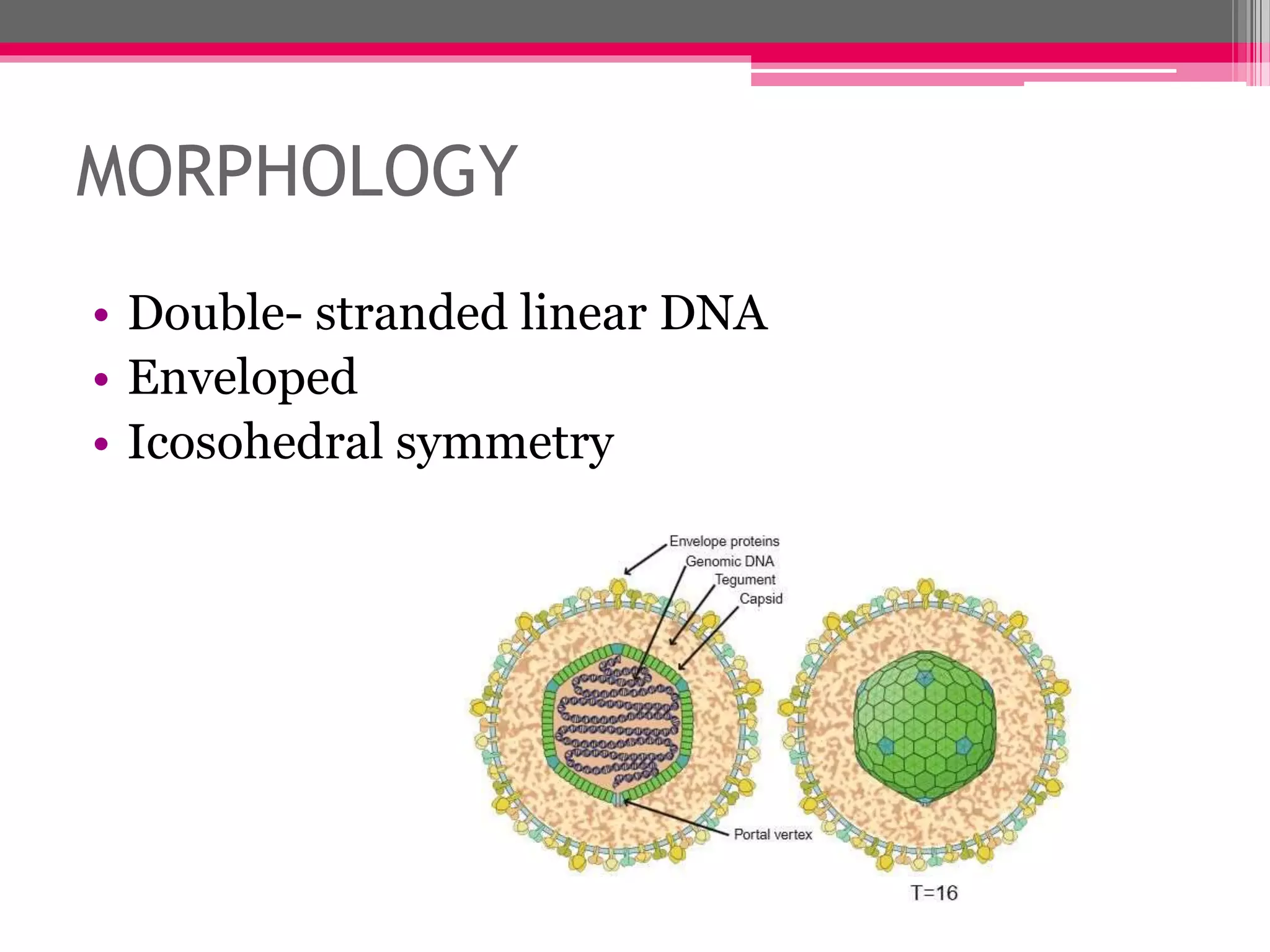
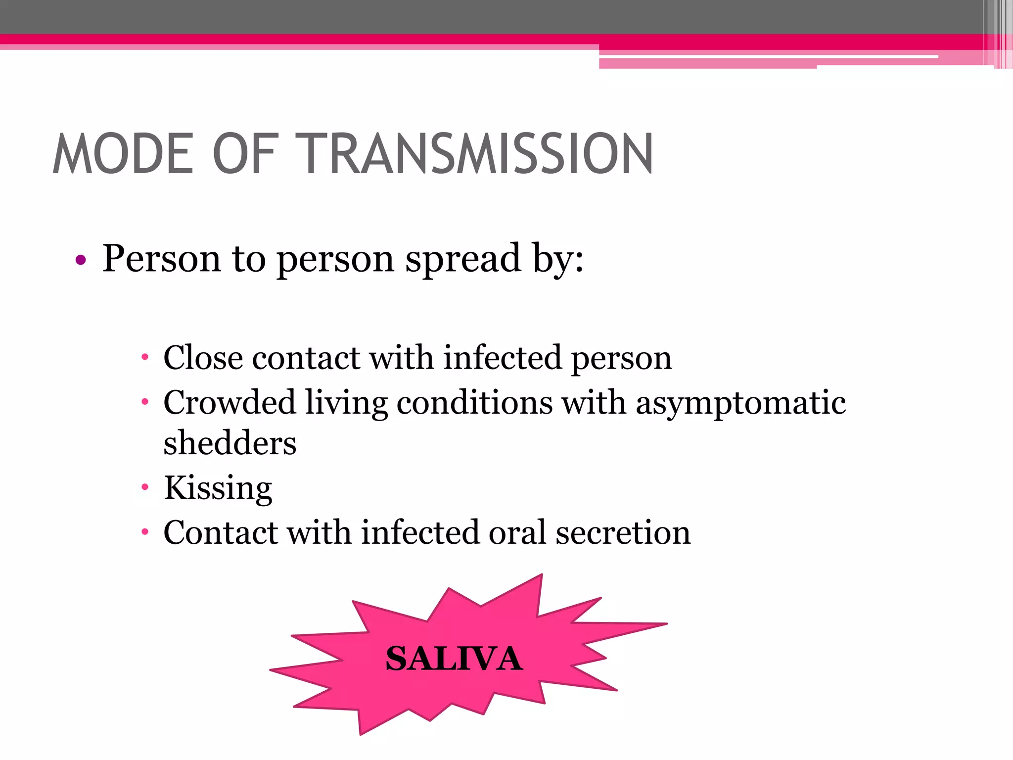
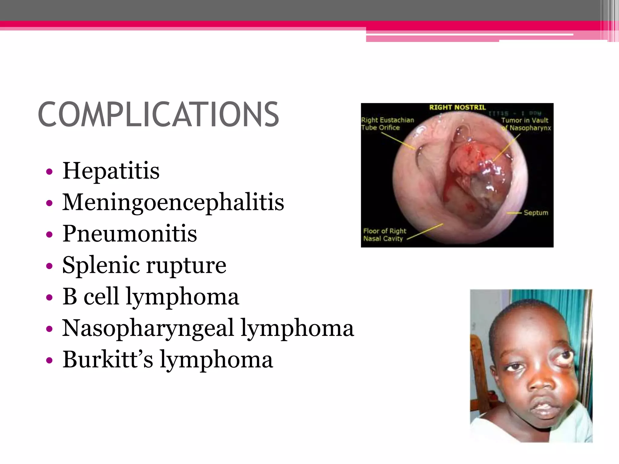
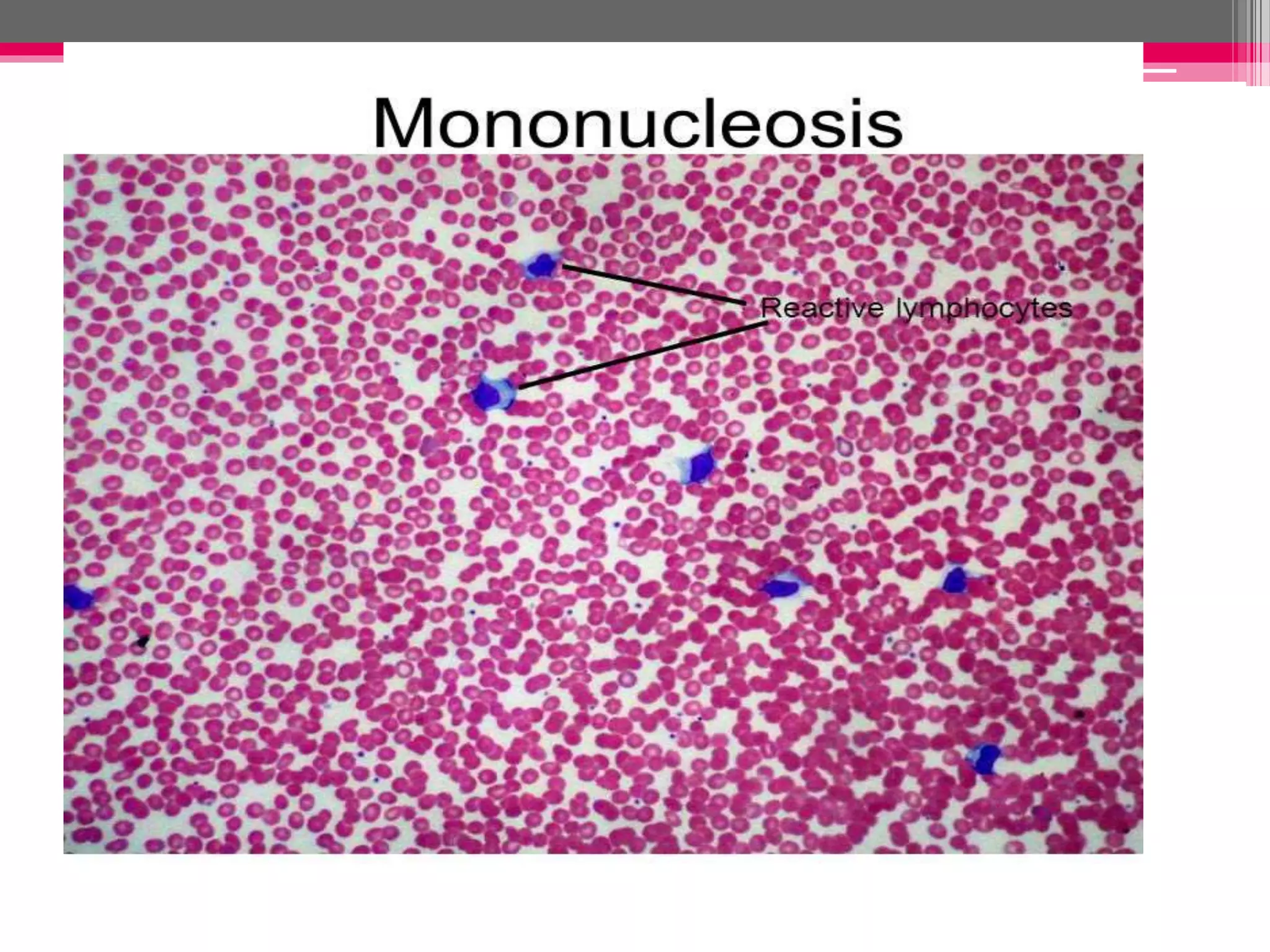
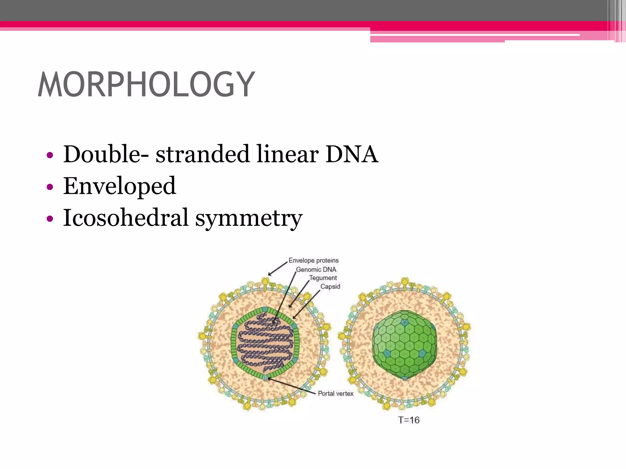
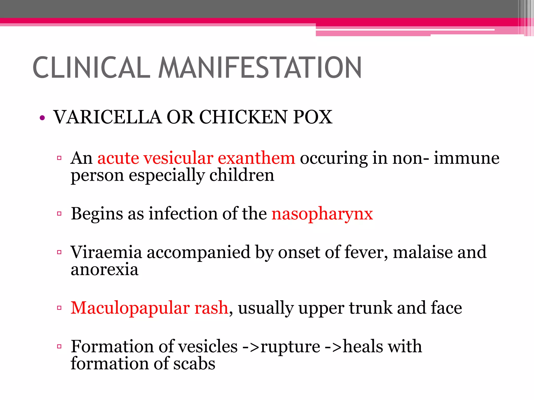
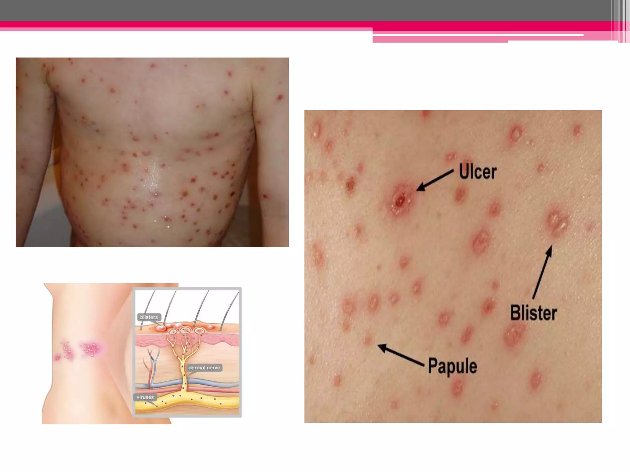
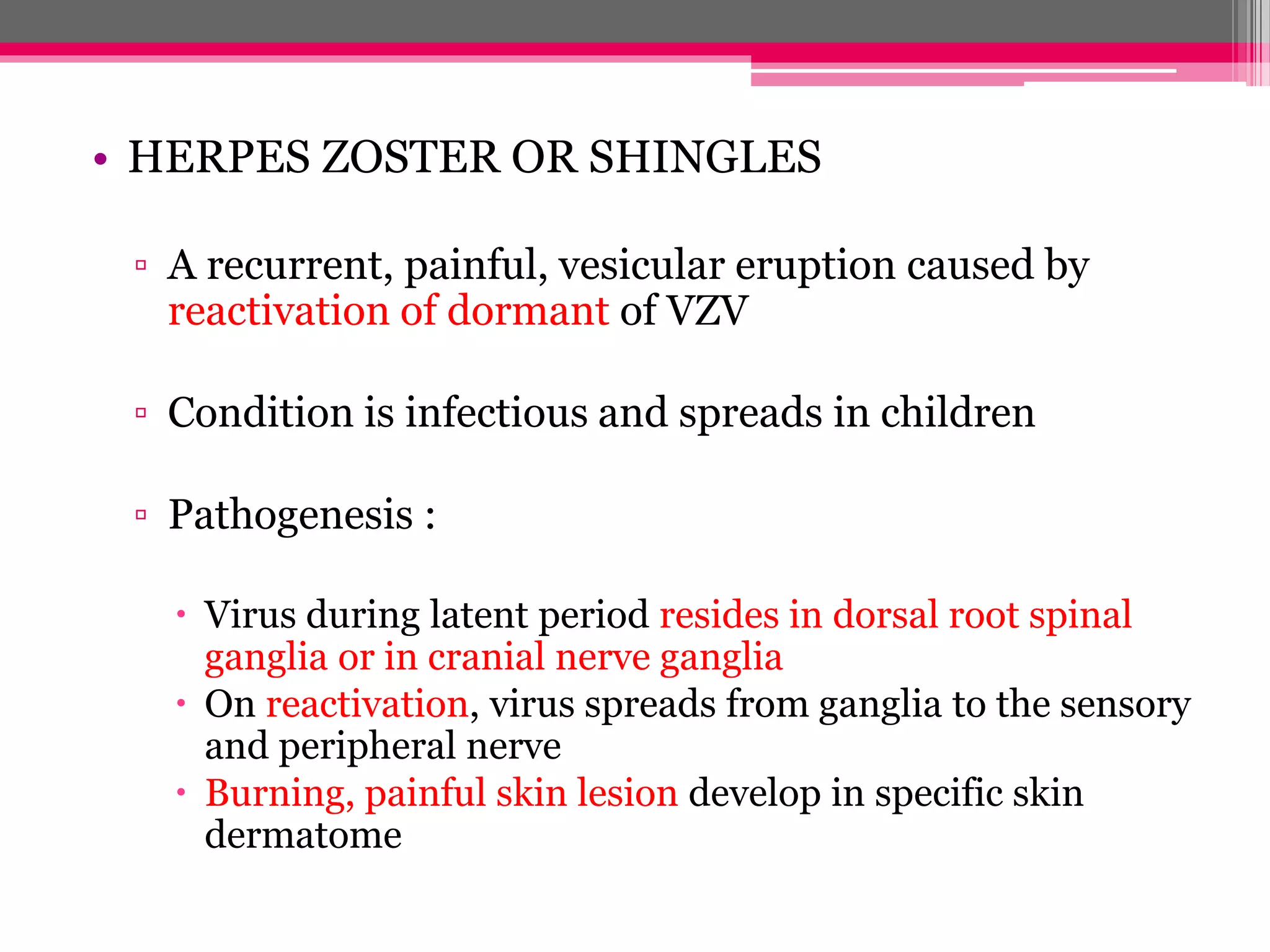
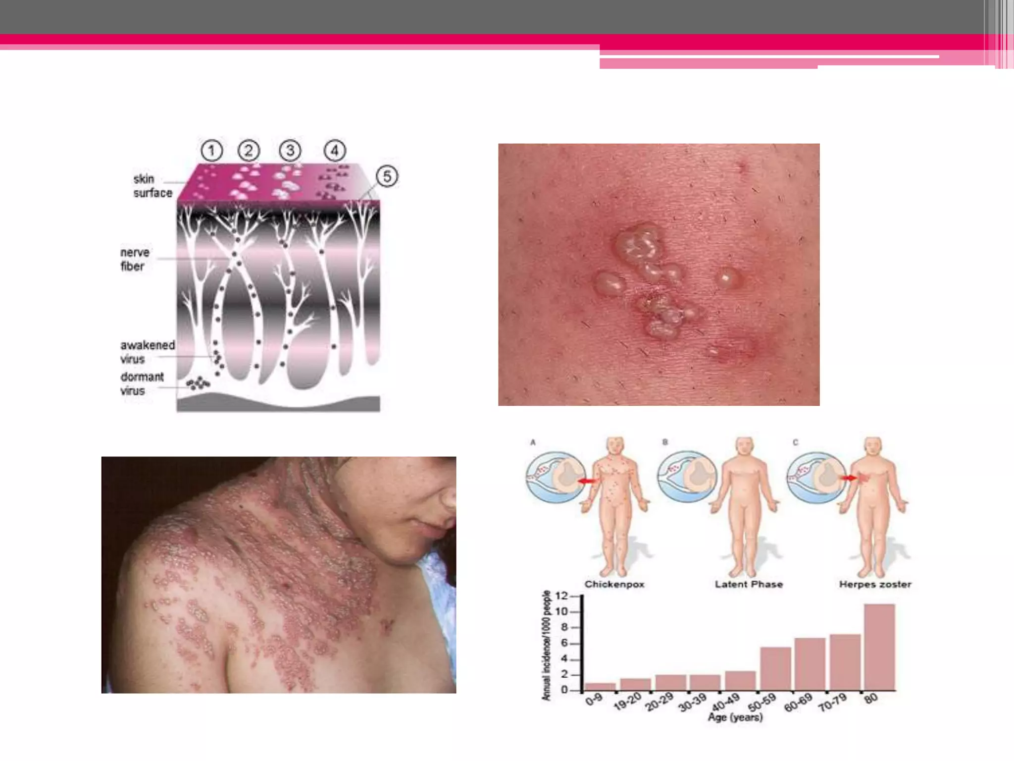
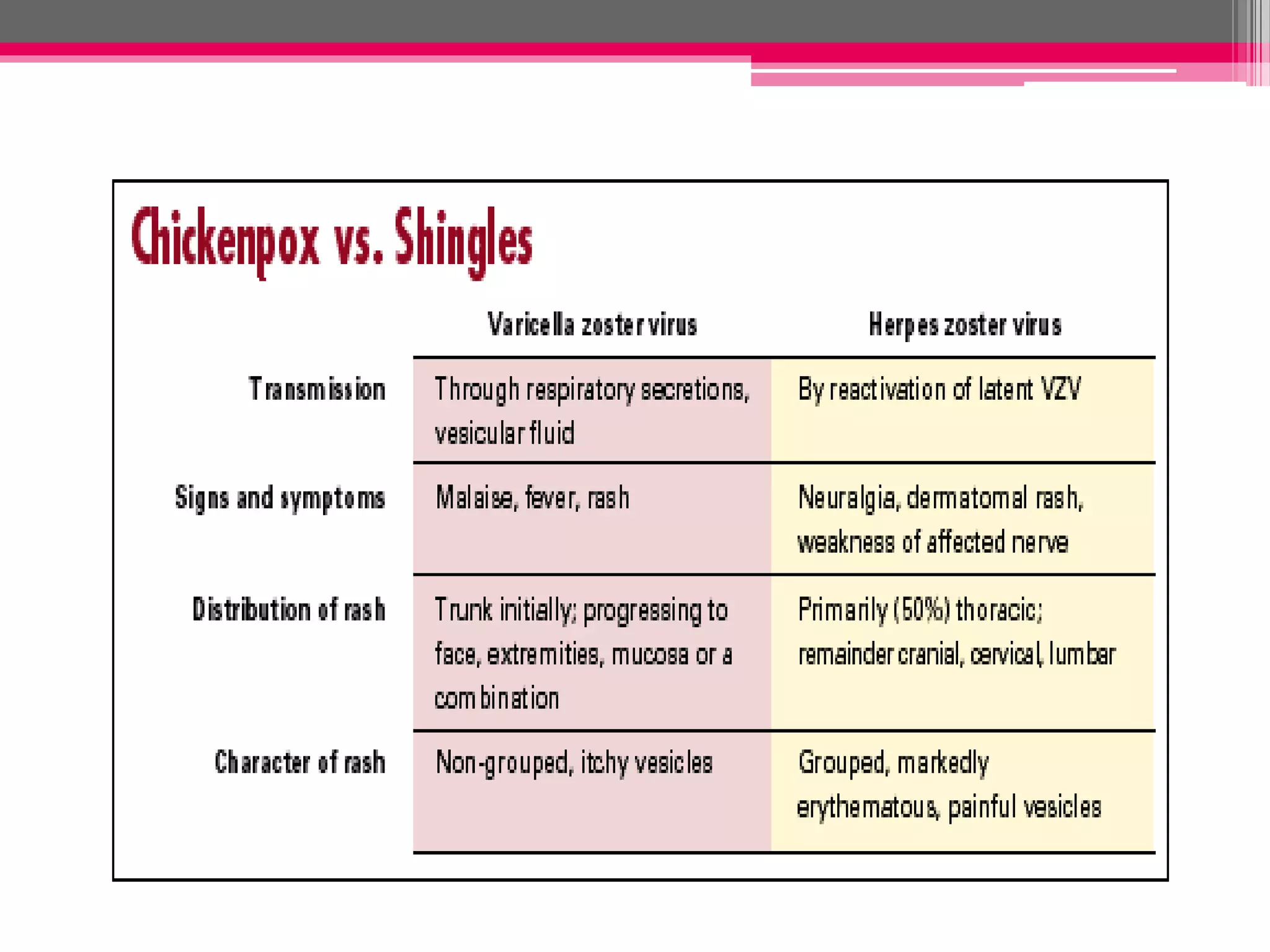
EPSTEIN BARR VIRUS (EBV)
AND VARICELLA ZOSTER
NUR IZZATUL NAJWA BINTI
SHARUPUDDIN
082015100036
LEARNING OUTCOMES :
All of students are able to :
• Describe the morphology of virus
• Understand the clinical manifestations of virus
• Describe the pathogenesis of virus
• List the mode of transmission of virus
• Know the complications of virus
• List the laboratory diagnosis of virus
Epstein-Barr virus (EBV) and Varicella Zoster virus
INTRODUCTION
• First discovered by Epstein and Barr who isolated
the virus from Burkitt’s lymphoma
• Has affinity for lymphoid tissue
• EBV infects B lymphocytes which have receptors
( CD21 molecules) specific for it
• 80% - 90% children acquire EBV infections by the
age of three years and once infected, it stays for life
MORPHOLOGY
• Double- stranded linear DNA
• Enveloped
• Icosohedral symmetry
CLINICAL MANIFESTATION
• Infectious mononucleosis (IM) or Glandular
fever
▫ Acute self limiting disease of children
▫ Charaterised by fever, sore throat,
lymphadenopathy and presence of abnormal
lymphocyes in peripheral blood
▫ Incubation period : 4 to 7 weeks
▫ Presence of Downey cell
• Infection to immunocompromised host
▫ Cause progressive lymphoproliferative disease in
immunodeficient children, transplant recipient
and aids patient
• EBV- associated malignancies
▫ Burkitt’s lymphoma
▫ Nasopharyngeal lymphoma
▫ B-cell lymphoma
PATHOGENESIS OF EBV INFECTION
MALIGNANCY
Mutation and t (8:14)
Sustained B- cell proliferation
Activates NF-kB and
JAK/STAT pathways
Virus codes for latent
membrane protein 1 (LMP1)
Promotes cell cycle
Activation of cyclin
D
EBNA 2 gene
MODE OF TRANSMISSION
• Person to person spread by:
 Close contact with infected person
 Crowded living conditions with asymptomatic
shedders
 Kissing
 Contact with infected oral secretion
SALIVA
COMPLICATIONS
• Hepatitis
• Meningoencephalitis
• Pneumonitis
• Splenic rupture
• B cell lymphoma
• Nasopharyngeal lymphoma
• Burkitt’s lymphoma
LABORATORY DIAGNOSIS
WHITE BLOOD CELL COUNT
• Mild increase Total Leukocyte Count (TLC)
• Atypical lymphocytes
▫ Larger
▫ Abundant blue cytoplasm – Duck skirting
▫ Oval, folded nucleus
PAUL BUNNEL TEST
• Presence of heterophile antibodies - IgM
INTRODUCTION
• Family of herpes virus
• Virus causes two diseases: varicella (chickenpox)
and herpes zoster (shingles)
• Varicella is the primary infection in a non-
immune individual
• Herpes zoster is reactivation of the latent virus
when immunity falls to ineffective level
MORPHOLOGY
• Double- stranded linear DNA
• Enveloped
• Icosohedral symmetry
CLINICAL MANIFESTATION
• VARICELLA OR CHICKEN POX
▫ An acute vesicular exanthem occuring in non- immune
person especially children
▫ Begins as infection of the nasopharynx
▫ Viraemia accompanied by onset of fever, malaise and
anorexia
▫ Maculopapular rash, usually upper trunk and face
▫ Formation of vesicles ->rupture ->heals with
formation of scabs
• HERPES ZOSTER OR SHINGLES
▫ A recurrent, painful, vesicular eruption caused by
reactivation of dormant of VZV
▫ Condition is infectious and spreads in children
▫ Pathogenesis :
 Virus during latent period resides in dorsal root spinal
ganglia or in cranial nerve ganglia
 On reactivation, virus spreads from ganglia to the sensory
and peripheral nerve
 Burning, painful skin lesion develop in specific skin
dermatome
MODE OF TRANSMISSION
• Varicella
▫ Aerosolized respiratory secretion
▫ Contact with ruptured vesicles
• Zoster
▫ Reactivation from dorsal root ganglion
LABORATORY DIAGNOSTIC
• Few drops on tops of rose petals ; a red base with
fluid filled vesicle
• Multinucleated giant cells
SUMMARY
REFERENCE
• TEXTBOOK OF PATHOLOGY, HARSH
MOHAN, 7TH EDITION
• CLINICAL MICROBIOLOGY MADE
RIDICULOUSLY SIMPLE, 4TH EDITION
• TEXTBOOK OF MICROBIOLOGY, PROF CP
BAVEJA, 4TH EDITION
Epstein barr virus (ebv) and varicella

Epstein barr virus (ebv) and varicella

  • 1.
    EPSTEIN BARR VIRUS(EBV) AND VARICELLA ZOSTER NUR IZZATUL NAJWA BINTI SHARUPUDDIN 082015100036
  • 2.
    LEARNING OUTCOMES : Allof students are able to : • Describe the morphology of virus • Understand the clinical manifestations of virus • Describe the pathogenesis of virus • List the mode of transmission of virus • Know the complications of virus • List the laboratory diagnosis of virus Epstein-Barr virus (EBV) and Varicella Zoster virus
  • 3.
    INTRODUCTION • First discoveredby Epstein and Barr who isolated the virus from Burkitt’s lymphoma • Has affinity for lymphoid tissue • EBV infects B lymphocytes which have receptors ( CD21 molecules) specific for it • 80% - 90% children acquire EBV infections by the age of three years and once infected, it stays for life
  • 4.
    MORPHOLOGY • Double- strandedlinear DNA • Enveloped • Icosohedral symmetry
  • 5.
    CLINICAL MANIFESTATION • Infectiousmononucleosis (IM) or Glandular fever ▫ Acute self limiting disease of children ▫ Charaterised by fever, sore throat, lymphadenopathy and presence of abnormal lymphocyes in peripheral blood ▫ Incubation period : 4 to 7 weeks ▫ Presence of Downey cell
  • 6.
    • Infection toimmunocompromised host ▫ Cause progressive lymphoproliferative disease in immunodeficient children, transplant recipient and aids patient • EBV- associated malignancies ▫ Burkitt’s lymphoma ▫ Nasopharyngeal lymphoma ▫ B-cell lymphoma
  • 7.
    PATHOGENESIS OF EBVINFECTION MALIGNANCY Mutation and t (8:14) Sustained B- cell proliferation Activates NF-kB and JAK/STAT pathways Virus codes for latent membrane protein 1 (LMP1) Promotes cell cycle Activation of cyclin D EBNA 2 gene
  • 9.
    MODE OF TRANSMISSION •Person to person spread by:  Close contact with infected person  Crowded living conditions with asymptomatic shedders  Kissing  Contact with infected oral secretion SALIVA
  • 10.
    COMPLICATIONS • Hepatitis • Meningoencephalitis •Pneumonitis • Splenic rupture • B cell lymphoma • Nasopharyngeal lymphoma • Burkitt’s lymphoma
  • 11.
    LABORATORY DIAGNOSIS WHITE BLOODCELL COUNT • Mild increase Total Leukocyte Count (TLC) • Atypical lymphocytes ▫ Larger ▫ Abundant blue cytoplasm – Duck skirting ▫ Oval, folded nucleus PAUL BUNNEL TEST • Presence of heterophile antibodies - IgM
  • 14.
    INTRODUCTION • Family ofherpes virus • Virus causes two diseases: varicella (chickenpox) and herpes zoster (shingles) • Varicella is the primary infection in a non- immune individual • Herpes zoster is reactivation of the latent virus when immunity falls to ineffective level
  • 15.
    MORPHOLOGY • Double- strandedlinear DNA • Enveloped • Icosohedral symmetry
  • 16.
    CLINICAL MANIFESTATION • VARICELLAOR CHICKEN POX ▫ An acute vesicular exanthem occuring in non- immune person especially children ▫ Begins as infection of the nasopharynx ▫ Viraemia accompanied by onset of fever, malaise and anorexia ▫ Maculopapular rash, usually upper trunk and face ▫ Formation of vesicles ->rupture ->heals with formation of scabs
  • 18.
    • HERPES ZOSTEROR SHINGLES ▫ A recurrent, painful, vesicular eruption caused by reactivation of dormant of VZV ▫ Condition is infectious and spreads in children ▫ Pathogenesis :  Virus during latent period resides in dorsal root spinal ganglia or in cranial nerve ganglia  On reactivation, virus spreads from ganglia to the sensory and peripheral nerve  Burning, painful skin lesion develop in specific skin dermatome
  • 21.
    MODE OF TRANSMISSION •Varicella ▫ Aerosolized respiratory secretion ▫ Contact with ruptured vesicles • Zoster ▫ Reactivation from dorsal root ganglion
  • 22.
    LABORATORY DIAGNOSTIC • Fewdrops on tops of rose petals ; a red base with fluid filled vesicle • Multinucleated giant cells
  • 24.
  • 25.
    REFERENCE • TEXTBOOK OFPATHOLOGY, HARSH MOHAN, 7TH EDITION • CLINICAL MICROBIOLOGY MADE RIDICULOUSLY SIMPLE, 4TH EDITION • TEXTBOOK OF MICROBIOLOGY, PROF CP BAVEJA, 4TH EDITION