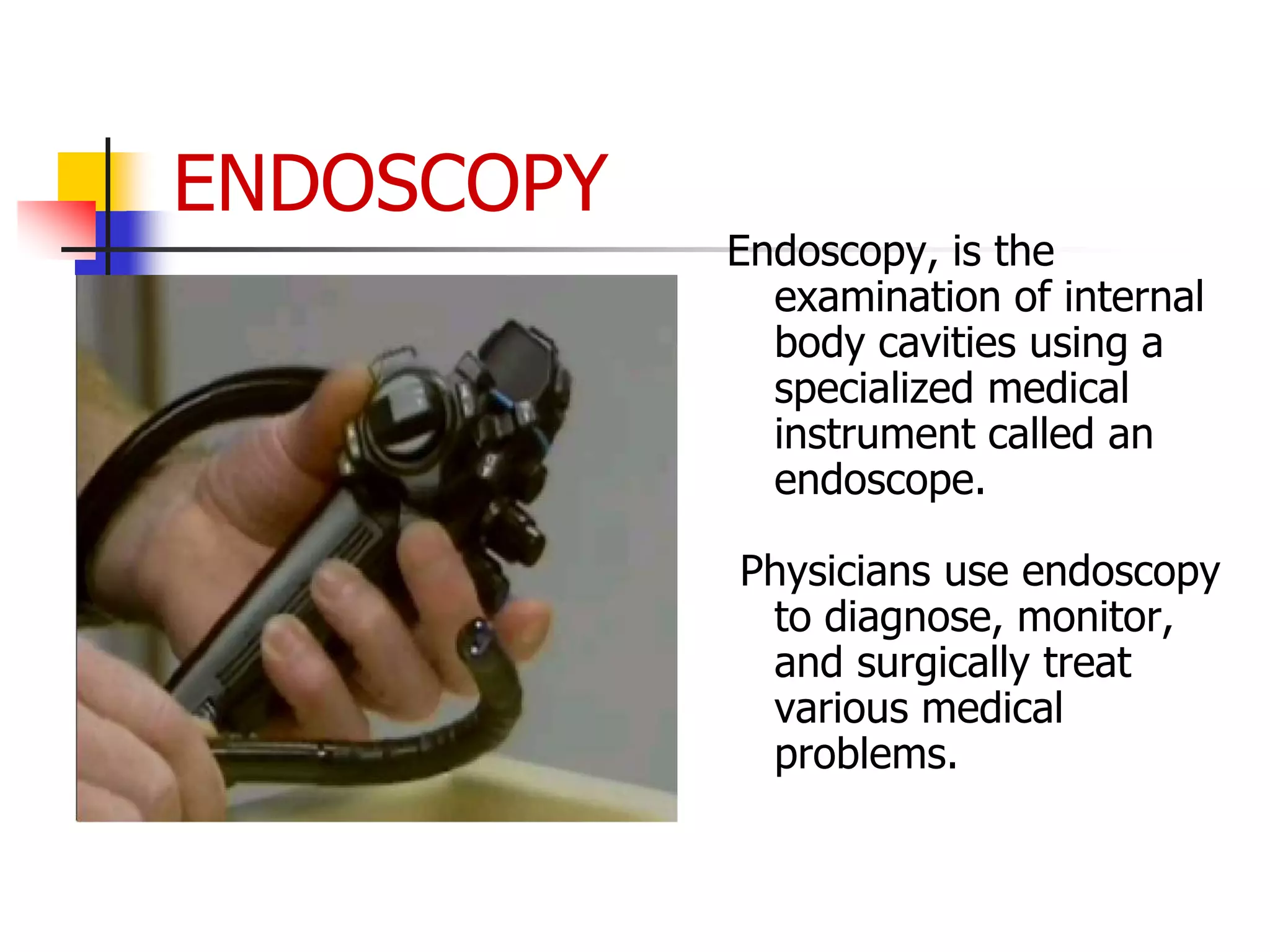
HISTORY OF ENDOSCOPY
 The word endoscopy is derived from the Greek
words "Endo" meaning "inside" and "skopeein"
meaning "to see". It is a word used in medicine to
describe the procedure used see inside various
parts of the body.
 Historically, it was known that it is possible to insert
tubes into body orifices, but to see clearly a method
was needed to illuminate the inside of the organ to
be seen.
 The earliest crude attempts used oil lamps, which
were later replaced by small electric filament bulbs.
These were not very bright and tended to produce a
lot of heat.
Science of Endoscopy
 Medical endoscopy really came into its own after the
invention and application of fibre-optic technology
to endoscopy.
 Fibre-optic endoscopes use bundles of thin glass
fibres to transmit light to and from the organ being
viewed. These fibres use the principle of total
internal reflection to transmit almost 100 % of the
light entering one end to the other end.
ENDOSCOPY
Endoscopy, is the
examination of internal
body cavities using a
specialized medical
instrument called an
endoscope.
Physicians use endoscopy
to diagnose, monitor,
and surgically treat
various medical
problems.
 An endoscope is a slender,
flexible tube equipped with
lenses and a light source.
Illumination is done by the
help of a number of optical
fibres.
 Reflected light rays are
collected by CCD( Charge
coupled device) and electrical
signals are produced, which
are fed to the video monitor
to get image.
 Thorough one channel of
endoscope water and air is
conducted to wash and dry
the surgical site.
EQUIPMENT
COMPONENTS
 The endoscope also has a
channel through which surgeons
can manipulate tiny instruments,
such as forceps, surgical
scissors, and suction devices.
 A variety of instruments can be
fitted to the endoscope for
different purposes.
 A surgeon introduces the
endoscope into the body either
through a body opening, such as
the mouth or the anus, or
through a small incision in the
skin.
ENDOSCOPY AND
IT’S PROCEDURE
How Do I Prepare for Endoscopy?
 Gut Preparation. Examining the upper
digestive tract (upper endoscopy or ERCP)
requires nothing more than fasting for 6-8
hours prior to the procedure.
 To examine the colon, it must be cleared of
stool. Therefore, a laxative or group
of laxatives is given on the day before the
procedure.
 Sedation. For most examinations with an
endoscope, a sedative is provided. This increases
the comfort of the individual undergoing the
examination. The sedative, which is administered
via an injection into the vein, produces relaxation
and light sleep.
 There are usually few if any recollections of the
procedure. Patients wake up within an hour, but
the effects of the medicines are more prolonged,
so it is not safe to drive until the next day.
Capsule endoscopy
Scientists recently devised a disposable
flash camera only slightly larger than a
vitamin pill.
In a procedure called capsule endoscopy,
the patient swallows the minicam, which
then takes pictures inside the small
intestine.
On its journey through the digestive tract,
the tiny tumbling camera transmits images
that are stored in a recorder that the
person wears around the waist. After 8
hours, the camera's battery runs out, and
the capsule is eliminated in the faeces.
Scientists then download the recorder's
images into a computer.
USES OF
ENDOSCOPY
 The endoscope
gives visual
evidence of the
problem, such as
ulceration or
inflammation
 It can be used to
collect a sample of
tissue; remove
problematic
tissue, such as
polyps
 It is used to take
photograph of the
hollow internal
organs
Uses of endoscopy:
 Stomach pain
 Ulcers, gastritis, or difficulty swallowing
 Digestive tract bleeding
 Changes in bowel habits (chronic
constipation or diarrhea)
 Polyps or growths in the colon
 In addition, your doctor may use an endoscope
to take a biopsy(removal of tissue) to look for
the presence of disease.
 Endoscopy may also be used to treat a digestive tract
problem. For example, the endoscope might not only
detect active bleeding from an ulcer, but devices can be
passed through the endoscope that can stop the bleeding.
In the colon, polyps can be removed through the scope to
prevent the development of colon cancer.
 Also, using ERCP, gallstones that have passed outside the
gallbladder and into the bile duct can often be removed.
 ERCP is special form of endoscopy called endoscopic
retrograde cholangiopancreaticography, or ERCP, allows
pictures of the pancreas,gallbladder, and related
structures to be taken
List of the major types of
endoscopy
 GASTROSCOPY: To see the gullet, stomach and upper small
intestine.
 COLONOSCOPY: To see the large intestine.
 LAPAROSCOPY: To see the "stomach cavity" and the organs
therein like uterus, fallopian tube.
 PROCTOSCOPY: This is used to check for piles and other
conditions of the anus and rectum. The picture on the right
illustrates this.
 CYSTOSCOPY: To see the urinary bladder.
 BRONCHOSCOPY: To see the air passages to the lungs.
 LARYNGOSCOPY: To see the larynx or voice box.
 NASOPHARYNGOSCOPY: To see the nose and related cavities.
 ARTHROSCOPY: To see inside joints such as the knee joint.
 THORACOSCOPY: To see inside the chest cavity.
 The gastrointestinal tract (GI tract):
 esophagus, stomach and duodenum (esophagogastro
duodenoscopy)
 small intestine (enteroscopy)
 large intestine/colon (colonoscopy, sigmoidoscopy)
 Magnification endoscopy
 bile duct
 endoscopic
retrogradecholangiopancreatography (ERCP),
duodenoscope-assisted cholangiopancreatoscopy,
intraoperative cholangioscopy
 rectum (rectoscopy) and anus (anoscopy), both also
referred to as (proctoscopy)
The respiratory tract
 The nose (rhinoscopy)
 The lower respiratory tract (bronchoscopy)
 The ear (otoscope)
 The urinary tract (cystoscopy)
 The female reproductive system (gynoscopy)
 The cervix (colposcopy)
 The uterus (hysteroscopy)
 The fallopian tubes (falloposcopy)
 Normally closed body cavities (through a small
incision):
 The abdominal or pelvic cavity (laparoscopy)
 The interior of a joint (arthroscopy)
 Organs of the chest
(thoracoscopy and mediastinoscopy)
Complications:
 Overall, endoscopy is very safe; however, the
procedure does have a few potential
complications, which may include:
 Perforation (tear in the gut wall)
 Reaction to sedation
 Infection
 Bleeding
 Pancreatitis as a result of ERCP
Limitations of Endoscopy
 Compared with other parts of the digestive tract, the
small intestine is difficult for doctors to access.
 While a camera-tipped tube slipped down the throat
can get images of the stomach and a tube inserted
at the other end of the tract reveals the large
intestine, no such device reaches into most of the
small intestine.
 So, doctors rely on externally generated images
vedio links
 https://www.youtube.com/watch?v=5h99Ib3qY
ss&ab_channel=BDENDOSCOPY
Thank you 

Endoscopy.pptx

  • 2.
    HISTORY OF ENDOSCOPY The word endoscopy is derived from the Greek words "Endo" meaning "inside" and "skopeein" meaning "to see". It is a word used in medicine to describe the procedure used see inside various parts of the body.  Historically, it was known that it is possible to insert tubes into body orifices, but to see clearly a method was needed to illuminate the inside of the organ to be seen.  The earliest crude attempts used oil lamps, which were later replaced by small electric filament bulbs. These were not very bright and tended to produce a lot of heat.
  • 3.
    Science of Endoscopy Medical endoscopy really came into its own after the invention and application of fibre-optic technology to endoscopy.  Fibre-optic endoscopes use bundles of thin glass fibres to transmit light to and from the organ being viewed. These fibres use the principle of total internal reflection to transmit almost 100 % of the light entering one end to the other end.
  • 4.
    ENDOSCOPY Endoscopy, is the examinationof internal body cavities using a specialized medical instrument called an endoscope. Physicians use endoscopy to diagnose, monitor, and surgically treat various medical problems.
  • 5.
     An endoscopeis a slender, flexible tube equipped with lenses and a light source. Illumination is done by the help of a number of optical fibres.  Reflected light rays are collected by CCD( Charge coupled device) and electrical signals are produced, which are fed to the video monitor to get image.  Thorough one channel of endoscope water and air is conducted to wash and dry the surgical site. EQUIPMENT COMPONENTS
  • 7.
     The endoscopealso has a channel through which surgeons can manipulate tiny instruments, such as forceps, surgical scissors, and suction devices.  A variety of instruments can be fitted to the endoscope for different purposes.  A surgeon introduces the endoscope into the body either through a body opening, such as the mouth or the anus, or through a small incision in the skin. ENDOSCOPY AND IT’S PROCEDURE
  • 11.
    How Do IPrepare for Endoscopy?  Gut Preparation. Examining the upper digestive tract (upper endoscopy or ERCP) requires nothing more than fasting for 6-8 hours prior to the procedure.  To examine the colon, it must be cleared of stool. Therefore, a laxative or group of laxatives is given on the day before the procedure.
  • 12.
     Sedation. Formost examinations with an endoscope, a sedative is provided. This increases the comfort of the individual undergoing the examination. The sedative, which is administered via an injection into the vein, produces relaxation and light sleep.  There are usually few if any recollections of the procedure. Patients wake up within an hour, but the effects of the medicines are more prolonged, so it is not safe to drive until the next day.
  • 13.
    Capsule endoscopy Scientists recentlydevised a disposable flash camera only slightly larger than a vitamin pill. In a procedure called capsule endoscopy, the patient swallows the minicam, which then takes pictures inside the small intestine. On its journey through the digestive tract, the tiny tumbling camera transmits images that are stored in a recorder that the person wears around the waist. After 8 hours, the camera's battery runs out, and the capsule is eliminated in the faeces. Scientists then download the recorder's images into a computer.
  • 14.
    USES OF ENDOSCOPY  Theendoscope gives visual evidence of the problem, such as ulceration or inflammation  It can be used to collect a sample of tissue; remove problematic tissue, such as polyps  It is used to take photograph of the hollow internal organs
  • 15.
    Uses of endoscopy: Stomach pain  Ulcers, gastritis, or difficulty swallowing  Digestive tract bleeding  Changes in bowel habits (chronic constipation or diarrhea)  Polyps or growths in the colon  In addition, your doctor may use an endoscope to take a biopsy(removal of tissue) to look for the presence of disease.
  • 16.
     Endoscopy mayalso be used to treat a digestive tract problem. For example, the endoscope might not only detect active bleeding from an ulcer, but devices can be passed through the endoscope that can stop the bleeding. In the colon, polyps can be removed through the scope to prevent the development of colon cancer.  Also, using ERCP, gallstones that have passed outside the gallbladder and into the bile duct can often be removed.  ERCP is special form of endoscopy called endoscopic retrograde cholangiopancreaticography, or ERCP, allows pictures of the pancreas,gallbladder, and related structures to be taken
  • 17.
    List of themajor types of endoscopy  GASTROSCOPY: To see the gullet, stomach and upper small intestine.  COLONOSCOPY: To see the large intestine.  LAPAROSCOPY: To see the "stomach cavity" and the organs therein like uterus, fallopian tube.  PROCTOSCOPY: This is used to check for piles and other conditions of the anus and rectum. The picture on the right illustrates this.  CYSTOSCOPY: To see the urinary bladder.  BRONCHOSCOPY: To see the air passages to the lungs.  LARYNGOSCOPY: To see the larynx or voice box.  NASOPHARYNGOSCOPY: To see the nose and related cavities.  ARTHROSCOPY: To see inside joints such as the knee joint.  THORACOSCOPY: To see inside the chest cavity.
  • 18.
     The gastrointestinaltract (GI tract):  esophagus, stomach and duodenum (esophagogastro duodenoscopy)  small intestine (enteroscopy)  large intestine/colon (colonoscopy, sigmoidoscopy)  Magnification endoscopy  bile duct  endoscopic retrogradecholangiopancreatography (ERCP), duodenoscope-assisted cholangiopancreatoscopy, intraoperative cholangioscopy  rectum (rectoscopy) and anus (anoscopy), both also referred to as (proctoscopy)
  • 19.
    The respiratory tract The nose (rhinoscopy)  The lower respiratory tract (bronchoscopy)  The ear (otoscope)  The urinary tract (cystoscopy)
  • 20.
     The femalereproductive system (gynoscopy)  The cervix (colposcopy)  The uterus (hysteroscopy)  The fallopian tubes (falloposcopy)  Normally closed body cavities (through a small incision):  The abdominal or pelvic cavity (laparoscopy)  The interior of a joint (arthroscopy)  Organs of the chest (thoracoscopy and mediastinoscopy)
  • 21.
    Complications:  Overall, endoscopyis very safe; however, the procedure does have a few potential complications, which may include:  Perforation (tear in the gut wall)  Reaction to sedation  Infection  Bleeding  Pancreatitis as a result of ERCP
  • 22.
    Limitations of Endoscopy Compared with other parts of the digestive tract, the small intestine is difficult for doctors to access.  While a camera-tipped tube slipped down the throat can get images of the stomach and a tube inserted at the other end of the tract reveals the large intestine, no such device reaches into most of the small intestine.  So, doctors rely on externally generated images
  • 23.
  • 24.