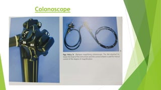
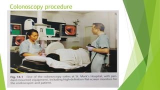
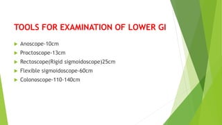
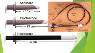
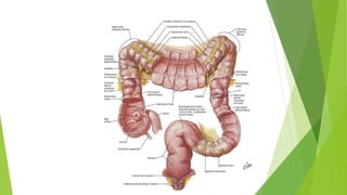
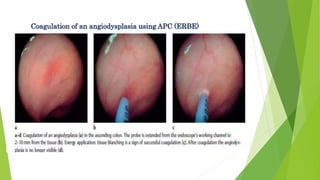
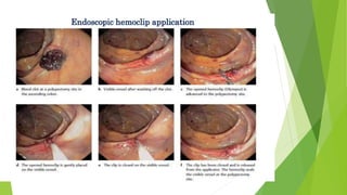
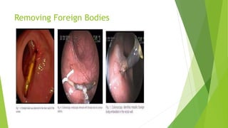
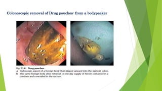
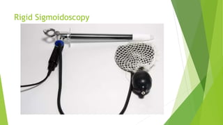
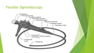
LOWER GI ENDOSCOPY IN
SURGICAL PRACTICE
BY:DR.GHAIYOOR AHMED
PGT-1
M.S. GENERAL SURGERY
WHAT IS ENDOSCOPY?
 Endoscopy is Greek word “Endo”means inside
“Skopeein”means to see
 Examination of the interior of a canal or hollow viscus
by means of a special instrument known as endoscope
 Direct viewing interior of an organ is very helpful in
determining the cause of disease and helps in
diagnosis
PARTS OF ENDOSCOPE
 A thin long tube - rigid or flexible
 A lens / lens system
 A light transmitting system
 Eyepiece
 Control system
 Channel through which tiny instruments such as scissors
can be manipulated
Colonoscope
TYPES OF ENDOSCOPY
 Upper GI Endoscopy
 Enteroscopy
 Lower GI Endoscopy(colonoscopy)
Endoscopy Principle
 It is a minimally invasive diagnostic procedure for
evaluation of interior surface of an organ
 Endoscope may be rigid or felixible tube inserted into
body cavity which looks inside the body using a
variety of small cameras attached to flexible or rigid
tube
 An endoscope equipped with lens and light source
 Illumination is done by help of numbers of optical
fibers
 Video endoscopy is performed byattaching microchip
camera at the insertion tube setup image viewed on a
video monitor
Colonoscopy procedure
TOOLS FOR EXAMINATION OF LOWER GI
 Anoscope-10cm
 Proctoscope-13cm
 Rectoscope(Rigid sigmoidoscope)25cm
 Flexible sigmoidoscope-60cm
 Colonoscope-110-140cm
COLONOSCOPY
Introduction:
Colonoscopy is a procedure to see inside the colon and
rectum.
It is helpful to diagnose unexplained changes in bowel
habbit,abdominal pain ,bleeding per rectum and weight loss.
It also helpful for looking early signs of colorectal cancer
INDICATION OF COLONOSCOPY
 Lower GI bleeding
 Screening and surveillance of colorectal polyp and cancer
a)colon cancer
b)surveillance after polypectomy,
c) colorectal cancer post resection surveillance
d)IBD
 Acute and chronic diarrhoea
 Therapeutic indication for colorectal cancer
Therapeutic Indication
a)excision and ablation of lesion
b)treatment of lower gi bleed
c)colonic decompression
d)dialation of colonic stenosis
e)foreign body removal
 Miscellaneous indication
a)abnormal radiological examination i.e RIF mass in usg
s/o colonic origin
b)unexplained abdominal pain
CONTRA INDICATION OF COLONOSCOPY
 Inadequate bowel preparation
 Uncoperative patients
 Patients refusal
 Inadequate sedation
 Known or suspected colonic perforation
 Clinically unstable patients
 Recent MI
 Peritonism
COMPLICATION AND RISK
 Risk of perforation
 Infection
 Injury of blood vessel causing bleeding
 More complication arise from therapeutic colonoscopy i.e
polypectomy
COLONOSCOPY PREPARATION
 Emptying the content of the colon is the key requirement
for successful colonscopy
 If bowel preparation is not adequate polyp and lesion can
be missed
 Advise low fiber diet few days before colonoscopy
 Avoid solid food day before colonoscopy
 On the day of colonoscopy only clear fluid is advise.
 Dissolve 1 packet of PEG in 2lts of fluid and asked pt to
consumed
 Give sodium picu sulphate enema.
 Pt will purge several time so bowel will get clean
 Colonoscopy can be done under iv sedation.
BASIC RULES OF COLONOSCOPY
 Don’t advanced the endoscope without the clear view of
the lumen
 Don’t advanced the endoscope if there is any resistance
 When in doubt pull back
 Use a little air as possible and as much air as necessary
 Pay attention to pts pain reaction
Abnormal Findings in Colonoscopy
Therapeutic use of Colonoscopy
Snare Polypectomy
Endoscopic Mucosectomy
SEMS- for Malignant Strictures
Haemostasis in Colorectal Pathologies
Removing Foreign Bodies
Sigmoidoscopy
 Sigmoidoscopy is the minimally
invasive medical examination of the large intestine from
the rectum to sigmoid colon.
Types of sigmoidoscopy:
 flexible sigmoidoscopy, which uses a flexible endoscope
 rigid sigmoidoscopy, which uses a rigid device. Flexible
sigmoidoscopy is generally the preferred procedure.
 A sigmoidoscopy is similar to, but not the same as,
a colonoscopy. A sigmoidoscopy only examines up to
the sigmoid, the most distal part of the colon, while
colonoscopy examines the whole large bowel
Flexible sigmoidoscopy
 With flexible sigmoidoscopy, the physician can
see intestinal bleeding, inflammation, abnormal growths,
and ulcers in the descending colon and rectum. the sites
which can be observed represent areas which are most
frequently affected by diseases such as colorectal cancer,
for example the rectum.
 Flexible sigmoidoscopy takes 10 to 20 minutes..
 Preparation same as Colonoscopy
Rigid sigmoidoscopy
 Rigid sigmoidoscopy may be useful in ano-rectal diseases such
as bleeding per rectum or inflammatory rectal disease,
particularly in the general practice and pediatrics.
 Position of rigid sigmoidoscopy- Sims' position. The bowels are
previously emptied with a suppository, and a digital rectal
examination is first performed. The sigmoidoscope is lubricated
and inserted with obturator in general direction of the navel.
The direction is then changed and the obturator is removed so
that the physician may penetrate further with direct vision.
A bellows is used to insufflate air to distend the rectum. Lateral
movements of the sigmoidoscope's tip negotiate the Houston
valve and the recto-sigmoid junction.
Rigid Sigmoidoscopy
Flexible Sigmoidoscopy

LOWER GI ENDOSCOPY IN SURGICAL PRACTICE.pptx

  • 1.
    LOWER GI ENDOSCOPYIN SURGICAL PRACTICE BY:DR.GHAIYOOR AHMED PGT-1 M.S. GENERAL SURGERY
  • 2.
    WHAT IS ENDOSCOPY? Endoscopy is Greek word “Endo”means inside “Skopeein”means to see  Examination of the interior of a canal or hollow viscus by means of a special instrument known as endoscope  Direct viewing interior of an organ is very helpful in determining the cause of disease and helps in diagnosis
  • 3.
    PARTS OF ENDOSCOPE A thin long tube - rigid or flexible  A lens / lens system  A light transmitting system  Eyepiece  Control system  Channel through which tiny instruments such as scissors can be manipulated
  • 4.
  • 5.
    TYPES OF ENDOSCOPY Upper GI Endoscopy  Enteroscopy  Lower GI Endoscopy(colonoscopy)
  • 6.
    Endoscopy Principle  Itis a minimally invasive diagnostic procedure for evaluation of interior surface of an organ  Endoscope may be rigid or felixible tube inserted into body cavity which looks inside the body using a variety of small cameras attached to flexible or rigid tube  An endoscope equipped with lens and light source  Illumination is done by help of numbers of optical fibers  Video endoscopy is performed byattaching microchip camera at the insertion tube setup image viewed on a video monitor
  • 7.
  • 8.
    TOOLS FOR EXAMINATIONOF LOWER GI  Anoscope-10cm  Proctoscope-13cm  Rectoscope(Rigid sigmoidoscope)25cm  Flexible sigmoidoscope-60cm  Colonoscope-110-140cm
  • 11.
    COLONOSCOPY Introduction: Colonoscopy is aprocedure to see inside the colon and rectum. It is helpful to diagnose unexplained changes in bowel habbit,abdominal pain ,bleeding per rectum and weight loss. It also helpful for looking early signs of colorectal cancer
  • 12.
    INDICATION OF COLONOSCOPY Lower GI bleeding  Screening and surveillance of colorectal polyp and cancer a)colon cancer b)surveillance after polypectomy, c) colorectal cancer post resection surveillance d)IBD  Acute and chronic diarrhoea  Therapeutic indication for colorectal cancer
  • 13.
    Therapeutic Indication a)excision andablation of lesion b)treatment of lower gi bleed c)colonic decompression d)dialation of colonic stenosis e)foreign body removal  Miscellaneous indication a)abnormal radiological examination i.e RIF mass in usg s/o colonic origin b)unexplained abdominal pain
  • 14.
    CONTRA INDICATION OFCOLONOSCOPY  Inadequate bowel preparation  Uncoperative patients  Patients refusal  Inadequate sedation  Known or suspected colonic perforation  Clinically unstable patients  Recent MI  Peritonism
  • 15.
    COMPLICATION AND RISK Risk of perforation  Infection  Injury of blood vessel causing bleeding  More complication arise from therapeutic colonoscopy i.e polypectomy
  • 16.
    COLONOSCOPY PREPARATION  Emptyingthe content of the colon is the key requirement for successful colonscopy  If bowel preparation is not adequate polyp and lesion can be missed  Advise low fiber diet few days before colonoscopy  Avoid solid food day before colonoscopy  On the day of colonoscopy only clear fluid is advise.  Dissolve 1 packet of PEG in 2lts of fluid and asked pt to consumed  Give sodium picu sulphate enema.
  • 17.
     Pt willpurge several time so bowel will get clean  Colonoscopy can be done under iv sedation.
  • 18.
    BASIC RULES OFCOLONOSCOPY  Don’t advanced the endoscope without the clear view of the lumen  Don’t advanced the endoscope if there is any resistance  When in doubt pull back  Use a little air as possible and as much air as necessary  Pay attention to pts pain reaction
  • 20.
  • 21.
    Therapeutic use ofColonoscopy
  • 22.
  • 23.
  • 24.
  • 25.
  • 28.
  • 31.
    Sigmoidoscopy  Sigmoidoscopy isthe minimally invasive medical examination of the large intestine from the rectum to sigmoid colon. Types of sigmoidoscopy:  flexible sigmoidoscopy, which uses a flexible endoscope  rigid sigmoidoscopy, which uses a rigid device. Flexible sigmoidoscopy is generally the preferred procedure.  A sigmoidoscopy is similar to, but not the same as, a colonoscopy. A sigmoidoscopy only examines up to the sigmoid, the most distal part of the colon, while colonoscopy examines the whole large bowel
  • 32.
    Flexible sigmoidoscopy  Withflexible sigmoidoscopy, the physician can see intestinal bleeding, inflammation, abnormal growths, and ulcers in the descending colon and rectum. the sites which can be observed represent areas which are most frequently affected by diseases such as colorectal cancer, for example the rectum.  Flexible sigmoidoscopy takes 10 to 20 minutes..  Preparation same as Colonoscopy
  • 33.
    Rigid sigmoidoscopy  Rigidsigmoidoscopy may be useful in ano-rectal diseases such as bleeding per rectum or inflammatory rectal disease, particularly in the general practice and pediatrics.  Position of rigid sigmoidoscopy- Sims' position. The bowels are previously emptied with a suppository, and a digital rectal examination is first performed. The sigmoidoscope is lubricated and inserted with obturator in general direction of the navel. The direction is then changed and the obturator is removed so that the physician may penetrate further with direct vision. A bellows is used to insufflate air to distend the rectum. Lateral movements of the sigmoidoscope's tip negotiate the Houston valve and the recto-sigmoid junction.
  • 34.
  • 35.