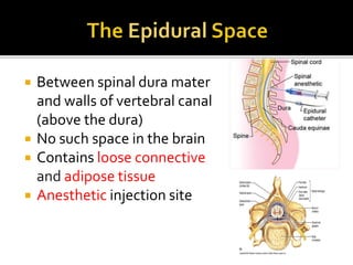
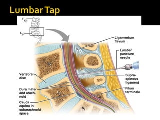
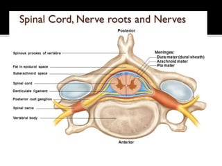
NUR IZZATUL NAJWA BINTI SHARUPUDDIN
082015100036
ATTHE END OFTHE SEMINAR, STUDENTS
SHOULD BE ABLETO :
 List and expain the spinal meninges
 Know the epidural space and its importance
 Upper end – medulla
oblongata
 Lower end – first
lumbar vertebra
 45cm
 Conus medullaris (L1)
 Filum terminale
(20cm)
 Specialized membranes isolate spinal cord
from surroundings
 Functions :
 protect spinal cord
 carry blood supply
 continuous with cranial meninges
Figure 13–3
Dura mater:
outer layer
Arachnoid mater:
middle layer
Pia mater:
inner layer
 Tough and fibrous
membrane
 Meningeal layer only
 Contains loose
areolar tissue &
internal vertebral
venous space
 Extend up 5 to S2
 Arachnoid = spiderlike
 Delicate impermeable
membrane
 Thin membrane
 Lies between pia and
dura mater
 Extend up to S2
 Thin and vascular membrane
 Closely covers spinal cord
 Continue as filum terminale
 Undergoes modification :
 Ligamentum denticulatum
▪ 21 pairs teeth like projection
▪ Keep spinal cord in position
 Linea splendens
▪ Thickening at anteromedian sulcus
 Between spinal dura mater
and walls of vertebral canal
(above the dura)
 No such space in the brain
 Contains loose connective
and adipose tissue
 Anesthetic injection site
 Subdural space:
 between arachnoid mater and dura mater
 Subarachnoid space:
 between arachnoid mater and pia mater
 filled with cerebrospinal fluid (CSF)
 LumbarTap withdraws CSF from inferior
lumbar region (below conus medularis) for
diagnostic purposes.
 BD CHAURASIA 25TH EDITION
 TEXTBOOK OF HUMAN NEUROANATOMY,
INDERBIR SINGH 7TH EDITION
Epidural space and spinal meninges

Epidural space and spinal meninges

  • 1.
    NUR IZZATUL NAJWABINTI SHARUPUDDIN 082015100036
  • 2.
    ATTHE END OFTHESEMINAR, STUDENTS SHOULD BE ABLETO :  List and expain the spinal meninges  Know the epidural space and its importance
  • 3.
     Upper end– medulla oblongata  Lower end – first lumbar vertebra  45cm  Conus medullaris (L1)  Filum terminale (20cm)
  • 4.
     Specialized membranesisolate spinal cord from surroundings  Functions :  protect spinal cord  carry blood supply  continuous with cranial meninges
  • 5.
    Figure 13–3 Dura mater: outerlayer Arachnoid mater: middle layer Pia mater: inner layer
  • 6.
     Tough andfibrous membrane  Meningeal layer only  Contains loose areolar tissue & internal vertebral venous space  Extend up 5 to S2
  • 7.
     Arachnoid =spiderlike  Delicate impermeable membrane  Thin membrane  Lies between pia and dura mater  Extend up to S2
  • 8.
     Thin andvascular membrane  Closely covers spinal cord  Continue as filum terminale  Undergoes modification :  Ligamentum denticulatum ▪ 21 pairs teeth like projection ▪ Keep spinal cord in position  Linea splendens ▪ Thickening at anteromedian sulcus
  • 9.
     Between spinaldura mater and walls of vertebral canal (above the dura)  No such space in the brain  Contains loose connective and adipose tissue  Anesthetic injection site
  • 10.
     Subdural space: between arachnoid mater and dura mater  Subarachnoid space:  between arachnoid mater and pia mater  filled with cerebrospinal fluid (CSF)  LumbarTap withdraws CSF from inferior lumbar region (below conus medularis) for diagnostic purposes.
  • 13.
     BD CHAURASIA25TH EDITION  TEXTBOOK OF HUMAN NEUROANATOMY, INDERBIR SINGH 7TH EDITION