Downloaded 147 times


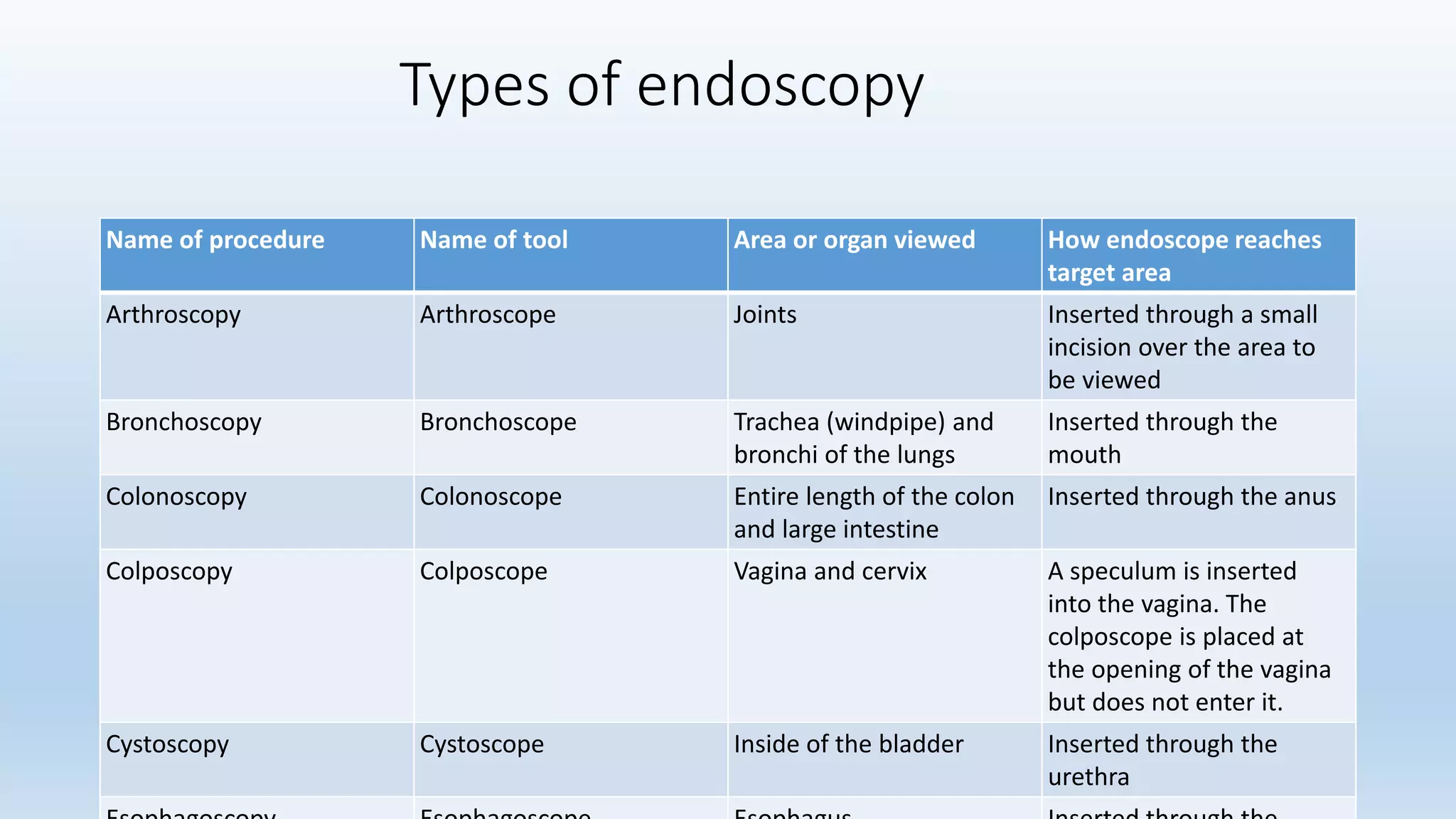

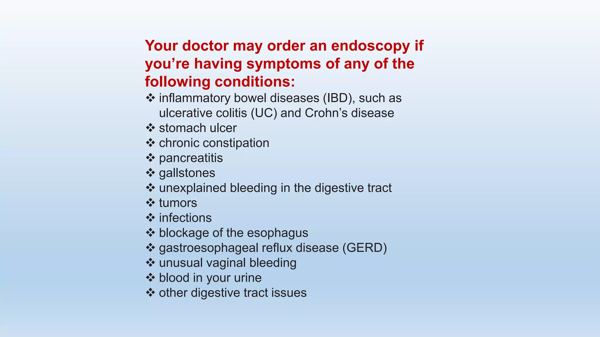

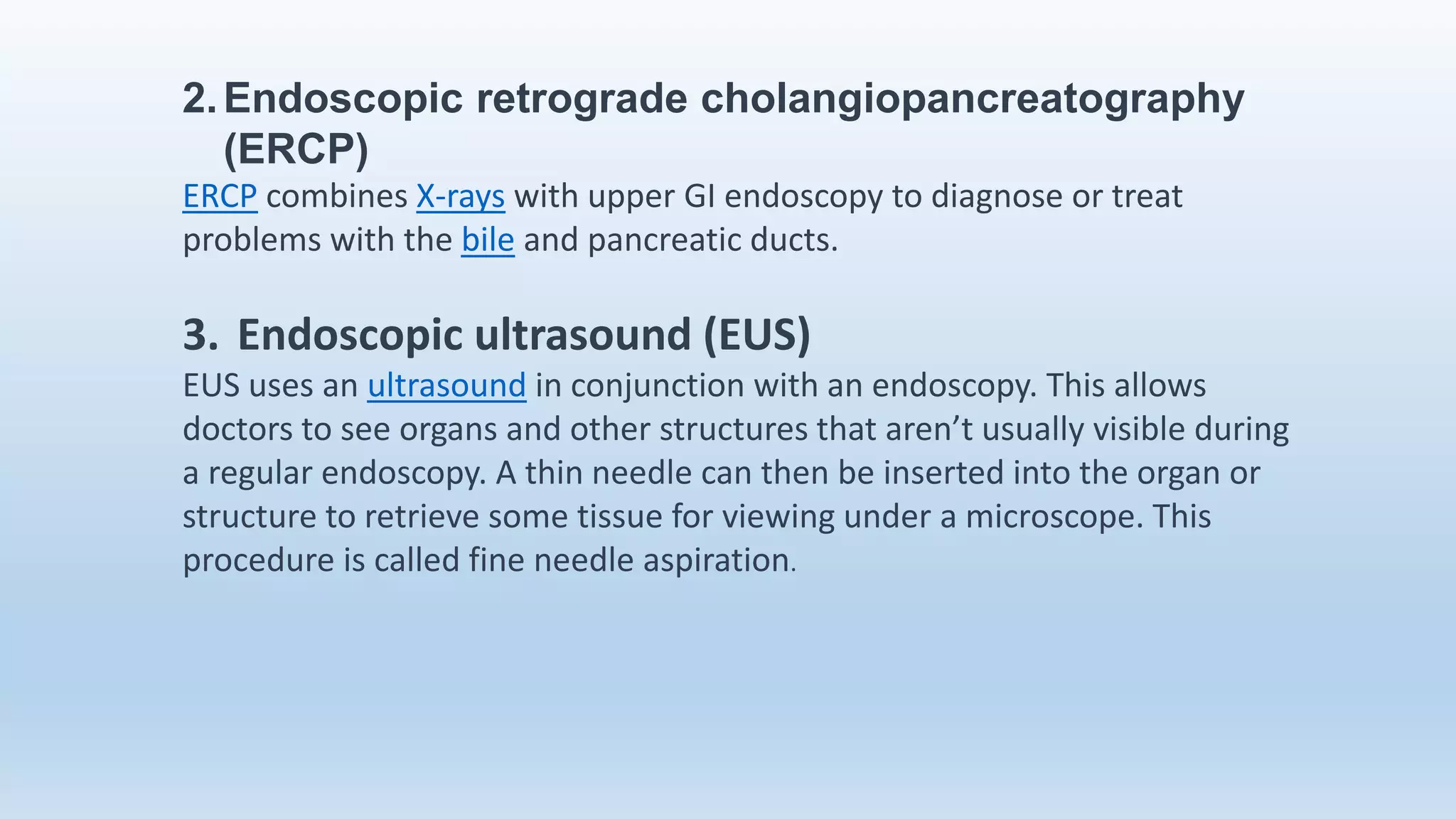


Endoscopy is a procedure that allows doctors to examine the inside of the body using an endoscope, a thin tube with a camera. Originally used only in the esophagus, stomach, and colon, endoscopy is now used to diagnose diseases in many areas including the ear, nose, throat, heart, urinary tract and joints. Doctors insert various tools through the endoscope to collect tissue samples or provide treatment. While endoscopy allows examination without large incisions, there are risks like damage to organs, perforation, pain and infection.


