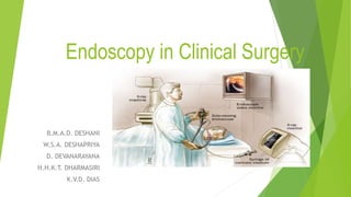
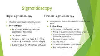
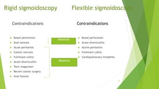
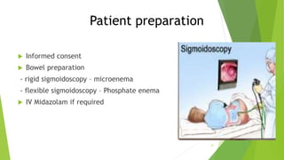
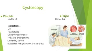
Endoscopy in Clinical Surgery
B.M.A.D. DESHANI
W.S.A. DESHAPRIYA
D. DEVANARAYANA
H.H.K.T. DHARMASIRI
K.V.D. DIAS
Endoscopy
 The process of inspecting both internal body cavities & epithelium
lined organs by the means of rigid or flexible instrument.
 This may be through a natural orifice e.g.: oesophagoscopy,
sigmoidoscopy
 Through a surgically created hole e.g. : arthroscopy, laparoscopy
Endoscope
 Instrument used to examine the interior of a hollow organ or cavity
of the body
2
endoscopes
 Rigid  Flexible
3
4
Uses of endoscopy
 Investigation of symptoms
e.g. : difficulty in swallowing, GI bleeding , abdominal pain, nausea , vomiting
 Diagnostic
e.g. : biopsy in anemia , bleeding, inflammation, cancers of GI tract
 Therapeutic
e.g. : widening of narrow esophagus , cauterization of bleeding vessel, clipping off a polyp
 Monitoring & screening
e.g. : Gastric carcinoma,colorecatal carcinoma
 Research
5
classification
 The GI tract
- Oesophagus ,stomach & duodenum – Oesophagogastroduodenoscopy
- Small intestine – Enteroscopy
- Colon – Colonoscopy . Sigmoidoscopy
- Bile duct – ERCP
- Rectum & anus – Proctoscopy
 The urinary tract – Cystoscopy
 The respiratory tract
- Upper respiratory tract - Rhinoscopy ,Laryngoscopy
- Lower respiratory tract - Broncoscopy
 The ear – Otoscopy
6
 The female reproductive system
- The cervix- Colposcopy
- The uterus – Hysteroscopy
- The fallopian tube – Falloposcopy
 Closed body cavities
- The abdominal , pelvic cavity – Laparoscopy
- Joint interior – Arthroscopy
 Intrathoracic– Thoracoscopy & Mediastinoscopy
 Vascular - Arterioscopy
7
Upper GI Endoscopy
 Gastroscopy/ oesophago-gastro-dudenoscopy (OGD)
 Endoscopic Retrograde Cholangio Pancreatography (ERCP)
 Enteroscopy
8
OGD
Indications
 Investigation of;
 Dysphagia
 Dyspepsia, reflux disease, upper abdominal pain
 Acute or chronic upper GI bleeding
 IDA (with colonoscopy)
 Therapeutic intervention of upper GI pathology;
 Balloon dilatation of benign strictures
 Endoluminal stenting of malignant strictures
 Injection, coagulation or banding of bleeding sources including ulcers, varices,
tumours & vascular malformation
9
Patient preparation & procedure
 Informed written consent
 6-8 hrs fasting (except in emergencies)
 IV access
 10% lignocaine spray to the pharynx
 IV Midazolam 2-5 mg
 Hyoscine bromide 10mg
 White balance
 Insert the mouthpiece
 Lubricate the OGD with Lignocaine gel
 Instruct the patient to cooperate
 Measure the distance from incisor teeth to the lesion
 Take biopsy if necessary
 Advice patient not to swallow until the effect of anaesthetic is over
10
Complications
 Perforation -highest in elderly with oesophageal pathology
 Bleeding -commonest after biopsy or therapeutic procedure
 Respiratory depression and arrest -over medication and sedatives
11
ERCP
 Indications
 Investigation of Billiiary dd- bile duct stones ,biliary strictures, biliary
tumours,biliary injuries,intrahepatic biliary disease
 Pancreatic dd- pancreatic duct strictures and abnormalities
 Therapeutic intrventions for pancreatico –biliary disease
 Stenting for common bile duct stones,strictures,tumours
 Sphincterotomy for the extraction of biliary stones
12
Patient preparation
 Informed written consent
 Fasting 4 hrs
 IV access
 Lignocaine spray to the pharynx
 IV Midazolam 5mg
 Analgesia occassionaly – Pethidine 50mg or fentanyl
13
Complications
 Perforation of oesophagus or duodenum
 Bleeding – usually controlled by balloon pressure
 Acute pancreatitis
 Septicemia
 Respiratory depression and arrest
14
Lower GI Endoscopy
 Colonoscopy
 Sigmoidoscopy
15
Colonoscopy
 Visualize the entire colon
 Indications
- investigation of rectal bleeding
- colorectal cancer screening and assessment
- evaluation and removal of colonic polyp
- assessment of colitis
- management of inflammatory bowel disease
- decompression of colonic volvulus
 Contraindications
- pregnancy 16
Patient preparation
 Informed consent
 IV access
 Fasting for 12 hours
 Instruct the patient to take - light breakfast day before the procedure
- clear fluids
 Bowel preparation – Klean prep
- 1 sachet is dissoloved in 1 L of water
- Given over a period of 1 hour
- Next hour patient is allowed to take clear fluids
- Repeat the procedure up to 4 sachet
 Adequate hydration
17
Procedure
 Position the patient left lateraly
 IV midazolam 2-3 mg
 Hyscosine bromide
 Connect to pulse oxymeter
 Lubricate the colonoscope with lignocaine
 Explain the following to the patient while doing the procedure ;
- feeling to pass flatus or faeces
- abdominal pain
18
Sigmoidoscopy
Rigid sigmoidoscopy
 Visualise upto recto-sigmoid junction
 Indications
 Ix of rectal bleeding, mucous
diarrhoea , tenesmus
 To obtain biopsy
 To aceess the true height of rectal
cancers (distance from anal verge)
 Conservative Rx of sigmoid volvulus
Flexible sigmoidoscopy
 Visualise upto splenic flexure(60 cm from the anal
verge)
 Indications
 Screening for colorectal cancers
 Pre op evaluation before anorectal surgery
 Surveillance of previously diagnosed
colorectal malignancy
 Removal of foreign bodies
 To take biopsy
 To perform therapeutic procedures
 (eg: balloon dilatation)
19
Rigid sigmoidoscopy Flexible sigmoidoscopy
Contraindications
 Bowel perforation
 Anal stenosis
 Acute peritonitis
 Colonic necrosis
 Fulminant colitis
 Acute diverticulitis
 Toxic megacolon
 Recent colonic surgery
 Anal fissures
Contraindications
 Bowel perforation
 Acute diverticulitis
 Active peritonitis
 Fulminant colitis
 Cardiopulmonary instability
20
Relative
Absolute
Patient preparation
 Informed consent
 Bowel preparation
- rigid sigmoidoscopy – microenema
- flexible sigmoidoscopy – Phosphate enema
 IV Midazolam if required
21
Cystoscopy
Under LA
Indications
-UTI
-Haematuria
-Urinary incontinence
-Prostatic enlargement
-Urinanary calculi
-Suspected malignancy in urinary tract
22
 Flexible  Rigid
Under GA
Patient preparation
 Informed consent
 Check a urine sample to find any infection
 Urinate and come prior to the procedure
 Preparation of genitalia using an antiseptic solution
 Cover the area with sterile drapes
Complications
 Dysuria
 Haematuria
 Frequency
 UTI
 Bleeding
 Perforation of bladder
23
Bronchoscopy
 Visualize the larynx ,trachea, segmental bronchi
24
Rigid
 Mainly for therapeutic
purposes
 Indications
- stent placement
- pulmonary abscess drainage
- foreign body removal
- tracheobronchial tree
cleansing
 Under GA
Flexible
 Mainly for Diagnostic purposes
 Indications
- chronic cough
- vocal cord problems
- pulmonary abscess
- mediastinal neoplasia
 Under LA
 Laryngoscopy
Use to visualize oral cavity ,oropharynx, vocal cords.
 Thoracoscopy
Introduced through an incision of chest to gain access to the thoracic
cavity
 Mediastinoscopy
Visualization of the content of the mediastinum
For obtaining a biopsy ( lung cancer, Lymph nodes )
25
Advantages
- No GA is needed
- Usually well tolerated
- Biopsies are taken under direct visualization
- Can be used for management of patients
Disadvantages
- Tissue biopsies are small due to small size of biopsy canal
26
Complications
 Bleeding
- From mucosal injury , perforation sites and biopsy sites
 Infection
- due to cross infection of HIV and HBV
- Prophylaxis antibiotics are important in high risk patients
 Perforation of hollow viscus
 Pulmonary aspiration
 Intestinal obstruction
27
How to clean the endoscope?
 By Teepol
 Should brush the inside of endoscope
 Then clean with running water
 Finally clean with cidex solution
28
Thank you
29

Endoscopy in Clinical Surgery and Practice

  • 1.
    Endoscopy in ClinicalSurgery B.M.A.D. DESHANI W.S.A. DESHAPRIYA D. DEVANARAYANA H.H.K.T. DHARMASIRI K.V.D. DIAS
  • 2.
    Endoscopy  The processof inspecting both internal body cavities & epithelium lined organs by the means of rigid or flexible instrument.  This may be through a natural orifice e.g.: oesophagoscopy, sigmoidoscopy  Through a surgically created hole e.g. : arthroscopy, laparoscopy Endoscope  Instrument used to examine the interior of a hollow organ or cavity of the body 2
  • 3.
  • 4.
  • 5.
    Uses of endoscopy Investigation of symptoms e.g. : difficulty in swallowing, GI bleeding , abdominal pain, nausea , vomiting  Diagnostic e.g. : biopsy in anemia , bleeding, inflammation, cancers of GI tract  Therapeutic e.g. : widening of narrow esophagus , cauterization of bleeding vessel, clipping off a polyp  Monitoring & screening e.g. : Gastric carcinoma,colorecatal carcinoma  Research 5
  • 6.
    classification  The GItract - Oesophagus ,stomach & duodenum – Oesophagogastroduodenoscopy - Small intestine – Enteroscopy - Colon – Colonoscopy . Sigmoidoscopy - Bile duct – ERCP - Rectum & anus – Proctoscopy  The urinary tract – Cystoscopy  The respiratory tract - Upper respiratory tract - Rhinoscopy ,Laryngoscopy - Lower respiratory tract - Broncoscopy  The ear – Otoscopy 6
  • 7.
     The femalereproductive system - The cervix- Colposcopy - The uterus – Hysteroscopy - The fallopian tube – Falloposcopy  Closed body cavities - The abdominal , pelvic cavity – Laparoscopy - Joint interior – Arthroscopy  Intrathoracic– Thoracoscopy & Mediastinoscopy  Vascular - Arterioscopy 7
  • 8.
    Upper GI Endoscopy Gastroscopy/ oesophago-gastro-dudenoscopy (OGD)  Endoscopic Retrograde Cholangio Pancreatography (ERCP)  Enteroscopy 8
  • 9.
    OGD Indications  Investigation of; Dysphagia  Dyspepsia, reflux disease, upper abdominal pain  Acute or chronic upper GI bleeding  IDA (with colonoscopy)  Therapeutic intervention of upper GI pathology;  Balloon dilatation of benign strictures  Endoluminal stenting of malignant strictures  Injection, coagulation or banding of bleeding sources including ulcers, varices, tumours & vascular malformation 9
  • 10.
    Patient preparation &procedure  Informed written consent  6-8 hrs fasting (except in emergencies)  IV access  10% lignocaine spray to the pharynx  IV Midazolam 2-5 mg  Hyoscine bromide 10mg  White balance  Insert the mouthpiece  Lubricate the OGD with Lignocaine gel  Instruct the patient to cooperate  Measure the distance from incisor teeth to the lesion  Take biopsy if necessary  Advice patient not to swallow until the effect of anaesthetic is over 10
  • 11.
    Complications  Perforation -highestin elderly with oesophageal pathology  Bleeding -commonest after biopsy or therapeutic procedure  Respiratory depression and arrest -over medication and sedatives 11
  • 12.
    ERCP  Indications  Investigationof Billiiary dd- bile duct stones ,biliary strictures, biliary tumours,biliary injuries,intrahepatic biliary disease  Pancreatic dd- pancreatic duct strictures and abnormalities  Therapeutic intrventions for pancreatico –biliary disease  Stenting for common bile duct stones,strictures,tumours  Sphincterotomy for the extraction of biliary stones 12
  • 13.
    Patient preparation  Informedwritten consent  Fasting 4 hrs  IV access  Lignocaine spray to the pharynx  IV Midazolam 5mg  Analgesia occassionaly – Pethidine 50mg or fentanyl 13
  • 14.
    Complications  Perforation ofoesophagus or duodenum  Bleeding – usually controlled by balloon pressure  Acute pancreatitis  Septicemia  Respiratory depression and arrest 14
  • 15.
    Lower GI Endoscopy Colonoscopy  Sigmoidoscopy 15
  • 16.
    Colonoscopy  Visualize theentire colon  Indications - investigation of rectal bleeding - colorectal cancer screening and assessment - evaluation and removal of colonic polyp - assessment of colitis - management of inflammatory bowel disease - decompression of colonic volvulus  Contraindications - pregnancy 16
  • 17.
    Patient preparation  Informedconsent  IV access  Fasting for 12 hours  Instruct the patient to take - light breakfast day before the procedure - clear fluids  Bowel preparation – Klean prep - 1 sachet is dissoloved in 1 L of water - Given over a period of 1 hour - Next hour patient is allowed to take clear fluids - Repeat the procedure up to 4 sachet  Adequate hydration 17
  • 18.
    Procedure  Position thepatient left lateraly  IV midazolam 2-3 mg  Hyscosine bromide  Connect to pulse oxymeter  Lubricate the colonoscope with lignocaine  Explain the following to the patient while doing the procedure ; - feeling to pass flatus or faeces - abdominal pain 18
  • 19.
    Sigmoidoscopy Rigid sigmoidoscopy  Visualiseupto recto-sigmoid junction  Indications  Ix of rectal bleeding, mucous diarrhoea , tenesmus  To obtain biopsy  To aceess the true height of rectal cancers (distance from anal verge)  Conservative Rx of sigmoid volvulus Flexible sigmoidoscopy  Visualise upto splenic flexure(60 cm from the anal verge)  Indications  Screening for colorectal cancers  Pre op evaluation before anorectal surgery  Surveillance of previously diagnosed colorectal malignancy  Removal of foreign bodies  To take biopsy  To perform therapeutic procedures  (eg: balloon dilatation) 19
  • 20.
    Rigid sigmoidoscopy Flexiblesigmoidoscopy Contraindications  Bowel perforation  Anal stenosis  Acute peritonitis  Colonic necrosis  Fulminant colitis  Acute diverticulitis  Toxic megacolon  Recent colonic surgery  Anal fissures Contraindications  Bowel perforation  Acute diverticulitis  Active peritonitis  Fulminant colitis  Cardiopulmonary instability 20 Relative Absolute
  • 21.
    Patient preparation  Informedconsent  Bowel preparation - rigid sigmoidoscopy – microenema - flexible sigmoidoscopy – Phosphate enema  IV Midazolam if required 21
  • 22.
    Cystoscopy Under LA Indications -UTI -Haematuria -Urinary incontinence -Prostaticenlargement -Urinanary calculi -Suspected malignancy in urinary tract 22  Flexible  Rigid Under GA
  • 23.
    Patient preparation  Informedconsent  Check a urine sample to find any infection  Urinate and come prior to the procedure  Preparation of genitalia using an antiseptic solution  Cover the area with sterile drapes Complications  Dysuria  Haematuria  Frequency  UTI  Bleeding  Perforation of bladder 23
  • 24.
    Bronchoscopy  Visualize thelarynx ,trachea, segmental bronchi 24 Rigid  Mainly for therapeutic purposes  Indications - stent placement - pulmonary abscess drainage - foreign body removal - tracheobronchial tree cleansing  Under GA Flexible  Mainly for Diagnostic purposes  Indications - chronic cough - vocal cord problems - pulmonary abscess - mediastinal neoplasia  Under LA
  • 25.
     Laryngoscopy Use tovisualize oral cavity ,oropharynx, vocal cords.  Thoracoscopy Introduced through an incision of chest to gain access to the thoracic cavity  Mediastinoscopy Visualization of the content of the mediastinum For obtaining a biopsy ( lung cancer, Lymph nodes ) 25
  • 26.
    Advantages - No GAis needed - Usually well tolerated - Biopsies are taken under direct visualization - Can be used for management of patients Disadvantages - Tissue biopsies are small due to small size of biopsy canal 26
  • 27.
    Complications  Bleeding - Frommucosal injury , perforation sites and biopsy sites  Infection - due to cross infection of HIV and HBV - Prophylaxis antibiotics are important in high risk patients  Perforation of hollow viscus  Pulmonary aspiration  Intestinal obstruction 27
  • 28.
    How to cleanthe endoscope?  By Teepol  Should brush the inside of endoscope  Then clean with running water  Finally clean with cidex solution 28
  • 29.