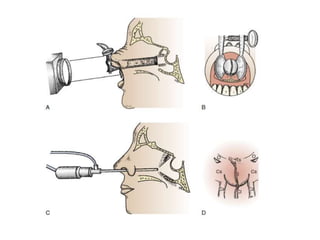
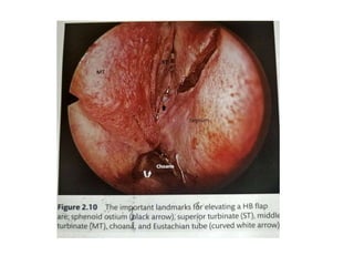
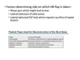
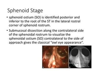
SlideShare a Scribd company logo
Endonasal Endoscopic
Anatomy
Presented By –
Dr. Rahul Jain
Moderated by : Dr V.C. Jha
HoD Deptt of Neurosurgery,
AIIMS Patna
Background
• Hermann Schloffer (1907) performed the first
transsphenoidal pituitary surgery via a lateral
rhinotomy approach
• Oscar Hirsch and Harvey Cushing attempted to
refine the technique in 1910.
• In 1965, Hardy was the first to use a microscope to
approach the sella, pioneering the transsphenoidal
approach.
• use of the endoscope was first described by Jho
and Carrau in 1997.
Advantages of an Endoscope
• The endoscope can physically enter into the
sphenoid sinus and provide a wide-angled
panoramic view with zooming capability.
• endoscope shows a diverging flask-shaped wide-
angled view.
• Angled views may be advantageous when large
suprasellar macroadenomas are to be removed or
direct visualization of the medial wall of a
cavernous sinus is required.
Microscope vs Endoscope
Parameter Microscope Endoscope
Fogging No Yes
Vision 3D 2D
Hemostasis Good Difficult
Dissection Good (Bimanual) Difficult
Exposure Tubular Panoramic
Extent of visualisation Narrow Wide
Extent of removal ++ ++++
C-Arm Yes Yes/No
Nasal Packing Yes Yes/No
Sinonasal Anatomy
Nasal Cavity is bordered
• medially – nasal septum (composed of the septal
cartilage, the perpendicular plate of the ethmoid bone,
and the vomer);
• superiorly by the cribriform plate of the ethmoid bone
and bridge of the nose (consisting of the nasal portion
of the frontal bone, nasal bone, and frontal process of
the maxilla);
• inferiorly by the floor of the nasal cavity (involving the
palatine process of the maxilla and the horizontal plate
of the palatine bone); and
• conchae or turbinates laterally (inferior, middle,
superior, and sometimes supreme turbinates).
• The superior and middle conchae (along with the
occasional supreme concha) are components of the
ethmoid bone, whereas the inferior concha is a
separate bone.
• The EE-TS procedure traverses the,region medial to
the middle turbinate, between the middle
turbinate and the nasal septum, on the way to the
sphenoid sinus then the pituitary fossa at the sella
turcica.
Osteomeatal Complex
• normal sinonasal anatomy located laterally to the
middle turbinate is referred to as the osteomeatal
complex (OMC) and comprises a key set of
structures for sinonasal function.
• consists of the middle turbinate, uncinate process,
hiatus semilunaris, ethmoid infundibulum, and
ethmoid bulla.
Hiatus semilunaris is
essentially a two-
dimensional (2D) crescent-
shaped opening leading
from the middle meatus
into the three-dimensional
(3D) funnel-shaped
ethmoid infundibulum to
which the frontal sinus,
anterior ethmoid sinus,
and maxillary sinus usually
drain.
• important point is that there are individual structural
variations, which can affect paranasal sinus physiology
and surgical anatomy.
• When the path of physiologic mucus flow is interrupted
mechanically or functionally, the paranasal sinuses can
retain stagnant mucus, which can subsequently
become infected and result in sinusitis.
• Anatomic variations may also include a pneumatized or
aerated turbinate, most frequently the middle
turbinate, which is referred to as concha bullosa and
can be enlarged.
• Despite these variations or the presence of any
nasal polyps, the main point is that structures of
the OMC should not be significantly disturbed en
route to the sphenoid sinus during EE-TS.
Ethmoidal Air Cells Variations
1. Agger Nasi cells
• Most anterior ethmoidal cells located just anterior
and lateral to the nasofrontal recess.
• For EE-TS, the surgeon should be aware that a
hyperpneumatized agger nasi cell can occasionally
present as a bulge that mimics the anterior view of
a turbinate.
2. Haller cells
• infraorbital ethmoid air cells or
maxilloethmoidal cells.
• closely related to the ethmoid
infundibulum along the medial
roof of the maxillary sinus.
• Because Haller cells are quite
lateral, these usually do not
present a problem during EE-TS,
although their proximity to the
ethmoid infundibulum can
result in inadvertent orbital
entry if not recognized.
3. Onodi Cells
• sphenoethmoidal cells
• posterior ethmoidal air cells that can project superiorly
into the sphenoid sinus toward the lateral side and can
potentially be confused with a septated region of the
sphenoid sinus.
• optic nerve and/or internal carotid artery (ICA) can
bulge into Onodi cells instead of the sphenoid sinus
proper or occasionally may have either partial or
complete bony dehiscence at the sphenoid sinus,
presenting risk for injury during surgery.
Nasal Turbinates
• inferior turbinate (IT) extends along the length of
the nasal cavity. Mucosal inferior turbinate
hypertrophy is its most common variation and may
narrow the nasal corridor space, but it usually
shrivels with the use of topical decongestants.
• middle turbinate (MT) has three attachments in the
sagittal, coronal and horizontal planes from an
anterior to posterior direction. MT presents with
many variations, its pneumatisation being the
commonest.
• A pneumatised MT, known as the concha bullosa,
needs to be opened (by means of conchoplasty), before
lateralising the MT. Sometimes, the MT may be
bulbous, owing to its bony structure, in which case,
conchoplasty cannot be performed, Neither can it be
lateralised easily to achieve wide nasal corridor.
• In such cases it is advisable to sacrifice the MT. We also
perform middle turbinectomy in cases which require
extended trans-sphenoidal route, transpterygoid route
or in paediatric patients, in whom adequate space is of
prime importance during the surgery.
• superior turbinate (ST) carries majority of the
olfactory fibres. Hence superior turbinectomy is
generally avoided to maintain post-op olfaction.
• In cases which require extended endoscopic trans-
sphenoid surgery, or in cases (where the sphenoid
is filled with pathology and the landmarks are
difficult to visualise, or in cases where space is
premium (for parking the endoscope, partial
superior turbinectomy is done and the posterior
ethmoids are opened.
Right partial superior turbincetomy done to expose posterior ethmoid cells
(white arrow)
Vasculature of Nasal Septum
• The blood supply originates from the ophthalmic
branch of the internal carotid artery (ICA) and
maxillary and facial branches of the external carotid
artery (ECA).
• The blood supply to the upper nasal septum is from
the anastomosis of anterior and posterior
ethmoidal arteries which are branches of
ophthalmic artery.
• Majority of nasal septum is supplied by
Sphenopalatine artery (SPA), branch of ECA.
• SPA perfuses the posterior and inferior division of
septa.
• SPA enters the nasal cavity through sphenopalatine
foramen where it divides into 2 major branches
namely septal and posterior lateral nasal artery.
• Septal artery exits SP foramen, courses through
rostrum of sphenoid and distributes the nasal
septum with 2-3 branches. Hence nasoseptal flap
should be wide enough including atleast 2-3
branches to maintain its viability.
Anatomic Landmarks in Stages of
EE-TS Pituitary Surgery
4 stages
1. Nasal stage
2. Sphenoid stage
3. Sellar stage
4. Reconstruction stage
Recognition of important landmarks during each of
these stages is the key to a safe exposure.
Nasal Stage
• endoscope is inserted in line with the floor of the
nasal cavity, parallel to the MT at an angle of 25°
inferiorly to initially visualize the choana.
• choana is the anatomic reference point.
• inferior margin of the MT leads to clival
indentation, which is about 1 cm, below the level of
sellar floor. This is quite a consistent surgical
landmark.
• Hadad Bassagasteguy (HB) flap is most commonly
used nasoseptal flap to seal off meninges from
nasal cavity and harvested prior to tumor
dissection. It is local, robust, easily harvestable flap.
• Based on sphenopalatine artery it is considered
gold standard for endoscopic skull base
reconstruction.
• Landmarks for harvesting HB flap – sphenoid
ostium, ST, MT, Eustachian tube, Choanal arch,
Mucocutaneous junction of septum.
• Factors determining side on which HB flap is taken:-
• Sharp spur which might lead to tear
• Lateral extension of sella tumor
• Lateral sphenoid CSF leak which requires sacrifice of septal
branch.
Sphenoid Stage
• sphenoid ostium (SO) is identified posterior and
inferior to the root of the ST in the lateral rostral
corner of sphenoid rostrum.
• Submucosal dissection along the contralateral side
of the sphenoidal rostrum to visualize the
sphenoidal ostium (SO) contralateral to the side of
approach gives the classical “owl eye appearance”.
• vomer is drilled and the rostrum of the sphenoid sinus
is removed
• The limits of the sphenoidotomy include: Cranially, the
superior limit of SO providing adequate visualization of
the planum sphenoidale, the optico-carotid recesses
and the optic protuberances; caudally, the
pterygo-sphenoid synchondrosis/vidian canal at 5 and 7
O’ clock position.
• The lateral limit is the crest marking the the junction of
the sphenoid and ethmoid sinuses with visualization of
the carotid artery (CA) protuberance.
Sellar Stage
• sphenoidal mucosa located only on the anterior
wall of the sella and the floor is coagulated with a
bipolar and excised.
• Anatomical landmarks are identified in the aerial
panoramic view and mimic a “fetal face”.
• Gentle drilling with a diamond burr under low
speed is done to thin the sellar floor to an egg shell
thickness.
• The anterior wall of the sella and its floor is
removed millimeter by millimeter circumferentially
till four blue lines (both the superiorly and inferiorly
located inter-cavernous sinuses and the laterally
located cavernous sinuses) are seen.
References
1. Schmidek and Sweet’s operative neurosurgical
techniques 7th Ed. Vol 1
2. Youmann’s and Winn Neurological surgery 8th Ed.
3. YR Yadav, BS Sharma – Neuroendoscopic Surgery.
4. Sharma BS, Sawarkar DP, Suri A. Endoscopic
pituitary surgery: Techniques, tips and tricks,
nuances, and complication avoidance. Neurol
India 2016;64:724-36
THANK YOU

More Related Content

What's hot

Csf rhinorrhoea endoscopic repair
Csf rhinorrhoea endoscopic repairCsf rhinorrhoea endoscopic repair
Csf rhinorrhoea endoscopic repair
Dr Shrikant Phatak
 
Endoscopic skull base surgery level iii
Endoscopic skull base surgery level iiiEndoscopic skull base surgery level iii
Endoscopic skull base surgery level iii
lpgupta
 
Imaging for Endoscopic Sinus Surgery
Imaging for Endoscopic Sinus SurgeryImaging for Endoscopic Sinus Surgery
Imaging for Endoscopic Sinus Surgery
Karnataka ENT Hospital & Research Center
 
middle fossa surgery
middle fossa surgerymiddle fossa surgery
middle fossa surgery
Surbhi narayan
 
Surgical anatomy of Infratemporal fossa. by Dr. Aditya Tiwari
Surgical anatomy of Infratemporal fossa. by Dr. Aditya TiwariSurgical anatomy of Infratemporal fossa. by Dr. Aditya Tiwari
Surgical anatomy of Infratemporal fossa. by Dr. Aditya Tiwari
Aditya Tiwari
 
Gene therapy Otolaryngology
Gene therapy  OtolaryngologyGene therapy  Otolaryngology
Gene therapy Otolaryngology
Balasubramanian Thiagarajan
 
Laryngeal transplantation
Laryngeal transplantationLaryngeal transplantation
Laryngeal transplantation
Dražen Shejbal
 
Temporal bone neoplasms
Temporal bone neoplasmsTemporal bone neoplasms
Temporal bone neoplasms
abhijeet89singh
 
Temporal bone
Temporal boneTemporal bone
Temporal bone
Thaiwat Tatsanawiwat
 
Facial nerve decompression
Facial nerve decompressionFacial nerve decompression
Facial nerve decompression
Mamoon Ameen
 
Endoscopic SKULL BASE surgery
Endoscopic SKULL BASE surgery Endoscopic SKULL BASE surgery
Endoscopic SKULL BASE surgery
Sundhar Krishnan
 
016 Transsphenoidal approch microscopic
016 Transsphenoidal approch microscopic016 Transsphenoidal approch microscopic
016 Transsphenoidal approch microscopic
Neurosurgery Vajira
 
Closed rhinoplasty
Closed rhinoplastyClosed rhinoplasty
Closed rhinoplasty
bhakabhob mahachitsattaya
 
Local flaps in ent
Local flaps in entLocal flaps in ent
Local flaps in ent
Ajay Manickam
 
Endoscopic middle ear surgery
Endoscopic middle ear surgeryEndoscopic middle ear surgery
Endoscopic middle ear surgery
Divya Raana
 
Sinus tympani prof dr bikash
Sinus tympani prof dr bikashSinus tympani prof dr bikash
Sinus tympani prof dr bikash
Bikash Shrestha
 
Frontal sinus surgical aproach
Frontal sinus surgical aproachFrontal sinus surgical aproach
Frontal sinus surgical aproach
Azadmeena7
 

What's hot (20)

Csf rhinorrhoea endoscopic repair
Csf rhinorrhoea endoscopic repairCsf rhinorrhoea endoscopic repair
Csf rhinorrhoea endoscopic repair
 
Endoscopic skull base surgery level iii
Endoscopic skull base surgery level iiiEndoscopic skull base surgery level iii
Endoscopic skull base surgery level iii
 
Imaging for Endoscopic Sinus Surgery
Imaging for Endoscopic Sinus SurgeryImaging for Endoscopic Sinus Surgery
Imaging for Endoscopic Sinus Surgery
 
middle fossa surgery
middle fossa surgerymiddle fossa surgery
middle fossa surgery
 
External rhinoplasty
External rhinoplastyExternal rhinoplasty
External rhinoplasty
 
Surgical anatomy of Infratemporal fossa. by Dr. Aditya Tiwari
Surgical anatomy of Infratemporal fossa. by Dr. Aditya TiwariSurgical anatomy of Infratemporal fossa. by Dr. Aditya Tiwari
Surgical anatomy of Infratemporal fossa. by Dr. Aditya Tiwari
 
Gene therapy Otolaryngology
Gene therapy  OtolaryngologyGene therapy  Otolaryngology
Gene therapy Otolaryngology
 
Laryngeal transplantation
Laryngeal transplantationLaryngeal transplantation
Laryngeal transplantation
 
Temporal bone neoplasms
Temporal bone neoplasmsTemporal bone neoplasms
Temporal bone neoplasms
 
Temporal bone
Temporal boneTemporal bone
Temporal bone
 
Facial nerve decompression
Facial nerve decompressionFacial nerve decompression
Facial nerve decompression
 
Endoscopic SKULL BASE surgery
Endoscopic SKULL BASE surgery Endoscopic SKULL BASE surgery
Endoscopic SKULL BASE surgery
 
Petrous apex 360°
Petrous apex 360°Petrous apex 360°
Petrous apex 360°
 
016 Transsphenoidal approch microscopic
016 Transsphenoidal approch microscopic016 Transsphenoidal approch microscopic
016 Transsphenoidal approch microscopic
 
Closed rhinoplasty
Closed rhinoplastyClosed rhinoplasty
Closed rhinoplasty
 
Local flaps in ent
Local flaps in entLocal flaps in ent
Local flaps in ent
 
Endoscopic middle ear surgery
Endoscopic middle ear surgeryEndoscopic middle ear surgery
Endoscopic middle ear surgery
 
Sinus tympani prof dr bikash
Sinus tympani prof dr bikashSinus tympani prof dr bikash
Sinus tympani prof dr bikash
 
Frontal sinus surgical aproach
Frontal sinus surgical aproachFrontal sinus surgical aproach
Frontal sinus surgical aproach
 
Petrous apex and skull base
Petrous apex and skull basePetrous apex and skull base
Petrous apex and skull base
 

Similar to Endoscopic Endonasal Anatomy.pptx

Imaging in ent
Imaging in entImaging in ent
Imaging in ent
trisha srivastava
 
Clinical Evaluation in Maxillofacial Trauma
Clinical Evaluation in Maxillofacial Trauma Clinical Evaluation in Maxillofacial Trauma
Clinical Evaluation in Maxillofacial Trauma
Arjun Shenoy
 
Functional endoscopic sinus surgery
Functional endoscopic sinus surgeryFunctional endoscopic sinus surgery
Functional endoscopic sinus surgery
Dʀ Smruti Ranjan Samal
 
PNS (Para-nasal-sinuses) anatomy and variants
PNS (Para-nasal-sinuses) anatomy and variantsPNS (Para-nasal-sinuses) anatomy and variants
PNS (Para-nasal-sinuses) anatomy and variants
Dr. Mohit Goel
 
Subtemporal Approach by Momen
Subtemporal Approach by MomenSubtemporal Approach by Momen
Subtemporal Approach by Momen
Momen Ali Khan
 
Surgical anatomy of Salivary glands - ORAL AND MAXILLOFACIAL SURGERY
Surgical anatomy of Salivary glands - ORAL AND MAXILLOFACIAL SURGERYSurgical anatomy of Salivary glands - ORAL AND MAXILLOFACIAL SURGERY
Surgical anatomy of Salivary glands - ORAL AND MAXILLOFACIAL SURGERY
ANIKET SARKAR
 
Basics of CT Scan interpretation of paranasal sinuses.pptx
Basics of CT Scan interpretation of paranasal sinuses.pptxBasics of CT Scan interpretation of paranasal sinuses.pptx
Basics of CT Scan interpretation of paranasal sinuses.pptx
RUTAYISIRE François Xavier
 
Imaging of paranasal sinuses
Imaging of paranasal sinusesImaging of paranasal sinuses
Imaging of paranasal sinuses
Archana Koshy
 
Anatomy temporal bone
Anatomy temporal bone Anatomy temporal bone
Anatomy temporal bone
KevinMungasia
 
Anatomy of temporal bone
Anatomy of temporal boneAnatomy of temporal bone
Anatomy of temporal bone
praneeth koduru
 
dhwani ct pns final (1).pptx all about it pns
dhwani ct pns final (1).pptx all about it pnsdhwani ct pns final (1).pptx all about it pns
dhwani ct pns final (1).pptx all about it pns
VivekMakadiya2
 
Radiology in ENT
Radiology in ENTRadiology in ENT
Radiology in ENT
Prasanna Datta
 
an overview of the paranasal sinuses.ppt
an overview of the paranasal sinuses.pptan overview of the paranasal sinuses.ppt
an overview of the paranasal sinuses.ppt
AnshikaRajput45
 
Paranasal sinuses anatomy and pathology dr ashok
Paranasal  sinuses  anatomy and pathology dr ashokParanasal  sinuses  anatomy and pathology dr ashok
Paranasal sinuses anatomy and pathology dr ashok
Ashok Sharma
 
Nasal septal anatomy and septoplasty
Nasal septal anatomy and septoplastyNasal septal anatomy and septoplasty
Nasal septal anatomy and septoplasty
Dr Safika Zaman
 
Fess part 2,3,4,5
Fess part 2,3,4,5Fess part 2,3,4,5
Fess part 2,3,4,5
drmhndalali
 
Endoscopic Transnasal transsphenoid pituitary Surgery (Erbil).pptx
Endoscopic Transnasal transsphenoid pituitary Surgery (Erbil).pptxEndoscopic Transnasal transsphenoid pituitary Surgery (Erbil).pptx
Endoscopic Transnasal transsphenoid pituitary Surgery (Erbil).pptx
ahmedmhoder
 
Lesions of the temporal bone & petrous ppt
Lesions of the  temporal bone & petrous  pptLesions of the  temporal bone & petrous  ppt
Lesions of the temporal bone & petrous ppt
DEBKUMAR BISWAS
 
Round window
Round windowRound window
temporalbone-141009084034-conversion-gate02 (1).pdf
temporalbone-141009084034-conversion-gate02 (1).pdftemporalbone-141009084034-conversion-gate02 (1).pdf
temporalbone-141009084034-conversion-gate02 (1).pdf
joanluciya
 

Similar to Endoscopic Endonasal Anatomy.pptx (20)

Imaging in ent
Imaging in entImaging in ent
Imaging in ent
 
Clinical Evaluation in Maxillofacial Trauma
Clinical Evaluation in Maxillofacial Trauma Clinical Evaluation in Maxillofacial Trauma
Clinical Evaluation in Maxillofacial Trauma
 
Functional endoscopic sinus surgery
Functional endoscopic sinus surgeryFunctional endoscopic sinus surgery
Functional endoscopic sinus surgery
 
PNS (Para-nasal-sinuses) anatomy and variants
PNS (Para-nasal-sinuses) anatomy and variantsPNS (Para-nasal-sinuses) anatomy and variants
PNS (Para-nasal-sinuses) anatomy and variants
 
Subtemporal Approach by Momen
Subtemporal Approach by MomenSubtemporal Approach by Momen
Subtemporal Approach by Momen
 
Surgical anatomy of Salivary glands - ORAL AND MAXILLOFACIAL SURGERY
Surgical anatomy of Salivary glands - ORAL AND MAXILLOFACIAL SURGERYSurgical anatomy of Salivary glands - ORAL AND MAXILLOFACIAL SURGERY
Surgical anatomy of Salivary glands - ORAL AND MAXILLOFACIAL SURGERY
 
Basics of CT Scan interpretation of paranasal sinuses.pptx
Basics of CT Scan interpretation of paranasal sinuses.pptxBasics of CT Scan interpretation of paranasal sinuses.pptx
Basics of CT Scan interpretation of paranasal sinuses.pptx
 
Imaging of paranasal sinuses
Imaging of paranasal sinusesImaging of paranasal sinuses
Imaging of paranasal sinuses
 
Anatomy temporal bone
Anatomy temporal bone Anatomy temporal bone
Anatomy temporal bone
 
Anatomy of temporal bone
Anatomy of temporal boneAnatomy of temporal bone
Anatomy of temporal bone
 
dhwani ct pns final (1).pptx all about it pns
dhwani ct pns final (1).pptx all about it pnsdhwani ct pns final (1).pptx all about it pns
dhwani ct pns final (1).pptx all about it pns
 
Radiology in ENT
Radiology in ENTRadiology in ENT
Radiology in ENT
 
an overview of the paranasal sinuses.ppt
an overview of the paranasal sinuses.pptan overview of the paranasal sinuses.ppt
an overview of the paranasal sinuses.ppt
 
Paranasal sinuses anatomy and pathology dr ashok
Paranasal  sinuses  anatomy and pathology dr ashokParanasal  sinuses  anatomy and pathology dr ashok
Paranasal sinuses anatomy and pathology dr ashok
 
Nasal septal anatomy and septoplasty
Nasal septal anatomy and septoplastyNasal septal anatomy and septoplasty
Nasal septal anatomy and septoplasty
 
Fess part 2,3,4,5
Fess part 2,3,4,5Fess part 2,3,4,5
Fess part 2,3,4,5
 
Endoscopic Transnasal transsphenoid pituitary Surgery (Erbil).pptx
Endoscopic Transnasal transsphenoid pituitary Surgery (Erbil).pptxEndoscopic Transnasal transsphenoid pituitary Surgery (Erbil).pptx
Endoscopic Transnasal transsphenoid pituitary Surgery (Erbil).pptx
 
Lesions of the temporal bone & petrous ppt
Lesions of the  temporal bone & petrous  pptLesions of the  temporal bone & petrous  ppt
Lesions of the temporal bone & petrous ppt
 
Round window
Round windowRound window
Round window
 
temporalbone-141009084034-conversion-gate02 (1).pdf
temporalbone-141009084034-conversion-gate02 (1).pdftemporalbone-141009084034-conversion-gate02 (1).pdf
temporalbone-141009084034-conversion-gate02 (1).pdf
 

More from Dr. Rahul Jain

Moya Moya disease (vasculopathy/angiopathy)
Moya Moya disease (vasculopathy/angiopathy)Moya Moya disease (vasculopathy/angiopathy)
Moya Moya disease (vasculopathy/angiopathy)
Dr. Rahul Jain
 
Cerebrovascular Vasospasm - Etiopathogenesis and Management
Cerebrovascular Vasospasm - Etiopathogenesis and ManagementCerebrovascular Vasospasm - Etiopathogenesis and Management
Cerebrovascular Vasospasm - Etiopathogenesis and Management
Dr. Rahul Jain
 
Chiari Malformations.pptx
Chiari Malformations.pptxChiari Malformations.pptx
Chiari Malformations.pptx
Dr. Rahul Jain
 
Journal Club - Extra axial Endoscopic Third Ventriculostomy.pptx
Journal Club - Extra axial Endoscopic Third Ventriculostomy.pptxJournal Club - Extra axial Endoscopic Third Ventriculostomy.pptx
Journal Club - Extra axial Endoscopic Third Ventriculostomy.pptx
Dr. Rahul Jain
 
Diffuse Midline Gliomas/ Diffuse Pontine Gliomas.pptx
Diffuse Midline Gliomas/ Diffuse Pontine Gliomas.pptxDiffuse Midline Gliomas/ Diffuse Pontine Gliomas.pptx
Diffuse Midline Gliomas/ Diffuse Pontine Gliomas.pptx
Dr. Rahul Jain
 
Trigeminal Neuralgia.pptx
Trigeminal Neuralgia.pptxTrigeminal Neuralgia.pptx
Trigeminal Neuralgia.pptx
Dr. Rahul Jain
 
Microscopes and Endoscopes in Neurosurgery.pptx
Microscopes and Endoscopes in Neurosurgery.pptxMicroscopes and Endoscopes in Neurosurgery.pptx
Microscopes and Endoscopes in Neurosurgery.pptx
Dr. Rahul Jain
 
Subaxial Cervical spine fusion.pptx
Subaxial Cervical spine fusion.pptxSubaxial Cervical spine fusion.pptx
Subaxial Cervical spine fusion.pptx
Dr. Rahul Jain
 
Carotid Occlusive Disease.pptx
Carotid Occlusive Disease.pptxCarotid Occlusive Disease.pptx
Carotid Occlusive Disease.pptx
Dr. Rahul Jain
 
Brain AVM (ArterioVenous Malformation) Managment.pptx
Brain AVM (ArterioVenous Malformation) Managment.pptxBrain AVM (ArterioVenous Malformation) Managment.pptx
Brain AVM (ArterioVenous Malformation) Managment.pptx
Dr. Rahul Jain
 
Intracranial Vascular Bypass.pptx
Intracranial Vascular Bypass.pptxIntracranial Vascular Bypass.pptx
Intracranial Vascular Bypass.pptx
Dr. Rahul Jain
 
Anterior Choroidal Artery.pptx
Anterior Choroidal Artery.pptxAnterior Choroidal Artery.pptx
Anterior Choroidal Artery.pptx
Dr. Rahul Jain
 
Decompressive Craniectomy.pptx
Decompressive Craniectomy.pptxDecompressive Craniectomy.pptx
Decompressive Craniectomy.pptx
Dr. Rahul Jain
 
Audilogical Assessment.pptx
Audilogical Assessment.pptxAudilogical Assessment.pptx
Audilogical Assessment.pptx
Dr. Rahul Jain
 
HYDROCEPHALUS.pptx
HYDROCEPHALUS.pptxHYDROCEPHALUS.pptx
HYDROCEPHALUS.pptx
Dr. Rahul Jain
 
CNS WHO 2021 tumor classification.pptx
CNS WHO 2021 tumor classification.pptxCNS WHO 2021 tumor classification.pptx
CNS WHO 2021 tumor classification.pptx
Dr. Rahul Jain
 
Cerebral Venous thrombosis.pptx
Cerebral Venous thrombosis.pptxCerebral Venous thrombosis.pptx
Cerebral Venous thrombosis.pptx
Dr. Rahul Jain
 
Vertebral Artery Anatomy with Endovascular.pptx
Vertebral Artery Anatomy with Endovascular.pptxVertebral Artery Anatomy with Endovascular.pptx
Vertebral Artery Anatomy with Endovascular.pptx
Dr. Rahul Jain
 
Minimal Invasive Surgery in CA Rectum
Minimal Invasive Surgery in CA RectumMinimal Invasive Surgery in CA Rectum
Minimal Invasive Surgery in CA Rectum
Dr. Rahul Jain
 
Liver anatomy
Liver anatomyLiver anatomy
Liver anatomy
Dr. Rahul Jain
 

More from Dr. Rahul Jain (20)

Moya Moya disease (vasculopathy/angiopathy)
Moya Moya disease (vasculopathy/angiopathy)Moya Moya disease (vasculopathy/angiopathy)
Moya Moya disease (vasculopathy/angiopathy)
 
Cerebrovascular Vasospasm - Etiopathogenesis and Management
Cerebrovascular Vasospasm - Etiopathogenesis and ManagementCerebrovascular Vasospasm - Etiopathogenesis and Management
Cerebrovascular Vasospasm - Etiopathogenesis and Management
 
Chiari Malformations.pptx
Chiari Malformations.pptxChiari Malformations.pptx
Chiari Malformations.pptx
 
Journal Club - Extra axial Endoscopic Third Ventriculostomy.pptx
Journal Club - Extra axial Endoscopic Third Ventriculostomy.pptxJournal Club - Extra axial Endoscopic Third Ventriculostomy.pptx
Journal Club - Extra axial Endoscopic Third Ventriculostomy.pptx
 
Diffuse Midline Gliomas/ Diffuse Pontine Gliomas.pptx
Diffuse Midline Gliomas/ Diffuse Pontine Gliomas.pptxDiffuse Midline Gliomas/ Diffuse Pontine Gliomas.pptx
Diffuse Midline Gliomas/ Diffuse Pontine Gliomas.pptx
 
Trigeminal Neuralgia.pptx
Trigeminal Neuralgia.pptxTrigeminal Neuralgia.pptx
Trigeminal Neuralgia.pptx
 
Microscopes and Endoscopes in Neurosurgery.pptx
Microscopes and Endoscopes in Neurosurgery.pptxMicroscopes and Endoscopes in Neurosurgery.pptx
Microscopes and Endoscopes in Neurosurgery.pptx
 
Subaxial Cervical spine fusion.pptx
Subaxial Cervical spine fusion.pptxSubaxial Cervical spine fusion.pptx
Subaxial Cervical spine fusion.pptx
 
Carotid Occlusive Disease.pptx
Carotid Occlusive Disease.pptxCarotid Occlusive Disease.pptx
Carotid Occlusive Disease.pptx
 
Brain AVM (ArterioVenous Malformation) Managment.pptx
Brain AVM (ArterioVenous Malformation) Managment.pptxBrain AVM (ArterioVenous Malformation) Managment.pptx
Brain AVM (ArterioVenous Malformation) Managment.pptx
 
Intracranial Vascular Bypass.pptx
Intracranial Vascular Bypass.pptxIntracranial Vascular Bypass.pptx
Intracranial Vascular Bypass.pptx
 
Anterior Choroidal Artery.pptx
Anterior Choroidal Artery.pptxAnterior Choroidal Artery.pptx
Anterior Choroidal Artery.pptx
 
Decompressive Craniectomy.pptx
Decompressive Craniectomy.pptxDecompressive Craniectomy.pptx
Decompressive Craniectomy.pptx
 
Audilogical Assessment.pptx
Audilogical Assessment.pptxAudilogical Assessment.pptx
Audilogical Assessment.pptx
 
HYDROCEPHALUS.pptx
HYDROCEPHALUS.pptxHYDROCEPHALUS.pptx
HYDROCEPHALUS.pptx
 
CNS WHO 2021 tumor classification.pptx
CNS WHO 2021 tumor classification.pptxCNS WHO 2021 tumor classification.pptx
CNS WHO 2021 tumor classification.pptx
 
Cerebral Venous thrombosis.pptx
Cerebral Venous thrombosis.pptxCerebral Venous thrombosis.pptx
Cerebral Venous thrombosis.pptx
 
Vertebral Artery Anatomy with Endovascular.pptx
Vertebral Artery Anatomy with Endovascular.pptxVertebral Artery Anatomy with Endovascular.pptx
Vertebral Artery Anatomy with Endovascular.pptx
 
Minimal Invasive Surgery in CA Rectum
Minimal Invasive Surgery in CA RectumMinimal Invasive Surgery in CA Rectum
Minimal Invasive Surgery in CA Rectum
 
Liver anatomy
Liver anatomyLiver anatomy
Liver anatomy
 

Recently uploaded

Pulmonary Thromboembolism - etilogy, types, medical- Surgical and nursing man...
Pulmonary Thromboembolism - etilogy, types, medical- Surgical and nursing man...Pulmonary Thromboembolism - etilogy, types, medical- Surgical and nursing man...
Pulmonary Thromboembolism - etilogy, types, medical- Surgical and nursing man...
VarunMahajani
 
Phone Us ❤85270-49040❤ #ℂall #gIRLS In Surat By Surat @ℂall @Girls Hotel With...
Phone Us ❤85270-49040❤ #ℂall #gIRLS In Surat By Surat @ℂall @Girls Hotel With...Phone Us ❤85270-49040❤ #ℂall #gIRLS In Surat By Surat @ℂall @Girls Hotel With...
Phone Us ❤85270-49040❤ #ℂall #gIRLS In Surat By Surat @ℂall @Girls Hotel With...
Savita Shen $i11
 
Physiology of Chemical Sensation of smell.pdf
Physiology of Chemical Sensation of smell.pdfPhysiology of Chemical Sensation of smell.pdf
Physiology of Chemical Sensation of smell.pdf
MedicoseAcademics
 
Lung Cancer: Artificial Intelligence, Synergetics, Complex System Analysis, S...
Lung Cancer: Artificial Intelligence, Synergetics, Complex System Analysis, S...Lung Cancer: Artificial Intelligence, Synergetics, Complex System Analysis, S...
Lung Cancer: Artificial Intelligence, Synergetics, Complex System Analysis, S...
Oleg Kshivets
 
Alcohol_Dr. Jeenal Mistry MD Pharmacology.pdf
Alcohol_Dr. Jeenal Mistry MD Pharmacology.pdfAlcohol_Dr. Jeenal Mistry MD Pharmacology.pdf
Alcohol_Dr. Jeenal Mistry MD Pharmacology.pdf
Dr Jeenal Mistry
 
Flu Vaccine Alert in Bangalore Karnataka
Flu Vaccine Alert in Bangalore KarnatakaFlu Vaccine Alert in Bangalore Karnataka
Flu Vaccine Alert in Bangalore Karnataka
addon Scans
 
micro teaching on communication m.sc nursing.pdf
micro teaching on communication m.sc nursing.pdfmicro teaching on communication m.sc nursing.pdf
micro teaching on communication m.sc nursing.pdf
Anurag Sharma
 
ARTIFICIAL INTELLIGENCE IN HEALTHCARE.pdf
ARTIFICIAL INTELLIGENCE IN  HEALTHCARE.pdfARTIFICIAL INTELLIGENCE IN  HEALTHCARE.pdf
ARTIFICIAL INTELLIGENCE IN HEALTHCARE.pdf
Anujkumaranit
 
Maxilla, Mandible & Hyoid Bone & Clinical Correlations by Dr. RIG.pptx
Maxilla, Mandible & Hyoid Bone & Clinical Correlations by Dr. RIG.pptxMaxilla, Mandible & Hyoid Bone & Clinical Correlations by Dr. RIG.pptx
Maxilla, Mandible & Hyoid Bone & Clinical Correlations by Dr. RIG.pptx
Dr. Rabia Inam Gandapore
 
Triangles of Neck and Clinical Correlation by Dr. RIG.pptx
Triangles of Neck and Clinical Correlation by Dr. RIG.pptxTriangles of Neck and Clinical Correlation by Dr. RIG.pptx
Triangles of Neck and Clinical Correlation by Dr. RIG.pptx
Dr. Rabia Inam Gandapore
 
Evaluation of antidepressant activity of clitoris ternatea in animals
Evaluation of antidepressant activity of clitoris ternatea in animalsEvaluation of antidepressant activity of clitoris ternatea in animals
Evaluation of antidepressant activity of clitoris ternatea in animals
Shweta
 
Report Back from SGO 2024: What’s the Latest in Cervical Cancer?
Report Back from SGO 2024: What’s the Latest in Cervical Cancer?Report Back from SGO 2024: What’s the Latest in Cervical Cancer?
Report Back from SGO 2024: What’s the Latest in Cervical Cancer?
bkling
 
Are There Any Natural Remedies To Treat Syphilis.pdf
Are There Any Natural Remedies To Treat Syphilis.pdfAre There Any Natural Remedies To Treat Syphilis.pdf
Are There Any Natural Remedies To Treat Syphilis.pdf
Little Cross Family Clinic
 
New Drug Discovery and Development .....
New Drug Discovery and Development .....New Drug Discovery and Development .....
New Drug Discovery and Development .....
NEHA GUPTA
 
Hemodialysis: Chapter 3, Dialysis Water Unit - Dr.Gawad
Hemodialysis: Chapter 3, Dialysis Water Unit - Dr.GawadHemodialysis: Chapter 3, Dialysis Water Unit - Dr.Gawad
Hemodialysis: Chapter 3, Dialysis Water Unit - Dr.Gawad
NephroTube - Dr.Gawad
 
Ozempic: Preoperative Management of Patients on GLP-1 Receptor Agonists
Ozempic: Preoperative Management of Patients on GLP-1 Receptor Agonists  Ozempic: Preoperative Management of Patients on GLP-1 Receptor Agonists
Ozempic: Preoperative Management of Patients on GLP-1 Receptor Agonists
Saeid Safari
 
TEST BANK for Operations Management, 14th Edition by William J. Stevenson, Ve...
TEST BANK for Operations Management, 14th Edition by William J. Stevenson, Ve...TEST BANK for Operations Management, 14th Edition by William J. Stevenson, Ve...
TEST BANK for Operations Management, 14th Edition by William J. Stevenson, Ve...
kevinkariuki227
 
BENIGN PROSTATIC HYPERPLASIA.BPH. BPHpdf
BENIGN PROSTATIC HYPERPLASIA.BPH. BPHpdfBENIGN PROSTATIC HYPERPLASIA.BPH. BPHpdf
BENIGN PROSTATIC HYPERPLASIA.BPH. BPHpdf
DR SETH JOTHAM
 
Ophthalmology Clinical Tests for OSCE exam
Ophthalmology Clinical Tests for OSCE examOphthalmology Clinical Tests for OSCE exam
Ophthalmology Clinical Tests for OSCE exam
KafrELShiekh University
 
Pharynx and Clinical Correlations BY Dr.Rabia Inam Gandapore.pptx
Pharynx and Clinical Correlations BY Dr.Rabia Inam Gandapore.pptxPharynx and Clinical Correlations BY Dr.Rabia Inam Gandapore.pptx
Pharynx and Clinical Correlations BY Dr.Rabia Inam Gandapore.pptx
Dr. Rabia Inam Gandapore
 

Recently uploaded (20)

Pulmonary Thromboembolism - etilogy, types, medical- Surgical and nursing man...
Pulmonary Thromboembolism - etilogy, types, medical- Surgical and nursing man...Pulmonary Thromboembolism - etilogy, types, medical- Surgical and nursing man...
Pulmonary Thromboembolism - etilogy, types, medical- Surgical and nursing man...
 
Phone Us ❤85270-49040❤ #ℂall #gIRLS In Surat By Surat @ℂall @Girls Hotel With...
Phone Us ❤85270-49040❤ #ℂall #gIRLS In Surat By Surat @ℂall @Girls Hotel With...Phone Us ❤85270-49040❤ #ℂall #gIRLS In Surat By Surat @ℂall @Girls Hotel With...
Phone Us ❤85270-49040❤ #ℂall #gIRLS In Surat By Surat @ℂall @Girls Hotel With...
 
Physiology of Chemical Sensation of smell.pdf
Physiology of Chemical Sensation of smell.pdfPhysiology of Chemical Sensation of smell.pdf
Physiology of Chemical Sensation of smell.pdf
 
Lung Cancer: Artificial Intelligence, Synergetics, Complex System Analysis, S...
Lung Cancer: Artificial Intelligence, Synergetics, Complex System Analysis, S...Lung Cancer: Artificial Intelligence, Synergetics, Complex System Analysis, S...
Lung Cancer: Artificial Intelligence, Synergetics, Complex System Analysis, S...
 
Alcohol_Dr. Jeenal Mistry MD Pharmacology.pdf
Alcohol_Dr. Jeenal Mistry MD Pharmacology.pdfAlcohol_Dr. Jeenal Mistry MD Pharmacology.pdf
Alcohol_Dr. Jeenal Mistry MD Pharmacology.pdf
 
Flu Vaccine Alert in Bangalore Karnataka
Flu Vaccine Alert in Bangalore KarnatakaFlu Vaccine Alert in Bangalore Karnataka
Flu Vaccine Alert in Bangalore Karnataka
 
micro teaching on communication m.sc nursing.pdf
micro teaching on communication m.sc nursing.pdfmicro teaching on communication m.sc nursing.pdf
micro teaching on communication m.sc nursing.pdf
 
ARTIFICIAL INTELLIGENCE IN HEALTHCARE.pdf
ARTIFICIAL INTELLIGENCE IN  HEALTHCARE.pdfARTIFICIAL INTELLIGENCE IN  HEALTHCARE.pdf
ARTIFICIAL INTELLIGENCE IN HEALTHCARE.pdf
 
Maxilla, Mandible & Hyoid Bone & Clinical Correlations by Dr. RIG.pptx
Maxilla, Mandible & Hyoid Bone & Clinical Correlations by Dr. RIG.pptxMaxilla, Mandible & Hyoid Bone & Clinical Correlations by Dr. RIG.pptx
Maxilla, Mandible & Hyoid Bone & Clinical Correlations by Dr. RIG.pptx
 
Triangles of Neck and Clinical Correlation by Dr. RIG.pptx
Triangles of Neck and Clinical Correlation by Dr. RIG.pptxTriangles of Neck and Clinical Correlation by Dr. RIG.pptx
Triangles of Neck and Clinical Correlation by Dr. RIG.pptx
 
Evaluation of antidepressant activity of clitoris ternatea in animals
Evaluation of antidepressant activity of clitoris ternatea in animalsEvaluation of antidepressant activity of clitoris ternatea in animals
Evaluation of antidepressant activity of clitoris ternatea in animals
 
Report Back from SGO 2024: What’s the Latest in Cervical Cancer?
Report Back from SGO 2024: What’s the Latest in Cervical Cancer?Report Back from SGO 2024: What’s the Latest in Cervical Cancer?
Report Back from SGO 2024: What’s the Latest in Cervical Cancer?
 
Are There Any Natural Remedies To Treat Syphilis.pdf
Are There Any Natural Remedies To Treat Syphilis.pdfAre There Any Natural Remedies To Treat Syphilis.pdf
Are There Any Natural Remedies To Treat Syphilis.pdf
 
New Drug Discovery and Development .....
New Drug Discovery and Development .....New Drug Discovery and Development .....
New Drug Discovery and Development .....
 
Hemodialysis: Chapter 3, Dialysis Water Unit - Dr.Gawad
Hemodialysis: Chapter 3, Dialysis Water Unit - Dr.GawadHemodialysis: Chapter 3, Dialysis Water Unit - Dr.Gawad
Hemodialysis: Chapter 3, Dialysis Water Unit - Dr.Gawad
 
Ozempic: Preoperative Management of Patients on GLP-1 Receptor Agonists
Ozempic: Preoperative Management of Patients on GLP-1 Receptor Agonists  Ozempic: Preoperative Management of Patients on GLP-1 Receptor Agonists
Ozempic: Preoperative Management of Patients on GLP-1 Receptor Agonists
 
TEST BANK for Operations Management, 14th Edition by William J. Stevenson, Ve...
TEST BANK for Operations Management, 14th Edition by William J. Stevenson, Ve...TEST BANK for Operations Management, 14th Edition by William J. Stevenson, Ve...
TEST BANK for Operations Management, 14th Edition by William J. Stevenson, Ve...
 
BENIGN PROSTATIC HYPERPLASIA.BPH. BPHpdf
BENIGN PROSTATIC HYPERPLASIA.BPH. BPHpdfBENIGN PROSTATIC HYPERPLASIA.BPH. BPHpdf
BENIGN PROSTATIC HYPERPLASIA.BPH. BPHpdf
 
Ophthalmology Clinical Tests for OSCE exam
Ophthalmology Clinical Tests for OSCE examOphthalmology Clinical Tests for OSCE exam
Ophthalmology Clinical Tests for OSCE exam
 
Pharynx and Clinical Correlations BY Dr.Rabia Inam Gandapore.pptx
Pharynx and Clinical Correlations BY Dr.Rabia Inam Gandapore.pptxPharynx and Clinical Correlations BY Dr.Rabia Inam Gandapore.pptx
Pharynx and Clinical Correlations BY Dr.Rabia Inam Gandapore.pptx
 

Endoscopic Endonasal Anatomy.pptx

  • 1. Endonasal Endoscopic Anatomy Presented By – Dr. Rahul Jain Moderated by : Dr V.C. Jha HoD Deptt of Neurosurgery, AIIMS Patna
  • 2. Background • Hermann Schloffer (1907) performed the first transsphenoidal pituitary surgery via a lateral rhinotomy approach • Oscar Hirsch and Harvey Cushing attempted to refine the technique in 1910. • In 1965, Hardy was the first to use a microscope to approach the sella, pioneering the transsphenoidal approach. • use of the endoscope was first described by Jho and Carrau in 1997.
  • 3. Advantages of an Endoscope • The endoscope can physically enter into the sphenoid sinus and provide a wide-angled panoramic view with zooming capability. • endoscope shows a diverging flask-shaped wide- angled view. • Angled views may be advantageous when large suprasellar macroadenomas are to be removed or direct visualization of the medial wall of a cavernous sinus is required.
  • 4. Microscope vs Endoscope Parameter Microscope Endoscope Fogging No Yes Vision 3D 2D Hemostasis Good Difficult Dissection Good (Bimanual) Difficult Exposure Tubular Panoramic Extent of visualisation Narrow Wide Extent of removal ++ ++++ C-Arm Yes Yes/No Nasal Packing Yes Yes/No
  • 5.
  • 6. Sinonasal Anatomy Nasal Cavity is bordered • medially – nasal septum (composed of the septal cartilage, the perpendicular plate of the ethmoid bone, and the vomer); • superiorly by the cribriform plate of the ethmoid bone and bridge of the nose (consisting of the nasal portion of the frontal bone, nasal bone, and frontal process of the maxilla); • inferiorly by the floor of the nasal cavity (involving the palatine process of the maxilla and the horizontal plate of the palatine bone); and • conchae or turbinates laterally (inferior, middle, superior, and sometimes supreme turbinates).
  • 7.
  • 8. • The superior and middle conchae (along with the occasional supreme concha) are components of the ethmoid bone, whereas the inferior concha is a separate bone. • The EE-TS procedure traverses the,region medial to the middle turbinate, between the middle turbinate and the nasal septum, on the way to the sphenoid sinus then the pituitary fossa at the sella turcica.
  • 9. Osteomeatal Complex • normal sinonasal anatomy located laterally to the middle turbinate is referred to as the osteomeatal complex (OMC) and comprises a key set of structures for sinonasal function. • consists of the middle turbinate, uncinate process, hiatus semilunaris, ethmoid infundibulum, and ethmoid bulla.
  • 10. Hiatus semilunaris is essentially a two- dimensional (2D) crescent- shaped opening leading from the middle meatus into the three-dimensional (3D) funnel-shaped ethmoid infundibulum to which the frontal sinus, anterior ethmoid sinus, and maxillary sinus usually drain.
  • 11.
  • 12. • important point is that there are individual structural variations, which can affect paranasal sinus physiology and surgical anatomy. • When the path of physiologic mucus flow is interrupted mechanically or functionally, the paranasal sinuses can retain stagnant mucus, which can subsequently become infected and result in sinusitis. • Anatomic variations may also include a pneumatized or aerated turbinate, most frequently the middle turbinate, which is referred to as concha bullosa and can be enlarged.
  • 13. • Despite these variations or the presence of any nasal polyps, the main point is that structures of the OMC should not be significantly disturbed en route to the sphenoid sinus during EE-TS.
  • 14. Ethmoidal Air Cells Variations 1. Agger Nasi cells • Most anterior ethmoidal cells located just anterior and lateral to the nasofrontal recess. • For EE-TS, the surgeon should be aware that a hyperpneumatized agger nasi cell can occasionally present as a bulge that mimics the anterior view of a turbinate.
  • 15. 2. Haller cells • infraorbital ethmoid air cells or maxilloethmoidal cells. • closely related to the ethmoid infundibulum along the medial roof of the maxillary sinus. • Because Haller cells are quite lateral, these usually do not present a problem during EE-TS, although their proximity to the ethmoid infundibulum can result in inadvertent orbital entry if not recognized.
  • 16. 3. Onodi Cells • sphenoethmoidal cells • posterior ethmoidal air cells that can project superiorly into the sphenoid sinus toward the lateral side and can potentially be confused with a septated region of the sphenoid sinus. • optic nerve and/or internal carotid artery (ICA) can bulge into Onodi cells instead of the sphenoid sinus proper or occasionally may have either partial or complete bony dehiscence at the sphenoid sinus, presenting risk for injury during surgery.
  • 17.
  • 18. Nasal Turbinates • inferior turbinate (IT) extends along the length of the nasal cavity. Mucosal inferior turbinate hypertrophy is its most common variation and may narrow the nasal corridor space, but it usually shrivels with the use of topical decongestants. • middle turbinate (MT) has three attachments in the sagittal, coronal and horizontal planes from an anterior to posterior direction. MT presents with many variations, its pneumatisation being the commonest.
  • 19. • A pneumatised MT, known as the concha bullosa, needs to be opened (by means of conchoplasty), before lateralising the MT. Sometimes, the MT may be bulbous, owing to its bony structure, in which case, conchoplasty cannot be performed, Neither can it be lateralised easily to achieve wide nasal corridor. • In such cases it is advisable to sacrifice the MT. We also perform middle turbinectomy in cases which require extended trans-sphenoidal route, transpterygoid route or in paediatric patients, in whom adequate space is of prime importance during the surgery.
  • 20. • superior turbinate (ST) carries majority of the olfactory fibres. Hence superior turbinectomy is generally avoided to maintain post-op olfaction. • In cases which require extended endoscopic trans- sphenoid surgery, or in cases (where the sphenoid is filled with pathology and the landmarks are difficult to visualise, or in cases where space is premium (for parking the endoscope, partial superior turbinectomy is done and the posterior ethmoids are opened.
  • 21. Right partial superior turbincetomy done to expose posterior ethmoid cells (white arrow)
  • 22. Vasculature of Nasal Septum • The blood supply originates from the ophthalmic branch of the internal carotid artery (ICA) and maxillary and facial branches of the external carotid artery (ECA). • The blood supply to the upper nasal septum is from the anastomosis of anterior and posterior ethmoidal arteries which are branches of ophthalmic artery.
  • 23. • Majority of nasal septum is supplied by Sphenopalatine artery (SPA), branch of ECA. • SPA perfuses the posterior and inferior division of septa. • SPA enters the nasal cavity through sphenopalatine foramen where it divides into 2 major branches namely septal and posterior lateral nasal artery.
  • 24. • Septal artery exits SP foramen, courses through rostrum of sphenoid and distributes the nasal septum with 2-3 branches. Hence nasoseptal flap should be wide enough including atleast 2-3 branches to maintain its viability.
  • 25.
  • 26. Anatomic Landmarks in Stages of EE-TS Pituitary Surgery 4 stages 1. Nasal stage 2. Sphenoid stage 3. Sellar stage 4. Reconstruction stage Recognition of important landmarks during each of these stages is the key to a safe exposure.
  • 27. Nasal Stage • endoscope is inserted in line with the floor of the nasal cavity, parallel to the MT at an angle of 25° inferiorly to initially visualize the choana. • choana is the anatomic reference point. • inferior margin of the MT leads to clival indentation, which is about 1 cm, below the level of sellar floor. This is quite a consistent surgical landmark.
  • 28.
  • 29. • Hadad Bassagasteguy (HB) flap is most commonly used nasoseptal flap to seal off meninges from nasal cavity and harvested prior to tumor dissection. It is local, robust, easily harvestable flap. • Based on sphenopalatine artery it is considered gold standard for endoscopic skull base reconstruction. • Landmarks for harvesting HB flap – sphenoid ostium, ST, MT, Eustachian tube, Choanal arch, Mucocutaneous junction of septum.
  • 30.
  • 31. • Factors determining side on which HB flap is taken:- • Sharp spur which might lead to tear • Lateral extension of sella tumor • Lateral sphenoid CSF leak which requires sacrifice of septal branch.
  • 32. Sphenoid Stage • sphenoid ostium (SO) is identified posterior and inferior to the root of the ST in the lateral rostral corner of sphenoid rostrum. • Submucosal dissection along the contralateral side of the sphenoidal rostrum to visualize the sphenoidal ostium (SO) contralateral to the side of approach gives the classical “owl eye appearance”.
  • 33. • vomer is drilled and the rostrum of the sphenoid sinus is removed • The limits of the sphenoidotomy include: Cranially, the superior limit of SO providing adequate visualization of the planum sphenoidale, the optico-carotid recesses and the optic protuberances; caudally, the pterygo-sphenoid synchondrosis/vidian canal at 5 and 7 O’ clock position. • The lateral limit is the crest marking the the junction of the sphenoid and ethmoid sinuses with visualization of the carotid artery (CA) protuberance.
  • 34. Sellar Stage • sphenoidal mucosa located only on the anterior wall of the sella and the floor is coagulated with a bipolar and excised. • Anatomical landmarks are identified in the aerial panoramic view and mimic a “fetal face”. • Gentle drilling with a diamond burr under low speed is done to thin the sellar floor to an egg shell thickness.
  • 35.
  • 36. • The anterior wall of the sella and its floor is removed millimeter by millimeter circumferentially till four blue lines (both the superiorly and inferiorly located inter-cavernous sinuses and the laterally located cavernous sinuses) are seen.
  • 37.
  • 38. References 1. Schmidek and Sweet’s operative neurosurgical techniques 7th Ed. Vol 1 2. Youmann’s and Winn Neurological surgery 8th Ed. 3. YR Yadav, BS Sharma – Neuroendoscopic Surgery. 4. Sharma BS, Sawarkar DP, Suri A. Endoscopic pituitary surgery: Techniques, tips and tricks, nuances, and complication avoidance. Neurol India 2016;64:724-36