SlideShare a Scribd company logo
1 of 121
CT Scan
Paranasal Sinus
Dr. Dhwani Shah
Senior Resident
Introduction
ā€¢ A fundamental knowledge of the anatomy of
structures of nose and paranasal sinuses is
essential for the understanding of diseases of
nose and preoperative planning of endoscopic
sinus surgery.
ā€¢ Computed tomography (CT) has become a
standard diagnostic tool in the evaluation of nose
and paranasal sinuses. Multidetector CT(MDCT)
allows assessment of the patency of sinonasal
passages, shows the effect of anatomic variants,
inflammatory diseases on patency.
Need of Imaging
ā€¢ To confirm Diagnosis
ā€¢ To facilitate Surgical planning (road map)
ā€¢ To know extent of disease
ā€¢ To know character of disease
ā€¢ Location of surgically relevant anatomic
structures
ā€¢ Precise identification of critical anatomic
variations
Why not Xray PNS?
ā€¢ Inadequate details
ā€¢ Overlapping of osseous framework
When to be done
ā€¢ After a course of antibiotics so that acute
inflammation is not mistaken for chronic
mucosal disease
ā€¢ 4-6 weeks following therapy as radiological
findings may lag behind clinical response(for
follow-up)
ā€¢ A basic paranasal scan includes bony and soft
tissue window of 3-5 mm cuts taken anterior to
posterior coronal plane and axial sections from
inferior to superior.
ā€¢ A Sagittal reconstruction is done to observe some
particular structures.
ā€¢ It is extremely important to review the CT images
in all three planes for better understanding of the
anatomy and identification of these anatomic
variations
ā€¢ Proper positioning of the patientā€™s head is
important.
ā€¢ For Axial cuts, patientā€™s hard palate is placed
perpendicular to ct scanner table.
ā€¢ For coronal cuts, gantry is perpendicular to
patientā€™s hard palate.
ā€¢ ask the patient to blow nose and clear out all
loose secretions prior to ct
(1) Coronal ā€“ easy as we operate in same plane
ā€¢ Following structures can be assessed going from anterior to
posterior.
ļ± Frontal sinus and nasal bones
ļ± Nasal septum
ļ± Inferior turbinate
ļ± Nasolacrimal duct
ļ± Anterior ethmoidal cells
ļ± Ethmoidal bulla
ļ± Middle turbinate
ļ± Uncinate process and its attachment
ļ± Haller cells
ļ± Maxillary sinus
ļ± Depth olfactory fossa
ļ± Posterior ethmoidal cells
ļ± Sphenoid sinus
(2)Axial
ā€“ Parallel to hard palate
ā€“ Inferior(hard palate) to superior(upper margin of frontal sinus)
ā€¢ Certain structures are well seen on axial scan.
ā€¢ Nasolacrimal duct
ā€¢ Anteroposterior deviation of nasal septum
ā€¢ Fossa of Rosenmueller
ā€¢ Ground lamella and its attachment
ā€¢ Lamina papyracea
ā€¢ Demarcation between anterior and posterior ethmoid air
cells
ā€¢ Retrobullar recess
ā€¢ Pterygopalatine fossa
ā€¢ Onodi cells
(3) Sagittal
ā€¢ Lateral nasal wall
ā€¢ Frontal recess area
ā€¢ Retrobullar and Suprabullar recess
ā€¢ Sphenoid sinus ostium
ā€¢ Extent of Onodi cellā€™s migration
What kind of CT PNS should be
ordered?
ā€¢ CT PNS, coronal cuts, bone window setting,
spaced at 3 mm cuts +/- soft tissue setting +/-
contrast (In case of CSF rhinorrhoea or optic
nerve injury ā€“ 1 mm cuts required)
ā€¢ Start from the first cut itself
ā€¢ Donā€™t jump
ā€¢ Read for 2 times
Scout/Gantry cut (thickness and
positioning)
Scout filmā€”3 mm slice thickness Scout filmā€”1 mm slice thickness
External nose and Nasal bones
ā€¢ The most anterior cut shows frontal sinus and
nasal bones
ā€¢ Great variations of pneumatization of frontal
sinus
ā€¢ Interfrontal sinus septum is in midline
inferiorly, but may deviate to any side
posterosuperiorly
ā€¢ Septaļƒ deep lateral recessesļƒ classical
scalloping of frontal sinus
Cut showing interfrontal septum, multiple frontal septa show
classical scalloping of frontal sinus ļƒ  which is lost in mucoceles
Nasal septum
Mucosal swellingļƒ The septal tubercle(*)
ā€¢ Septal tubercle consists of erectile tissue, is a
phylogenetic remnant of vomeronasal organ
Agger nasi cells (A), nasolacrimal duct
(arrow), middle turbinate not yet visualized
The olfactory fossa. Anterior attachment of
middle turbinate seen at the junction of
medial and lateral lamella of cribriform plate
Ethmoid sinus and olfactory fossa
ā€¢ The level of the cribriform plate and the depth
of the olfactory fossa should be assessed and
classified according to the Keros classification.
ā€¢ The height of the ethmoidal fovea above the
level of the cribriform plate is noted.
(1) Horizontal lamella
(2) Lateral lamella (0.2 mm)
(3) Orbital plate of frontal bone(0.5 mm)
(4) Anterior ethmoidal artery
ā€¢ Fovea ethmoidalis:
ā€“ Makes up the roof of ethmoid bone labyrinth
ā€“ Is a part of frontal bone that separates the
ethmoidal cells from the anterior cranial fossa
The ethmoidal bulla
Osteomeatal unit
ā€¢ Components of OMC:
ā€“ Uncinate process
ā€“ Hiatus semilunaris
ā€“ Bulla ethmoidalis
ā€¢ Any septal spur impinging or compromising
OMC
ā€¢ Turbinate hypertrophy or concha bullosa
ā€¢ Mode of attachment or deviations of Uncinate
ā€¢ Large bulla ethmoidalis
ā€¢ Presence of haller cells
Factors affecting OMC patency
Maxillary sinus
ā€¢ The maxillary sinus changes shape from
triangular to ovoid, in more posterior cuts.
ā€¢ CT in the coronal plane demonstrates the
maxillary sinus to be narrow anteriorly, widest
in mid portion and narrow again posteriorly.
Maxillary sinus appears triangular in
anterior cuts (arrow: infraorbital nerve)
Accessory ostium in the left posterior fontanelle
Anterior ethmoidal artery. Beaking of lamina papyracea
seen on left. Orbitocranial canal seen on the right
ā€¢ 2-3 mm behind bulla, Anterior Ethmoidal
artery is seen as a classical ā€œbeakingā€ of
medial orbital wall
ā€¢ The artery may lie close to the skull base or
may cross low within the anterior ethmoids in
which case the orbitocranial canal with its
bony mesentery is clearly seen
The ground lamella (arrow). Intermediate
attachment of middle turbinate
Posterior attachment of middle turbinate
ā€¢ The middle turbinate is attached to the lamina
papyracea by its ground lamella separating
the anterior ethmoid cells from the posterior
ethmoid cells
ā€¢ superior turbinate is visualized in the more
posterior cuts and any variations in it, e.g.
pneumatization, paradoxical curvature should
be looked for
Posterior ethmoidal cells (asterix).
Inferior orbital fissure (arrow)-> opens laterally into Infra
Temporal Fossa
Orbit
ā€¢ The orbit changes from a circular outline to a
triangular or pyramidal shape
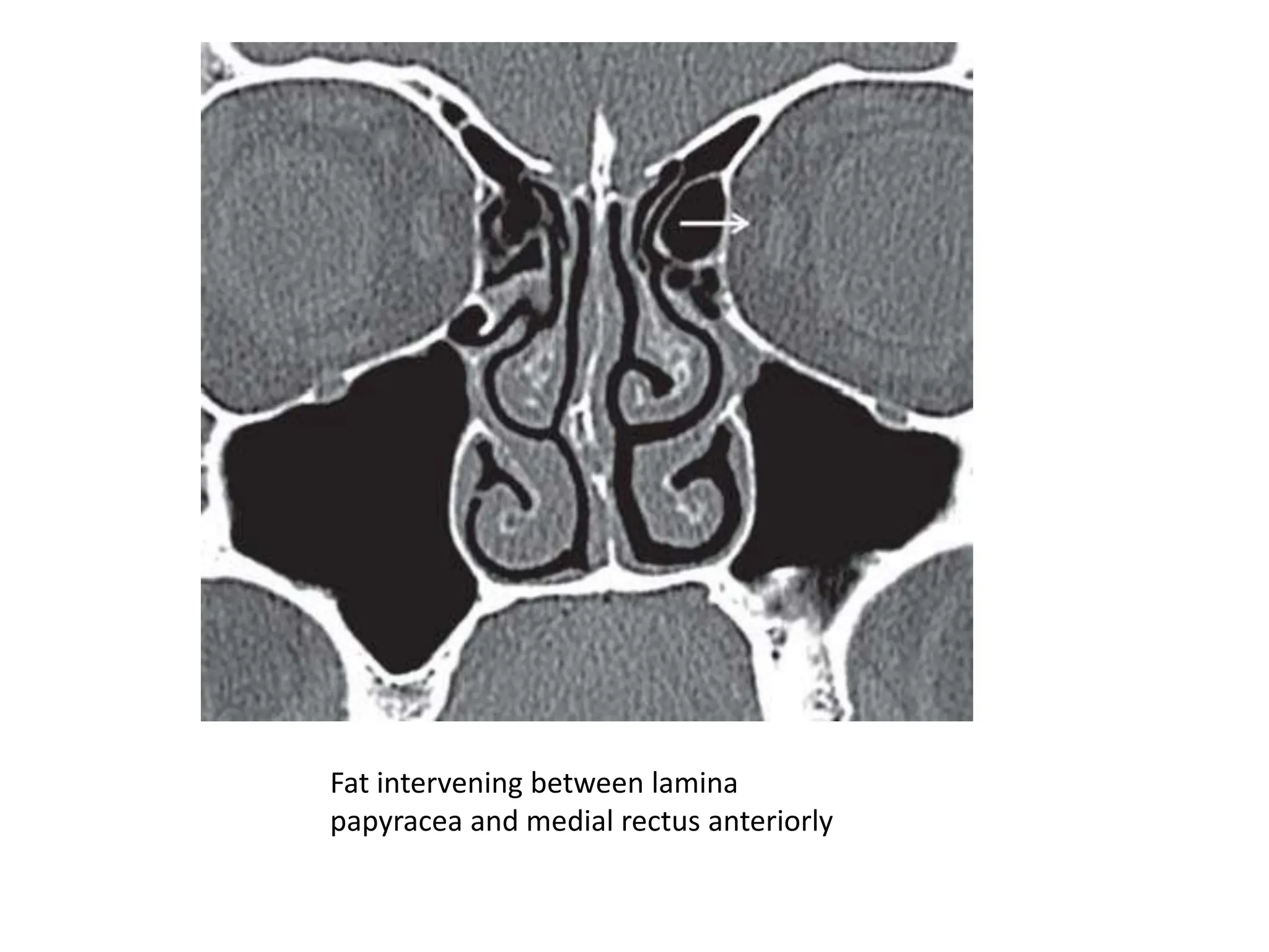
Fat intervening between lamina
papyracea and medial rectus anteriorly
Medial rectus in direct contact with lamina
papyracea posteriorly
ā€¢ It is important to know that the medial rectus
is separated from the lamina papyracea by a
pad of fat anteriorly. However, more
posteriorly in the orbit this pad of fat is absent
and the medial rectus is in direct relation to
the lamina papyracea and therefore more
prone to injury
Bilateral sphenoid ostia
ā€¢ In the more anterior cuts both the posterior
ethmoid cells and the sphenoid sinus are
seen. The superolateral cell is the posterior
ethmoid cell whereas the inferomedial cell is
the sphenoid sinus.
ā€¢ The more subsequent cuts show the sphenoid
anatomy more clearly.
Orbital apex (arrow), sphenoid dominance
(left), pterygoid processes (P)
ā€¢ The retort-shaped orbital apex is seen on
either side of the sphenoid sinus in the
anterior cuts
ā€¢ The pterygoid processes extend downwards
and are perforated by two canals. The first is
the foramen rotundum, which is seen just
below the orbital apex. Inferomedial to this
foramen is the opening of the vidian canal
Vidian canal (thin arrow), foramen rotundum
(thick arrow), and optic nerve (curved arrow)
Sphenopalatine foramen ( arrow)
ā€¢ A canal may be seen below the sphenoid sinus
between the pterygopalatine fossa and the
posterior choana
ā€¢ This is the sphenopalatine foramen, which
opens above the posterior end of the middle
turbinate.
Torus tubaris (T), fossa of Rosenmueller
(arrow), adenoids (A)
ā€¢ Coronal sections of the nasopharynx show the
eustachian tube opening, the torus tubaris,
the fossa of Rosenmueller and the adenoids, if
present
Foramen ovale
ā€¢ The foramen ovale is seen laterally in the
greater wing of sphenoid
ā€¢ Widening or destruction of the foramen
should be looked for in a case of
nasopharyngeal angiofibroma or a carcinoma
of nasopharynx respectively.
Axial Cuts
Nasolacrimal duct (arrow)
Fossa of Rosenmueller (arrow),
EOā€”eustachian tube opening
The ground lamella (arrow)
G: ground lamella, B: bulla,
R: retrobullar recess, U: uncinate process
The orbit in axial section
Lamina papyracea in lower section of the orbit
Crista galli (thick arrow),
foramen cecum (thin arrow)
Sagittal cuts
FS-frontal sinus, B-ethmoidal bulla, A- agger
nasi, PE-post. Ethmoidal cells, SB-suprabullar cells
(1) uncinate process (blue), (2) anterior wall of bulla (green), (3) ground
lamella (yellow), (4) anterior wall of sphenoid (black)
ā€¢ The four lamellae that the endoscopic surgeon
has to cross in an anteroposterior direction
are well seen in a single cut on the sagittal
section.
ā€“ Uncinate Process
ā€“ Ant wall of bulla
ā€“ Post wall of bulla
ā€“ Ant wall of sphenoid
Anatomical Variations
Septal deviations
ā€¢ Can present as a spur at the junction of
cartilage with vomer or along the length of
the septum
ā€¢ Compromised Osteomeatal complex(OMC)
Septal deviations
Septal spur with hypertrophied
Inferior turbinate
Pneumatization of the septum
Agger nasi
ā€¢ 1-3 in no.
ā€¢ Pneumatises lacrimal bone and frontonasal
process of maxilla
ā€¢ Displaces anterior attachment of Middle
turbinate postero-superiorly if prominent
Various attachments of Uncinate
Process
Attaching laterally to lamina papyracea
Attaching to cribriform plate
Attaching medially to middle turbinate
Lying free in middle meatus
Pneumatized uncinate process
Drainage of frontal recessā€”medial to infundibulum on the
right and into the infundibulum on the left
Middle turbinate variations
Concha bullosa
Interlamellar cell of Grunwald (when
superior meatus pneumatizes
vertical lamella of MT)
Paradoxically curved middle turbinate The turbinate sinus
Bulla Ethmoidalis
ā€¢ Most consistent and prominent anterior
ethmoid cell
ā€¢ Shows variations in pneumatization
(hypoplastic, rarely non-pneumatized,
extensively pneumatized)
ā€¢ Suprabullar recess: space between the upper
margin of the bulla and skull base
ā€“ Opens into the frontal recess
ā€¢ Retrobullar space: space between ground
lamella and bulla
ā€¢ Sinus lateralis
ā€¢ Hiatus semilunaris superioris
ā€¢ Hiatus semilunaris inferioris
Ethmoid air cells classification
according to their migration
Patterns of migration of ethmoidal air cells into: (1) lacrimal bone (agger nasi), (2) inferior to
orbit (Hallerā€™s cell), (3) frontal bone (frontal cells), (4) supraorbital cell, (5) middle turbinate
(concha bullosa), (6) crista galli, (7) above sphenoid (Onodi)
ā€¢ Anteriorlyā€”into the lacrimal bone and frontonasal
process of maxilla as the agger nasi cells.
ā€¢ Inferolaterallyā€”into the roof of maxillary sinus as
Haller cell
ā€¢ Anterosuperiorlyā€”into the frontal bone to form
the frontal sinus.
ā€¢ Superiorlyā€”above the ethmoidal bulla over the
orbit and behind frontal sinus to form supraorbital
cell.
ā€¢ Into the middle turbinateā€”concha bullosa
ā€¢ Pneumatized crista galli
ā€¢ Posteriorlyā€”above sphenoid sinus as Onodi cell.
ā€¢ Superiorlyā€”into the frontal recess to form the
different types of frontal cells.
`
Hallerā€™s cell
Supraorbital cell
Pneumatization of crista galli
Onodi cell with dehiscent optic nerve
ā€¢ Frontal cells classification
Type I: A single cell above the agger nasi cell.
Type II: Two or more cells above the agger nasi cell.
Type III: (Frontal bulla) A cell which extends well into the
frontal sinus and simulates the frontal sinus itself on
endoscopy
Type IV: An isolated ā€œloner cellā€ within the frontal sinus.
Ground lamella
Ground lamella spliting into septae
Missedā€ ground lamella
Superior turbinate
Pneumatized superior turbinate
Paradoxically curved superior
turbinate
The olfactory fossa
Keros classification
Type I ā€” 1-3 mm
Type II ā€” 4-7 mm
Type III ā€” 8-17 mm
Patterns of sphenoid pneumatization
Intersphenoid septum attaching
to optic nerve
Intersphenoid septum attaching to
internal carotid artery
Delano classification
Pneumatized anterior clinoid process
Lateral recesses of the sphenoid
Pneumatized interfrontal septum
Septa within maxillary sinus Dehiscent infraorbital nerve
Some pathological diseases
of PNS
ANTROCHOANAL POLYP
SINONASAL POLYPOSIS
INVERTED PAPILLOMA
AFRS
JUVENILE NASOPHARYNGEAL
ANGIOFIBROMA
LEFT MAXILLARY CARCINOMA
dhwani ct pns final (1).pptx all about it pns

More Related Content

Similar to dhwani ct pns final (1).pptx all about it pns

temporalbone-141009084034-conversion-gate02 (1).pdf
temporalbone-141009084034-conversion-gate02 (1).pdftemporalbone-141009084034-conversion-gate02 (1).pdf
temporalbone-141009084034-conversion-gate02 (1).pdfjoanluciya
Ā 
Temporal bone radiology
Temporal bone radiologyTemporal bone radiology
Temporal bone radiologySatish Naga
Ā 
Anatomy of temporal bone
Anatomy of temporal boneAnatomy of temporal bone
Anatomy of temporal bonepraneeth koduru
Ā 
Anatomy temporal bone
Anatomy temporal bone Anatomy temporal bone
Anatomy temporal bone KevinMungasia
Ā 
Surgical anatomy of Salivary glands - ORAL AND MAXILLOFACIAL SURGERY
Surgical anatomy of Salivary glands - ORAL AND MAXILLOFACIAL SURGERYSurgical anatomy of Salivary glands - ORAL AND MAXILLOFACIAL SURGERY
Surgical anatomy of Salivary glands - ORAL AND MAXILLOFACIAL SURGERYANIKET SARKAR
Ā 
Anatomy of orbit by pushkar dhir
Anatomy of orbit by pushkar dhirAnatomy of orbit by pushkar dhir
Anatomy of orbit by pushkar dhirPushkar Dhir
Ā 
Anatomy of external ear -- ent
Anatomy of external ear  -- entAnatomy of external ear  -- ent
Anatomy of external ear -- entNISCHAL SHRESTHA
Ā 
Anatomy of ear
Anatomy of ear Anatomy of ear
Anatomy of ear amit jha
Ā 
Carotid in neck , eca, ima &
Carotid in neck , eca, ima &Carotid in neck , eca, ima &
Carotid in neck , eca, ima &Malarvizhi R
Ā 
Orbit Anatomy and Surgical Spaces
Orbit Anatomy and Surgical Spaces Orbit Anatomy and Surgical Spaces
Orbit Anatomy and Surgical Spaces Vikas Khatri
Ā 
By pd anatomy of the orbit
By pd anatomy of the orbitBy pd anatomy of the orbit
By pd anatomy of the orbitPushkar Dhir
Ā 
LATERAL WALL OF NOSE.pptx
LATERAL WALL OF NOSE.pptxLATERAL WALL OF NOSE.pptx
LATERAL WALL OF NOSE.pptxBrijalPatel35
Ā 
landmarks and interpretation in extraoral radiography
landmarks and interpretation in extraoral radiographylandmarks and interpretation in extraoral radiography
landmarks and interpretation in extraoral radiographygaurav katheriya
Ā 
Failed fess spectrum of ct findings in the frontal recess
Failed fess spectrum of ct findings in the frontal recessFailed fess spectrum of ct findings in the frontal recess
Failed fess spectrum of ct findings in the frontal recesshimanshuslides
Ā 
Mastoid diseases imaging
Mastoid diseases imagingMastoid diseases imaging
Mastoid diseases imagingMilan Silwal
Ā 

Similar to dhwani ct pns final (1).pptx all about it pns (20)

temporalbone-141009084034-conversion-gate02 (1).pdf
temporalbone-141009084034-conversion-gate02 (1).pdftemporalbone-141009084034-conversion-gate02 (1).pdf
temporalbone-141009084034-conversion-gate02 (1).pdf
Ā 
Temporal bone radiology
Temporal bone radiologyTemporal bone radiology
Temporal bone radiology
Ā 
Anatomy of temporal bone
Anatomy of temporal boneAnatomy of temporal bone
Anatomy of temporal bone
Ā 
Anatomy temporal bone
Anatomy temporal bone Anatomy temporal bone
Anatomy temporal bone
Ā 
Surgical anatomy of Salivary glands - ORAL AND MAXILLOFACIAL SURGERY
Surgical anatomy of Salivary glands - ORAL AND MAXILLOFACIAL SURGERYSurgical anatomy of Salivary glands - ORAL AND MAXILLOFACIAL SURGERY
Surgical anatomy of Salivary glands - ORAL AND MAXILLOFACIAL SURGERY
Ā 
Orbital anatomy
Orbital anatomyOrbital anatomy
Orbital anatomy
Ā 
Sinus anatomy and variants
Sinus anatomy and variantsSinus anatomy and variants
Sinus anatomy and variants
Ā 
Anatomy of the orbit
Anatomy of the orbit Anatomy of the orbit
Anatomy of the orbit
Ā 
Anatomy of orbit by pushkar dhir
Anatomy of orbit by pushkar dhirAnatomy of orbit by pushkar dhir
Anatomy of orbit by pushkar dhir
Ā 
Anatomy of external ear -- ent
Anatomy of external ear  -- entAnatomy of external ear  -- ent
Anatomy of external ear -- ent
Ā 
Anatomy of ear
Anatomy of ear Anatomy of ear
Anatomy of ear
Ā 
Temporal bone
Temporal boneTemporal bone
Temporal bone
Ā 
Carotid in neck , eca, ima &
Carotid in neck , eca, ima &Carotid in neck , eca, ima &
Carotid in neck , eca, ima &
Ā 
Orbit Anatomy and Surgical Spaces
Orbit Anatomy and Surgical Spaces Orbit Anatomy and Surgical Spaces
Orbit Anatomy and Surgical Spaces
Ā 
By pd anatomy of the orbit
By pd anatomy of the orbitBy pd anatomy of the orbit
By pd anatomy of the orbit
Ā 
LATERAL WALL OF NOSE.pptx
LATERAL WALL OF NOSE.pptxLATERAL WALL OF NOSE.pptx
LATERAL WALL OF NOSE.pptx
Ā 
landmarks and interpretation in extraoral radiography
landmarks and interpretation in extraoral radiographylandmarks and interpretation in extraoral radiography
landmarks and interpretation in extraoral radiography
Ā 
The Orbit
The OrbitThe Orbit
The Orbit
Ā 
Failed fess spectrum of ct findings in the frontal recess
Failed fess spectrum of ct findings in the frontal recessFailed fess spectrum of ct findings in the frontal recess
Failed fess spectrum of ct findings in the frontal recess
Ā 
Mastoid diseases imaging
Mastoid diseases imagingMastoid diseases imaging
Mastoid diseases imaging
Ā 

More from VivekMakadiya2

Paper_VivekMakadiya.ppt.pptxEndoscopic cartilage type 1 tympanoplasty
Paper_VivekMakadiya.ppt.pptxEndoscopic cartilage type 1 tympanoplastyPaper_VivekMakadiya.ppt.pptxEndoscopic cartilage type 1 tympanoplasty
Paper_VivekMakadiya.ppt.pptxEndoscopic cartilage type 1 tympanoplastyVivekMakadiya2
Ā 
OTOACOUSTIC EMMISIONS.pptx
OTOACOUSTIC EMMISIONS.pptxOTOACOUSTIC EMMISIONS.pptx
OTOACOUSTIC EMMISIONS.pptxVivekMakadiya2
Ā 
Krishna - poster =A CASE STUDY OF TONSILLAR TERATOMA.pptx
Krishna - poster =A CASE STUDY OF TONSILLAR TERATOMA.pptxKrishna - poster =A CASE STUDY OF TONSILLAR TERATOMA.pptx
Krishna - poster =A CASE STUDY OF TONSILLAR TERATOMA.pptxVivekMakadiya2
Ā 
Anatomy of vestibular system.pptx
Anatomy of vestibular system.pptxAnatomy of vestibular system.pptx
Anatomy of vestibular system.pptxVivekMakadiya2
Ā 
Acute and Chronic sinusitis.pptx
Acute and Chronic sinusitis.pptxAcute and Chronic sinusitis.pptx
Acute and Chronic sinusitis.pptxVivekMakadiya2
Ā 
noisepollution 1.pptx
noisepollution 1.pptxnoisepollution 1.pptx
noisepollution 1.pptxVivekMakadiya2
Ā 
diseasesofexternalear
diseasesofexternaleardiseasesofexternalear
diseasesofexternalearVivekMakadiya2
Ā 

More from VivekMakadiya2 (10)

Paper_VivekMakadiya.ppt.pptxEndoscopic cartilage type 1 tympanoplasty
Paper_VivekMakadiya.ppt.pptxEndoscopic cartilage type 1 tympanoplastyPaper_VivekMakadiya.ppt.pptxEndoscopic cartilage type 1 tympanoplasty
Paper_VivekMakadiya.ppt.pptxEndoscopic cartilage type 1 tympanoplasty
Ā 
OTOACOUSTIC EMMISIONS.pptx
OTOACOUSTIC EMMISIONS.pptxOTOACOUSTIC EMMISIONS.pptx
OTOACOUSTIC EMMISIONS.pptx
Ā 
Krishna - poster =A CASE STUDY OF TONSILLAR TERATOMA.pptx
Krishna - poster =A CASE STUDY OF TONSILLAR TERATOMA.pptxKrishna - poster =A CASE STUDY OF TONSILLAR TERATOMA.pptx
Krishna - poster =A CASE STUDY OF TONSILLAR TERATOMA.pptx
Ā 
Anatomy of vestibular system.pptx
Anatomy of vestibular system.pptxAnatomy of vestibular system.pptx
Anatomy of vestibular system.pptx
Ā 
JNA.pptx
JNA.pptxJNA.pptx
JNA.pptx
Ā 
Acute and Chronic sinusitis.pptx
Acute and Chronic sinusitis.pptxAcute and Chronic sinusitis.pptx
Acute and Chronic sinusitis.pptx
Ā 
noisepollution 1.pptx
noisepollution 1.pptxnoisepollution 1.pptx
noisepollution 1.pptx
Ā 
ECA & ICA.pptx
ECA & ICA.pptxECA & ICA.pptx
ECA & ICA.pptx
Ā 
hyperthyroidism
hyperthyroidismhyperthyroidism
hyperthyroidism
Ā 
diseasesofexternalear
diseasesofexternaleardiseasesofexternalear
diseasesofexternalear
Ā 

Recently uploaded

Call Girls Madhapur 7001305949 all area service COD available Any Time
Call Girls Madhapur 7001305949 all area service COD available Any TimeCall Girls Madhapur 7001305949 all area service COD available Any Time
Call Girls Madhapur 7001305949 all area service COD available Any Timedelhimodelshub1
Ā 
Russian Call Girls Hyderabad Indira 9907093804 Independent Escort Service Hyd...
Russian Call Girls Hyderabad Indira 9907093804 Independent Escort Service Hyd...Russian Call Girls Hyderabad Indira 9907093804 Independent Escort Service Hyd...
Russian Call Girls Hyderabad Indira 9907093804 Independent Escort Service Hyd...delhimodelshub1
Ā 
Russian Escorts Delhi | 9711199171 | all area service available
Russian Escorts Delhi | 9711199171 | all area service availableRussian Escorts Delhi | 9711199171 | all area service available
Russian Escorts Delhi | 9711199171 | all area service availablesandeepkumar69420
Ā 
Call Girls Service Chandigarh Grishma ā¤ļøšŸ‘ 9907093804 šŸ‘„šŸ«¦ Independent Escort Se...
Call Girls Service Chandigarh Grishma ā¤ļøšŸ‘ 9907093804 šŸ‘„šŸ«¦ Independent Escort Se...Call Girls Service Chandigarh Grishma ā¤ļøšŸ‘ 9907093804 šŸ‘„šŸ«¦ Independent Escort Se...
Call Girls Service Chandigarh Grishma ā¤ļøšŸ‘ 9907093804 šŸ‘„šŸ«¦ Independent Escort Se...High Profile Call Girls Chandigarh Aarushi
Ā 
VIP Call Girls Hyderabad Megha 9907093804 Independent Escort Service Hyderabad
VIP Call Girls Hyderabad Megha 9907093804 Independent Escort Service HyderabadVIP Call Girls Hyderabad Megha 9907093804 Independent Escort Service Hyderabad
VIP Call Girls Hyderabad Megha 9907093804 Independent Escort Service Hyderabaddelhimodelshub1
Ā 
hyderabad call girl.pdfRussian Call Girls in Hyderabad Amrita 9907093804 Inde...
hyderabad call girl.pdfRussian Call Girls in Hyderabad Amrita 9907093804 Inde...hyderabad call girl.pdfRussian Call Girls in Hyderabad Amrita 9907093804 Inde...
hyderabad call girl.pdfRussian Call Girls in Hyderabad Amrita 9907093804 Inde...delhimodelshub1
Ā 
Call Girls Hyderabad Kirti 9907093804 Independent Escort Service Hyderabad
Call Girls Hyderabad Kirti 9907093804 Independent Escort Service HyderabadCall Girls Hyderabad Kirti 9907093804 Independent Escort Service Hyderabad
Call Girls Hyderabad Kirti 9907093804 Independent Escort Service Hyderabaddelhimodelshub1
Ā 
Call Girls Hyderabad Krisha 9907093804 Independent Escort Service Hyderabad
Call Girls Hyderabad Krisha 9907093804 Independent Escort Service HyderabadCall Girls Hyderabad Krisha 9907093804 Independent Escort Service Hyderabad
Call Girls Hyderabad Krisha 9907093804 Independent Escort Service Hyderabaddelhimodelshub1
Ā 
Call Girls in Hyderabad Lavanya 9907093804 Independent Escort Service Hyderabad
Call Girls in Hyderabad Lavanya 9907093804 Independent Escort Service HyderabadCall Girls in Hyderabad Lavanya 9907093804 Independent Escort Service Hyderabad
Call Girls in Hyderabad Lavanya 9907093804 Independent Escort Service Hyderabaddelhimodelshub1
Ā 
Russian Escorts Aishbagh Road * 9548273370 Naughty Call Girls Service in Lucknow
Russian Escorts Aishbagh Road * 9548273370 Naughty Call Girls Service in LucknowRussian Escorts Aishbagh Road * 9548273370 Naughty Call Girls Service in Lucknow
Russian Escorts Aishbagh Road * 9548273370 Naughty Call Girls Service in Lucknowgragteena
Ā 
Call Girl Service ITPL - [ Cash on Delivery ] Contact 7001305949 Escorts Service
Call Girl Service ITPL - [ Cash on Delivery ] Contact 7001305949 Escorts ServiceCall Girl Service ITPL - [ Cash on Delivery ] Contact 7001305949 Escorts Service
Call Girl Service ITPL - [ Cash on Delivery ] Contact 7001305949 Escorts Servicenarwatsonia7
Ā 
Call Girl Chandigarh Mallika ā¤ļøšŸ‘ 9907093804 šŸ‘„šŸ«¦ Independent Escort Service Cha...
Call Girl Chandigarh Mallika ā¤ļøšŸ‘ 9907093804 šŸ‘„šŸ«¦ Independent Escort Service Cha...Call Girl Chandigarh Mallika ā¤ļøšŸ‘ 9907093804 šŸ‘„šŸ«¦ Independent Escort Service Cha...
Call Girl Chandigarh Mallika ā¤ļøšŸ‘ 9907093804 šŸ‘„šŸ«¦ Independent Escort Service Cha...High Profile Call Girls Chandigarh Aarushi
Ā 
Russian Call Girls in Raipur 9873940964 Book Hot And Sexy Girls
Russian Call Girls in Raipur 9873940964 Book Hot And Sexy GirlsRussian Call Girls in Raipur 9873940964 Book Hot And Sexy Girls
Russian Call Girls in Raipur 9873940964 Book Hot And Sexy Girlsddev2574
Ā 
No Advance 9053900678 Chandigarh Call Girls , Indian Call Girls For Full Ni...
No Advance 9053900678 Chandigarh  Call Girls , Indian Call Girls  For Full Ni...No Advance 9053900678 Chandigarh  Call Girls , Indian Call Girls  For Full Ni...
No Advance 9053900678 Chandigarh Call Girls , Indian Call Girls For Full Ni...Vip call girls In Chandigarh
Ā 

Recently uploaded (20)

Call Girls Madhapur 7001305949 all area service COD available Any Time
Call Girls Madhapur 7001305949 all area service COD available Any TimeCall Girls Madhapur 7001305949 all area service COD available Any Time
Call Girls Madhapur 7001305949 all area service COD available Any Time
Ā 
Call Girls Guwahati Aaradhya šŸ‘‰ 7001305949šŸ‘ˆ šŸŽ¶ Independent Escort Service Guwahati
Call Girls Guwahati Aaradhya šŸ‘‰ 7001305949šŸ‘ˆ šŸŽ¶ Independent Escort Service GuwahatiCall Girls Guwahati Aaradhya šŸ‘‰ 7001305949šŸ‘ˆ šŸŽ¶ Independent Escort Service Guwahati
Call Girls Guwahati Aaradhya šŸ‘‰ 7001305949šŸ‘ˆ šŸŽ¶ Independent Escort Service Guwahati
Ā 
Russian Call Girls Hyderabad Indira 9907093804 Independent Escort Service Hyd...
Russian Call Girls Hyderabad Indira 9907093804 Independent Escort Service Hyd...Russian Call Girls Hyderabad Indira 9907093804 Independent Escort Service Hyd...
Russian Call Girls Hyderabad Indira 9907093804 Independent Escort Service Hyd...
Ā 
Russian Escorts Delhi | 9711199171 | all area service available
Russian Escorts Delhi | 9711199171 | all area service availableRussian Escorts Delhi | 9711199171 | all area service available
Russian Escorts Delhi | 9711199171 | all area service available
Ā 
Call Girls Service Chandigarh Grishma ā¤ļøšŸ‘ 9907093804 šŸ‘„šŸ«¦ Independent Escort Se...
Call Girls Service Chandigarh Grishma ā¤ļøšŸ‘ 9907093804 šŸ‘„šŸ«¦ Independent Escort Se...Call Girls Service Chandigarh Grishma ā¤ļøšŸ‘ 9907093804 šŸ‘„šŸ«¦ Independent Escort Se...
Call Girls Service Chandigarh Grishma ā¤ļøšŸ‘ 9907093804 šŸ‘„šŸ«¦ Independent Escort Se...
Ā 
VIP Call Girls Lucknow Isha šŸ” 9719455033 šŸ” šŸŽ¶ Independent Escort Service Lucknow
VIP Call Girls Lucknow Isha šŸ” 9719455033 šŸ” šŸŽ¶ Independent Escort Service LucknowVIP Call Girls Lucknow Isha šŸ” 9719455033 šŸ” šŸŽ¶ Independent Escort Service Lucknow
VIP Call Girls Lucknow Isha šŸ” 9719455033 šŸ” šŸŽ¶ Independent Escort Service Lucknow
Ā 
Call Girl Lucknow Gauri šŸ” 8923113531 šŸ” šŸŽ¶ Independent Escort Service Lucknow
Call Girl Lucknow Gauri šŸ” 8923113531  šŸ” šŸŽ¶ Independent Escort Service LucknowCall Girl Lucknow Gauri šŸ” 8923113531  šŸ” šŸŽ¶ Independent Escort Service Lucknow
Call Girl Lucknow Gauri šŸ” 8923113531 šŸ” šŸŽ¶ Independent Escort Service Lucknow
Ā 
VIP Call Girls Hyderabad Megha 9907093804 Independent Escort Service Hyderabad
VIP Call Girls Hyderabad Megha 9907093804 Independent Escort Service HyderabadVIP Call Girls Hyderabad Megha 9907093804 Independent Escort Service Hyderabad
VIP Call Girls Hyderabad Megha 9907093804 Independent Escort Service Hyderabad
Ā 
hyderabad call girl.pdfRussian Call Girls in Hyderabad Amrita 9907093804 Inde...
hyderabad call girl.pdfRussian Call Girls in Hyderabad Amrita 9907093804 Inde...hyderabad call girl.pdfRussian Call Girls in Hyderabad Amrita 9907093804 Inde...
hyderabad call girl.pdfRussian Call Girls in Hyderabad Amrita 9907093804 Inde...
Ā 
Model Call Girl in Subhash Nagar Delhi reach out to us at šŸ”9953056974šŸ”
Model Call Girl in Subhash Nagar Delhi reach out to us at šŸ”9953056974šŸ”Model Call Girl in Subhash Nagar Delhi reach out to us at šŸ”9953056974šŸ”
Model Call Girl in Subhash Nagar Delhi reach out to us at šŸ”9953056974šŸ”
Ā 
Call Girls Hyderabad Kirti 9907093804 Independent Escort Service Hyderabad
Call Girls Hyderabad Kirti 9907093804 Independent Escort Service HyderabadCall Girls Hyderabad Kirti 9907093804 Independent Escort Service Hyderabad
Call Girls Hyderabad Kirti 9907093804 Independent Escort Service Hyderabad
Ā 
Call Girls Hyderabad Krisha 9907093804 Independent Escort Service Hyderabad
Call Girls Hyderabad Krisha 9907093804 Independent Escort Service HyderabadCall Girls Hyderabad Krisha 9907093804 Independent Escort Service Hyderabad
Call Girls Hyderabad Krisha 9907093804 Independent Escort Service Hyderabad
Ā 
Call Girl Dehradun Aashi šŸ” 7001305949 šŸ” šŸ’ƒ Independent Escort Service Dehradun
Call Girl Dehradun Aashi šŸ” 7001305949 šŸ” šŸ’ƒ Independent Escort Service DehradunCall Girl Dehradun Aashi šŸ” 7001305949 šŸ” šŸ’ƒ Independent Escort Service Dehradun
Call Girl Dehradun Aashi šŸ” 7001305949 šŸ” šŸ’ƒ Independent Escort Service Dehradun
Ā 
Call Girls in Hyderabad Lavanya 9907093804 Independent Escort Service Hyderabad
Call Girls in Hyderabad Lavanya 9907093804 Independent Escort Service HyderabadCall Girls in Hyderabad Lavanya 9907093804 Independent Escort Service Hyderabad
Call Girls in Hyderabad Lavanya 9907093804 Independent Escort Service Hyderabad
Ā 
Russian Escorts Aishbagh Road * 9548273370 Naughty Call Girls Service in Lucknow
Russian Escorts Aishbagh Road * 9548273370 Naughty Call Girls Service in LucknowRussian Escorts Aishbagh Road * 9548273370 Naughty Call Girls Service in Lucknow
Russian Escorts Aishbagh Road * 9548273370 Naughty Call Girls Service in Lucknow
Ā 
College Call Girls Dehradun Kavya šŸ” 7001305949 šŸ” šŸ“ Independent Escort Service...
College Call Girls Dehradun Kavya šŸ” 7001305949 šŸ” šŸ“ Independent Escort Service...College Call Girls Dehradun Kavya šŸ” 7001305949 šŸ” šŸ“ Independent Escort Service...
College Call Girls Dehradun Kavya šŸ” 7001305949 šŸ” šŸ“ Independent Escort Service...
Ā 
Call Girl Service ITPL - [ Cash on Delivery ] Contact 7001305949 Escorts Service
Call Girl Service ITPL - [ Cash on Delivery ] Contact 7001305949 Escorts ServiceCall Girl Service ITPL - [ Cash on Delivery ] Contact 7001305949 Escorts Service
Call Girl Service ITPL - [ Cash on Delivery ] Contact 7001305949 Escorts Service
Ā 
Call Girl Chandigarh Mallika ā¤ļøšŸ‘ 9907093804 šŸ‘„šŸ«¦ Independent Escort Service Cha...
Call Girl Chandigarh Mallika ā¤ļøšŸ‘ 9907093804 šŸ‘„šŸ«¦ Independent Escort Service Cha...Call Girl Chandigarh Mallika ā¤ļøšŸ‘ 9907093804 šŸ‘„šŸ«¦ Independent Escort Service Cha...
Call Girl Chandigarh Mallika ā¤ļøšŸ‘ 9907093804 šŸ‘„šŸ«¦ Independent Escort Service Cha...
Ā 
Russian Call Girls in Raipur 9873940964 Book Hot And Sexy Girls
Russian Call Girls in Raipur 9873940964 Book Hot And Sexy GirlsRussian Call Girls in Raipur 9873940964 Book Hot And Sexy Girls
Russian Call Girls in Raipur 9873940964 Book Hot And Sexy Girls
Ā 
No Advance 9053900678 Chandigarh Call Girls , Indian Call Girls For Full Ni...
No Advance 9053900678 Chandigarh  Call Girls , Indian Call Girls  For Full Ni...No Advance 9053900678 Chandigarh  Call Girls , Indian Call Girls  For Full Ni...
No Advance 9053900678 Chandigarh Call Girls , Indian Call Girls For Full Ni...
Ā 

dhwani ct pns final (1).pptx all about it pns

  • 1. CT Scan Paranasal Sinus Dr. Dhwani Shah Senior Resident
  • 2. Introduction ā€¢ A fundamental knowledge of the anatomy of structures of nose and paranasal sinuses is essential for the understanding of diseases of nose and preoperative planning of endoscopic sinus surgery. ā€¢ Computed tomography (CT) has become a standard diagnostic tool in the evaluation of nose and paranasal sinuses. Multidetector CT(MDCT) allows assessment of the patency of sinonasal passages, shows the effect of anatomic variants, inflammatory diseases on patency.
  • 3. Need of Imaging ā€¢ To confirm Diagnosis ā€¢ To facilitate Surgical planning (road map) ā€¢ To know extent of disease ā€¢ To know character of disease ā€¢ Location of surgically relevant anatomic structures ā€¢ Precise identification of critical anatomic variations
  • 4. Why not Xray PNS? ā€¢ Inadequate details ā€¢ Overlapping of osseous framework
  • 5. When to be done ā€¢ After a course of antibiotics so that acute inflammation is not mistaken for chronic mucosal disease ā€¢ 4-6 weeks following therapy as radiological findings may lag behind clinical response(for follow-up)
  • 6. ā€¢ A basic paranasal scan includes bony and soft tissue window of 3-5 mm cuts taken anterior to posterior coronal plane and axial sections from inferior to superior. ā€¢ A Sagittal reconstruction is done to observe some particular structures. ā€¢ It is extremely important to review the CT images in all three planes for better understanding of the anatomy and identification of these anatomic variations
  • 7. ā€¢ Proper positioning of the patientā€™s head is important. ā€¢ For Axial cuts, patientā€™s hard palate is placed perpendicular to ct scanner table. ā€¢ For coronal cuts, gantry is perpendicular to patientā€™s hard palate. ā€¢ ask the patient to blow nose and clear out all loose secretions prior to ct
  • 8. (1) Coronal ā€“ easy as we operate in same plane ā€¢ Following structures can be assessed going from anterior to posterior. ļ± Frontal sinus and nasal bones ļ± Nasal septum ļ± Inferior turbinate ļ± Nasolacrimal duct ļ± Anterior ethmoidal cells ļ± Ethmoidal bulla ļ± Middle turbinate ļ± Uncinate process and its attachment ļ± Haller cells ļ± Maxillary sinus ļ± Depth olfactory fossa ļ± Posterior ethmoidal cells ļ± Sphenoid sinus
  • 9. (2)Axial ā€“ Parallel to hard palate ā€“ Inferior(hard palate) to superior(upper margin of frontal sinus) ā€¢ Certain structures are well seen on axial scan. ā€¢ Nasolacrimal duct ā€¢ Anteroposterior deviation of nasal septum ā€¢ Fossa of Rosenmueller ā€¢ Ground lamella and its attachment ā€¢ Lamina papyracea ā€¢ Demarcation between anterior and posterior ethmoid air cells ā€¢ Retrobullar recess ā€¢ Pterygopalatine fossa ā€¢ Onodi cells
  • 10. (3) Sagittal ā€¢ Lateral nasal wall ā€¢ Frontal recess area ā€¢ Retrobullar and Suprabullar recess ā€¢ Sphenoid sinus ostium ā€¢ Extent of Onodi cellā€™s migration
  • 11. What kind of CT PNS should be ordered? ā€¢ CT PNS, coronal cuts, bone window setting, spaced at 3 mm cuts +/- soft tissue setting +/- contrast (In case of CSF rhinorrhoea or optic nerve injury ā€“ 1 mm cuts required)
  • 12. ā€¢ Start from the first cut itself ā€¢ Donā€™t jump ā€¢ Read for 2 times
  • 13. Scout/Gantry cut (thickness and positioning) Scout filmā€”3 mm slice thickness Scout filmā€”1 mm slice thickness
  • 14. External nose and Nasal bones
  • 15. ā€¢ The most anterior cut shows frontal sinus and nasal bones ā€¢ Great variations of pneumatization of frontal sinus ā€¢ Interfrontal sinus septum is in midline inferiorly, but may deviate to any side posterosuperiorly ā€¢ Septaļƒ deep lateral recessesļƒ classical scalloping of frontal sinus
  • 16. Cut showing interfrontal septum, multiple frontal septa show classical scalloping of frontal sinus ļƒ  which is lost in mucoceles
  • 18. ā€¢ Septal tubercle consists of erectile tissue, is a phylogenetic remnant of vomeronasal organ
  • 19. Agger nasi cells (A), nasolacrimal duct (arrow), middle turbinate not yet visualized
  • 20. The olfactory fossa. Anterior attachment of middle turbinate seen at the junction of medial and lateral lamella of cribriform plate
  • 21. Ethmoid sinus and olfactory fossa ā€¢ The level of the cribriform plate and the depth of the olfactory fossa should be assessed and classified according to the Keros classification. ā€¢ The height of the ethmoidal fovea above the level of the cribriform plate is noted.
  • 22. (1) Horizontal lamella (2) Lateral lamella (0.2 mm) (3) Orbital plate of frontal bone(0.5 mm) (4) Anterior ethmoidal artery
  • 23. ā€¢ Fovea ethmoidalis: ā€“ Makes up the roof of ethmoid bone labyrinth ā€“ Is a part of frontal bone that separates the ethmoidal cells from the anterior cranial fossa
  • 24.
  • 27. ā€¢ Components of OMC: ā€“ Uncinate process ā€“ Hiatus semilunaris ā€“ Bulla ethmoidalis
  • 28. ā€¢ Any septal spur impinging or compromising OMC ā€¢ Turbinate hypertrophy or concha bullosa ā€¢ Mode of attachment or deviations of Uncinate ā€¢ Large bulla ethmoidalis ā€¢ Presence of haller cells Factors affecting OMC patency
  • 29. Maxillary sinus ā€¢ The maxillary sinus changes shape from triangular to ovoid, in more posterior cuts. ā€¢ CT in the coronal plane demonstrates the maxillary sinus to be narrow anteriorly, widest in mid portion and narrow again posteriorly.
  • 30. Maxillary sinus appears triangular in anterior cuts (arrow: infraorbital nerve)
  • 31. Accessory ostium in the left posterior fontanelle
  • 32. Anterior ethmoidal artery. Beaking of lamina papyracea seen on left. Orbitocranial canal seen on the right
  • 33. ā€¢ 2-3 mm behind bulla, Anterior Ethmoidal artery is seen as a classical ā€œbeakingā€ of medial orbital wall ā€¢ The artery may lie close to the skull base or may cross low within the anterior ethmoids in which case the orbitocranial canal with its bony mesentery is clearly seen
  • 34.
  • 35. The ground lamella (arrow). Intermediate attachment of middle turbinate
  • 36. Posterior attachment of middle turbinate
  • 37. ā€¢ The middle turbinate is attached to the lamina papyracea by its ground lamella separating the anterior ethmoid cells from the posterior ethmoid cells ā€¢ superior turbinate is visualized in the more posterior cuts and any variations in it, e.g. pneumatization, paradoxical curvature should be looked for
  • 38. Posterior ethmoidal cells (asterix). Inferior orbital fissure (arrow)-> opens laterally into Infra Temporal Fossa
  • 39. Orbit ā€¢ The orbit changes from a circular outline to a triangular or pyramidal shape
  • 40. Fat intervening between lamina papyracea and medial rectus anteriorly
  • 41. Medial rectus in direct contact with lamina papyracea posteriorly
  • 42. ā€¢ It is important to know that the medial rectus is separated from the lamina papyracea by a pad of fat anteriorly. However, more posteriorly in the orbit this pad of fat is absent and the medial rectus is in direct relation to the lamina papyracea and therefore more prone to injury
  • 44. ā€¢ In the more anterior cuts both the posterior ethmoid cells and the sphenoid sinus are seen. The superolateral cell is the posterior ethmoid cell whereas the inferomedial cell is the sphenoid sinus. ā€¢ The more subsequent cuts show the sphenoid anatomy more clearly.
  • 45. Orbital apex (arrow), sphenoid dominance (left), pterygoid processes (P)
  • 46. ā€¢ The retort-shaped orbital apex is seen on either side of the sphenoid sinus in the anterior cuts ā€¢ The pterygoid processes extend downwards and are perforated by two canals. The first is the foramen rotundum, which is seen just below the orbital apex. Inferomedial to this foramen is the opening of the vidian canal
  • 47. Vidian canal (thin arrow), foramen rotundum (thick arrow), and optic nerve (curved arrow)
  • 49. ā€¢ A canal may be seen below the sphenoid sinus between the pterygopalatine fossa and the posterior choana ā€¢ This is the sphenopalatine foramen, which opens above the posterior end of the middle turbinate.
  • 50. Torus tubaris (T), fossa of Rosenmueller (arrow), adenoids (A)
  • 51. ā€¢ Coronal sections of the nasopharynx show the eustachian tube opening, the torus tubaris, the fossa of Rosenmueller and the adenoids, if present
  • 53. ā€¢ The foramen ovale is seen laterally in the greater wing of sphenoid ā€¢ Widening or destruction of the foramen should be looked for in a case of nasopharyngeal angiofibroma or a carcinoma of nasopharynx respectively.
  • 56. Fossa of Rosenmueller (arrow), EOā€”eustachian tube opening
  • 58. G: ground lamella, B: bulla, R: retrobullar recess, U: uncinate process
  • 59. The orbit in axial section
  • 60. Lamina papyracea in lower section of the orbit
  • 61.
  • 62. Crista galli (thick arrow), foramen cecum (thin arrow)
  • 64.
  • 65. FS-frontal sinus, B-ethmoidal bulla, A- agger nasi, PE-post. Ethmoidal cells, SB-suprabullar cells
  • 66. (1) uncinate process (blue), (2) anterior wall of bulla (green), (3) ground lamella (yellow), (4) anterior wall of sphenoid (black)
  • 67. ā€¢ The four lamellae that the endoscopic surgeon has to cross in an anteroposterior direction are well seen in a single cut on the sagittal section. ā€“ Uncinate Process ā€“ Ant wall of bulla ā€“ Post wall of bulla ā€“ Ant wall of sphenoid
  • 69. Septal deviations ā€¢ Can present as a spur at the junction of cartilage with vomer or along the length of the septum ā€¢ Compromised Osteomeatal complex(OMC)
  • 70. Septal deviations Septal spur with hypertrophied Inferior turbinate
  • 73. ā€¢ 1-3 in no. ā€¢ Pneumatises lacrimal bone and frontonasal process of maxilla ā€¢ Displaces anterior attachment of Middle turbinate postero-superiorly if prominent
  • 74. Various attachments of Uncinate Process Attaching laterally to lamina papyracea
  • 76. Attaching medially to middle turbinate
  • 77. Lying free in middle meatus
  • 79. Drainage of frontal recessā€”medial to infundibulum on the right and into the infundibulum on the left
  • 80. Middle turbinate variations Concha bullosa Interlamellar cell of Grunwald (when superior meatus pneumatizes vertical lamella of MT)
  • 81. Paradoxically curved middle turbinate The turbinate sinus
  • 82. Bulla Ethmoidalis ā€¢ Most consistent and prominent anterior ethmoid cell ā€¢ Shows variations in pneumatization (hypoplastic, rarely non-pneumatized, extensively pneumatized)
  • 83. ā€¢ Suprabullar recess: space between the upper margin of the bulla and skull base ā€“ Opens into the frontal recess ā€¢ Retrobullar space: space between ground lamella and bulla ā€¢ Sinus lateralis ā€¢ Hiatus semilunaris superioris ā€¢ Hiatus semilunaris inferioris
  • 84.
  • 85.
  • 86. Ethmoid air cells classification according to their migration
  • 87. Patterns of migration of ethmoidal air cells into: (1) lacrimal bone (agger nasi), (2) inferior to orbit (Hallerā€™s cell), (3) frontal bone (frontal cells), (4) supraorbital cell, (5) middle turbinate (concha bullosa), (6) crista galli, (7) above sphenoid (Onodi)
  • 88. ā€¢ Anteriorlyā€”into the lacrimal bone and frontonasal process of maxilla as the agger nasi cells. ā€¢ Inferolaterallyā€”into the roof of maxillary sinus as Haller cell ā€¢ Anterosuperiorlyā€”into the frontal bone to form the frontal sinus. ā€¢ Superiorlyā€”above the ethmoidal bulla over the orbit and behind frontal sinus to form supraorbital cell. ā€¢ Into the middle turbinateā€”concha bullosa ā€¢ Pneumatized crista galli ā€¢ Posteriorlyā€”above sphenoid sinus as Onodi cell. ā€¢ Superiorlyā€”into the frontal recess to form the different types of frontal cells.
  • 92. Onodi cell with dehiscent optic nerve
  • 93. ā€¢ Frontal cells classification
  • 94. Type I: A single cell above the agger nasi cell.
  • 95. Type II: Two or more cells above the agger nasi cell.
  • 96. Type III: (Frontal bulla) A cell which extends well into the frontal sinus and simulates the frontal sinus itself on endoscopy
  • 97. Type IV: An isolated ā€œloner cellā€ within the frontal sinus.
  • 98. Ground lamella Ground lamella spliting into septae
  • 100. Superior turbinate Pneumatized superior turbinate Paradoxically curved superior turbinate
  • 102. Keros classification Type I ā€” 1-3 mm
  • 103. Type II ā€” 4-7 mm
  • 104. Type III ā€” 8-17 mm
  • 105. Patterns of sphenoid pneumatization
  • 106. Intersphenoid septum attaching to optic nerve Intersphenoid septum attaching to internal carotid artery
  • 108.
  • 109.
  • 111. Lateral recesses of the sphenoid
  • 113. Septa within maxillary sinus Dehiscent infraorbital nerve
  • 118. AFRS