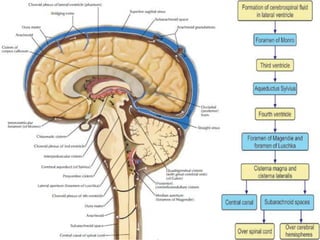
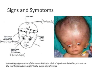
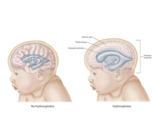
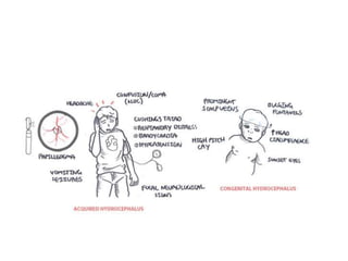
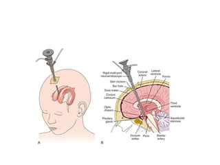
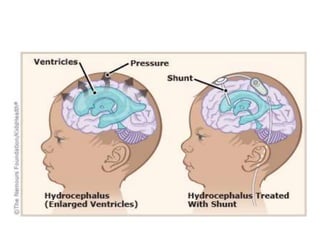
HYDROCEPHALUS
Dr. Rahul Jain
SR Neurosurgery
DEPTT OF NEUROSURGERY
AIIMS PATNA
CSF Physiology
• It circulates within the subarachnoid space, between
the arachnoid and the pial membranes.
• Function as a shock absorber for the CNS, may also
serve an immunological function analogous to the
lymphatic system.
• 80% of CSF is produced by the choroid plexuses,
located in both lateral ventricles (accounts for ≈95% of
CSF produced in the choroid plexuses) and in the 4th
ventricle.
• CSF is also produced by the ependymal lining of the
ventricles, and in the spine, in the dura of the nerve
root sleeves
• Average Total CSF volume in the body is 150 ml. CSF
is produced at rate of 0.3 ml/min (450 ml/24 hrs).
• Absorbed primarily by arachnoid villi (granulations)
that extend into the dural venous sinuses. Other
sites of absorption include the choroid plexuses
and glymphatics. The rate of absorption is pressure-
dependent.
Hydrocephalus
• An abnormal accumulation of cerebrospinal fluid
within the ventricles of the brain.
• Estimated prevalence: 1–1.5%.
• Hydrocephalus (HCP) is either due to subnormal
CSF reabsorption or, rarely, CSF overproduction.
• subnormal CSF reabsorption. Two main functional
subdivisions:
• 1. obstructive hydrocephalus (AKA non-communicating):
block proximal to the arachnoid granulations (AG).
• 2. communicating hydrocephalus (AKA non-obstructive):
defect in CSF reabsorption by the Arachnoid
granulations.
• CSF overproduction: rare. As with some choroid
plexus papillomas.
Signs and Symptoms
sun-setting appearance of the eyes - this latter clinical sign is attributed to pressure on
the mid-brain tectum by CSF in the supra-pineal recess
• Prior to closure of the cranial sutures and
obliteration of the fontanelle, hydrocephalus
results in disproportionate head growth.
• Thus, over the first 2–3 years of life, measurement
of the occipito-frontal circumference and plotting
this on a centile chart provides a simple and
sensitive test.
• Clinical symptoms are often subtle and include
general irritability, poor feeding and slow
attainment of milestones.
Other Signs
• Macewen’s sign: cracked pot sound on percussing
over-dilated ventricles
• 6th nerve (abducens) palsy: the long intracranial
course is postulated to render this nerve very
sensitive to pressure.
• beaten copper skull (some refer to beaten silver
appearance) on plain skull X-ray
Radiological Diagnosis
1. the size of both
temporal horns (TH)
is ≥ 2mm in width
2. Evans ratio or index:
ratio of FH to
maximal biparietal
diameter (BPD)
measured in the
same CT slice: > 0.3
suggests
hydrocephalus
Other radiological features
1. ballooning of frontal
horns of lateral
ventricles (“Mickey
Mouse” ventricles)
2. periventricular low
density on CT
3. sagittal MRI may
show upward bowing
of the corpus
callosum
Management
Medical treatment
• HCP remains a surgically treated condition.
Acetazolamide (Carbonic Anhydrase Inhibitor) may
be helpful for temporizing the condition.
• watch for electrolyte imbalance and acetazolamide
side effects: lethargy, tachypnea, diarrhea,
paresthesias (e.g. tingling in the fingertips)
SURGICAL
• Goal - Normal sized ventricles are not the goal of
therapy, Goals are optimum neurologic function
(which usually requires normal intracranial
pressure) and a good cosmetic result.
Surgical options
Options include
• third ventriculostomy: currently, endoscopic method is
preferred
• Shunting: VP shunts, VA shunt, ventriculopleural, LP
shunt
• eliminating the obstruction: e.g. opening a stenosed
Sylvian aqueduct. Often higher morbidity and lower
success rate than simple CSF diversion with shunts,
except perhaps in the case of tumor
• choroid plexectomy
QUESTIONS
Question 1
Which one of the following is the most common
cause of congenital hydrocephalus?
A. Craniosynostosis
B. Intra uterine meningitis
C. Aqueductal stenosis
D. Malformations of great vein of Galen
Question 2
Which of the following is expected after
subarachnoid hemorrhage over the convexities of
the brain?
1. increased reabsorption of CSF by arachnoid villi
2. noncommunicating hydrocephalus
3. communicating hydrocephalus
4. increased production of CSF
Question 3
The following statements concern cerebrospinal fluid
(CSF) formation: (which is correct)
(a) None of the fluid originates from the brain substance.
(b) lt ls largely formed by the choroid plexuses.
(c) It ls passively secreted by the ependymal cells
covering the choroid plexuses.
(d) It is produced continuously at a rate of about 5
ml/min.
(e) It is drained into the subarachnoid space fromthe
lymphatic vessels of the brain and spinal cord.
Question 4

HYDROCEPHALUS.pptx

  • 1.
    HYDROCEPHALUS Dr. Rahul Jain SRNeurosurgery DEPTT OF NEUROSURGERY AIIMS PATNA
  • 2.
    CSF Physiology • Itcirculates within the subarachnoid space, between the arachnoid and the pial membranes. • Function as a shock absorber for the CNS, may also serve an immunological function analogous to the lymphatic system. • 80% of CSF is produced by the choroid plexuses, located in both lateral ventricles (accounts for ≈95% of CSF produced in the choroid plexuses) and in the 4th ventricle. • CSF is also produced by the ependymal lining of the ventricles, and in the spine, in the dura of the nerve root sleeves
  • 3.
    • Average TotalCSF volume in the body is 150 ml. CSF is produced at rate of 0.3 ml/min (450 ml/24 hrs). • Absorbed primarily by arachnoid villi (granulations) that extend into the dural venous sinuses. Other sites of absorption include the choroid plexuses and glymphatics. The rate of absorption is pressure- dependent.
  • 5.
    Hydrocephalus • An abnormalaccumulation of cerebrospinal fluid within the ventricles of the brain. • Estimated prevalence: 1–1.5%. • Hydrocephalus (HCP) is either due to subnormal CSF reabsorption or, rarely, CSF overproduction. • subnormal CSF reabsorption. Two main functional subdivisions: • 1. obstructive hydrocephalus (AKA non-communicating): block proximal to the arachnoid granulations (AG).
  • 6.
    • 2. communicatinghydrocephalus (AKA non-obstructive): defect in CSF reabsorption by the Arachnoid granulations. • CSF overproduction: rare. As with some choroid plexus papillomas.
  • 8.
    Signs and Symptoms sun-settingappearance of the eyes - this latter clinical sign is attributed to pressure on the mid-brain tectum by CSF in the supra-pineal recess
  • 10.
    • Prior toclosure of the cranial sutures and obliteration of the fontanelle, hydrocephalus results in disproportionate head growth. • Thus, over the first 2–3 years of life, measurement of the occipito-frontal circumference and plotting this on a centile chart provides a simple and sensitive test. • Clinical symptoms are often subtle and include general irritability, poor feeding and slow attainment of milestones.
  • 12.
    Other Signs • Macewen’ssign: cracked pot sound on percussing over-dilated ventricles • 6th nerve (abducens) palsy: the long intracranial course is postulated to render this nerve very sensitive to pressure. • beaten copper skull (some refer to beaten silver appearance) on plain skull X-ray
  • 13.
    Radiological Diagnosis 1. thesize of both temporal horns (TH) is ≥ 2mm in width 2. Evans ratio or index: ratio of FH to maximal biparietal diameter (BPD) measured in the same CT slice: > 0.3 suggests hydrocephalus
  • 14.
    Other radiological features 1.ballooning of frontal horns of lateral ventricles (“Mickey Mouse” ventricles) 2. periventricular low density on CT 3. sagittal MRI may show upward bowing of the corpus callosum
  • 15.
    Management Medical treatment • HCPremains a surgically treated condition. Acetazolamide (Carbonic Anhydrase Inhibitor) may be helpful for temporizing the condition. • watch for electrolyte imbalance and acetazolamide side effects: lethargy, tachypnea, diarrhea, paresthesias (e.g. tingling in the fingertips)
  • 16.
    SURGICAL • Goal -Normal sized ventricles are not the goal of therapy, Goals are optimum neurologic function (which usually requires normal intracranial pressure) and a good cosmetic result.
  • 17.
    Surgical options Options include •third ventriculostomy: currently, endoscopic method is preferred • Shunting: VP shunts, VA shunt, ventriculopleural, LP shunt • eliminating the obstruction: e.g. opening a stenosed Sylvian aqueduct. Often higher morbidity and lower success rate than simple CSF diversion with shunts, except perhaps in the case of tumor • choroid plexectomy
  • 20.
  • 21.
    Question 1 Which oneof the following is the most common cause of congenital hydrocephalus? A. Craniosynostosis B. Intra uterine meningitis C. Aqueductal stenosis D. Malformations of great vein of Galen
  • 22.
    Question 2 Which ofthe following is expected after subarachnoid hemorrhage over the convexities of the brain? 1. increased reabsorption of CSF by arachnoid villi 2. noncommunicating hydrocephalus 3. communicating hydrocephalus 4. increased production of CSF
  • 23.
    Question 3 The followingstatements concern cerebrospinal fluid (CSF) formation: (which is correct) (a) None of the fluid originates from the brain substance. (b) lt ls largely formed by the choroid plexuses. (c) It ls passively secreted by the ependymal cells covering the choroid plexuses. (d) It is produced continuously at a rate of about 5 ml/min. (e) It is drained into the subarachnoid space fromthe lymphatic vessels of the brain and spinal cord.
  • 24.