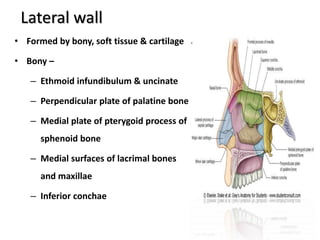
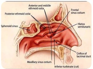
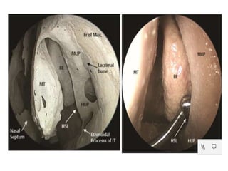
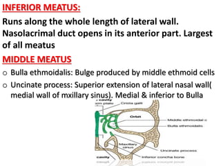
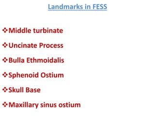
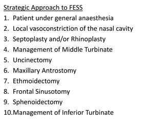
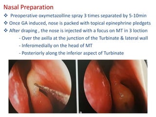
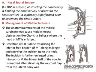
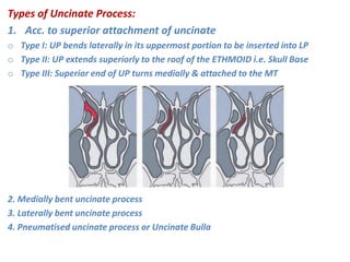
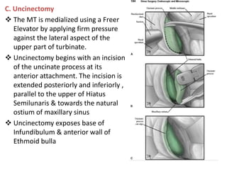
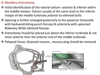
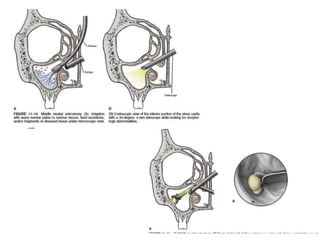
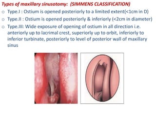
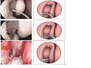
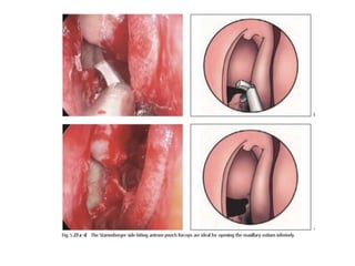
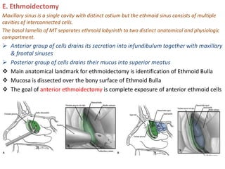
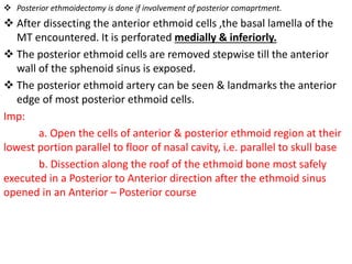
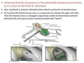
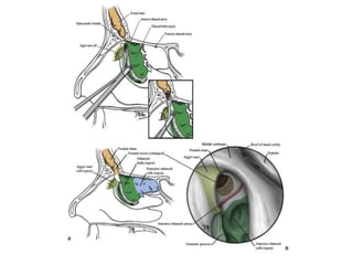
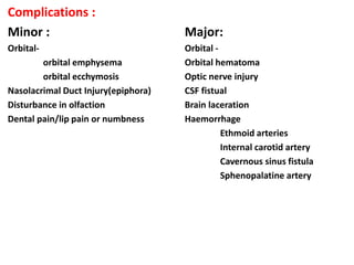
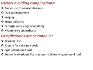
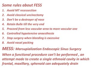
Definition and Terminology
PD is characterized by idiopathic progressive expansion of one or more paranasal sinuses beyond the normal margins, without evidence of mucous membrane changes. The expansion may involve the complete sinus or a part of it.3
The medical literature offers various labels to describe enlargement of the sinus by air, including frontal sinus hypertrophy, PD, pneumosinus frontalis, aerocele, pneumocele, sinus ectasia, hyperpneumatization, pneumatocele, air cyst, and others.1,4,5
The varying terminology used to describe abnormal expansion of the frontal sinus has caused some confusion about the etiology and diagnosis of the condition.1 Urken et al4 classified the deformity into three groups—hypersinus, pneumocele, and PD—as follows:
Hypersinus or hyperpneumatization was defined as an enlarged frontal sinus that has developed beyond the upper limits of normal. The walls are normal, and the hyperaerated sinus does not extend over the normal limits of the frontal bone. The patient is asymptomatic, and the condition requires no intervention.1
Pneumocele refers to an aerated sinus with variable thinning of the sinus walls. The thinning, focal or generalized, differentiates pneumocele from PD. It is a pathological abnormality.1
PD is a condition where the sinus abnormally expands beyond the normal limits of the frontal bone. The bony walls of the sinus are of normal thickness, but are displaced, causing frontal bossing. There is no evidence of erosion, and the mucosa is of normal appearance. The frontal sinus is most commonly affected, and the ethmoidal, sphenoidal, or unilateral maxillary sinus may be involved.6,7
Etiology
The etiology of primary PD has been the source of great debate for many years. It is still unknown, but eight possible mechanisms have been proposed as follows: a spontaneously draining mucocele, the presence of a gas-forming microorganism, the presence of a one-way valve, congenital abnormality, hormonal change, local growth disturbances, osteoclastic and osteoblastic activity, and trauma.1,7-9
Generally, frontonasal duct obstruction of any cause and the subsequent increase in sinus pressure seem to be the most important factors in the pathogenesis of PD.1,3,4 In this case, the ostium was inspected and found to be macroscopically normal, and we did not find a clear etiology.
Review of the literature reveals that age at presentation varies from puberty to the elderly, but PD has not been reported in children. This may be due to the age at which the normal paranasal sinus develops, as well as the gradual onset of PD.5,6
Diagnosis
Diagnosis is made by clinical examination, and confirmation by radiography (plain film or CT), when the characteristic enlargement of the sinus is seen.7,8
Clinical symptoms are typically related to the displaced structures. In the case of outward expansion, the typical signs are frontal bossing and prominence of the supraorbital ridge.
surgical anatomy of nose is a humble attempt to make the anatomy of nose simpler and easy for medical students and fellow physicians. at the end of the presentation the students will be able to identify all the structures.
Imaging of paranasal sinuses (including anatomy and varaints)pk1 pdf pptDr pradeep Kumar
This is very good powerpoint presentation of imaging anatomy and variants of paranasal sinuses and imaging pathology as well as multiple pathological imaging findings and images.it will helps for radiologist and radiology resident and even ent resident. our references is CT and mri whole body by Haaga and various internet sources. THANKS.
USMLE RESP 02 nose and paranasal sinuses anatomy medical .pdfAHMED ASHOUR
The nose and paranasal sinuses are interconnected structures in the upper respiratory system that play essential roles in the respiratory and olfactory processes.
Disorders of the nose and paranasal sinuses can include sinusitis (inflammation of the sinuses), nasal polyps, deviated septum, and various infections.
Proper care and treatment are essential to maintain respiratory function and overall health.
Detailed discussion on tumors and other pathologies of paranasal sinus and their management. Surgical anatomy and approaches are also discussed. Complications of PNS surgeries are discussed briefly
Ct anatomy of paranasal sinuses( PNS) pk.pdf pptDr pradeep Kumar
This presentation includes cross sectional anatomy like axial,saggital and coronal images of paranasal sinuses and most important variation of paranasal sinus.This help alot. Must read topic for radiology resident. Thanks
New Drug Discovery and Development .....NEHA GUPTA
The "New Drug Discovery and Development" process involves the identification, design, testing, and manufacturing of novel pharmaceutical compounds with the aim of introducing new and improved treatments for various medical conditions. This comprehensive endeavor encompasses various stages, including target identification, preclinical studies, clinical trials, regulatory approval, and post-market surveillance. It involves multidisciplinary collaboration among scientists, researchers, clinicians, regulatory experts, and pharmaceutical companies to bring innovative therapies to market and address unmet medical needs.
263778731218 Abortion Clinic /Pills In Harare ,sisternakatoto
263778731218 Abortion Clinic /Pills In Harare ,ABORTION WOMEN’S CLINIC +27730423979 IN women clinic we believe that every woman should be able to make choices in her pregnancy. Our job is to provide compassionate care, safety,affordable and confidential services. That’s why we have won the trust from all generations of women all over the world. we use non surgical method(Abortion pills) to terminate…Dr.LISA +27730423979women Clinic is committed to providing the highest quality of obstetrical and gynecological care to women of all ages. Our dedicated staff aim to treat each patient and her health concerns with compassion and respect.Our dedicated group ABORTION WOMEN’S CLINIC +27730423979 IN women clinic we believe that every woman should be able to make choices in her pregnancy. Our job is to provide compassionate care, safety,affordable and confidential services. That’s why we have won the trust from all generations of women all over the world. we use non surgical method(Abortion pills) to terminate…Dr.LISA +27730423979women Clinic is committed to providing the highest quality of obstetrical and gynecological care to women of all ages. Our dedicated staff aim to treat each patient and her health concerns with compassion and respect.Our dedicated group of receptionists, nurses, and physicians have worked together as a teamof receptionists, nurses, and physicians have worked together as a team wwww.lisywomensclinic.co.za/
Prix Galien International 2024 Forum ProgramLevi Shapiro
June 20, 2024, Prix Galien International and Jerusalem Ethics Forum in ROME. Detailed agenda including panels:
- ADVANCES IN CARDIOLOGY: A NEW PARADIGM IS COMING
- WOMEN’S HEALTH: FERTILITY PRESERVATION
- WHAT’S NEW IN THE TREATMENT OF INFECTIOUS,
ONCOLOGICAL AND INFLAMMATORY SKIN DISEASES?
- ARTIFICIAL INTELLIGENCE AND ETHICS
- GENE THERAPY
- BEYOND BORDERS: GLOBAL INITIATIVES FOR DEMOCRATIZING LIFE SCIENCE TECHNOLOGIES AND PROMOTING ACCESS TO HEALTHCARE
- ETHICAL CHALLENGES IN LIFE SCIENCES
- Prix Galien International Awards Ceremony
Knee anatomy and clinical tests 2024.pdfvimalpl1234
This includes all relevant anatomy and clinical tests compiled from standard textbooks, Campbell,netter etc..It is comprehensive and best suited for orthopaedicians and orthopaedic residents.
Tom Selleck Health: A Comprehensive Look at the Iconic Actor’s Wellness Journeygreendigital
Tom Selleck, an enduring figure in Hollywood. has captivated audiences for decades with his rugged charm, iconic moustache. and memorable roles in television and film. From his breakout role as Thomas Magnum in Magnum P.I. to his current portrayal of Frank Reagan in Blue Bloods. Selleck's career has spanned over 50 years. But beyond his professional achievements. fans have often been curious about Tom Selleck Health. especially as he has aged in the public eye.
Follow us on: Pinterest
Introduction
Many have been interested in Tom Selleck health. not only because of his enduring presence on screen but also because of the challenges. and lifestyle choices he has faced and made over the years. This article delves into the various aspects of Tom Selleck health. exploring his fitness regimen, diet, mental health. and the challenges he has encountered as he ages. We'll look at how he maintains his well-being. the health issues he has faced, and his approach to ageing .
Early Life and Career
Childhood and Athletic Beginnings
Tom Selleck was born on January 29, 1945, in Detroit, Michigan, and grew up in Sherman Oaks, California. From an early age, he was involved in sports, particularly basketball. which played a significant role in his physical development. His athletic pursuits continued into college. where he attended the University of Southern California (USC) on a basketball scholarship. This early involvement in sports laid a strong foundation for his physical health and disciplined lifestyle.
Transition to Acting
Selleck's transition from an athlete to an actor came with its physical demands. His first significant role in "Magnum P.I." required him to perform various stunts and maintain a fit appearance. This role, which he played from 1980 to 1988. necessitated a rigorous fitness routine to meet the show's demands. setting the stage for his long-term commitment to health and wellness.
Fitness Regimen
Workout Routine
Tom Selleck health and fitness regimen has evolved. adapting to his changing roles and age. During his "Magnum, P.I." days. Selleck's workouts were intense and focused on building and maintaining muscle mass. His routine included weightlifting, cardiovascular exercises. and specific training for the stunts he performed on the show.
Selleck adjusted his fitness routine as he aged to suit his body's needs. Today, his workouts focus on maintaining flexibility, strength, and cardiovascular health. He incorporates low-impact exercises such as swimming, walking, and light weightlifting. This balanced approach helps him stay fit without putting undue strain on his joints and muscles.
Importance of Flexibility and Mobility
In recent years, Selleck has emphasized the importance of flexibility and mobility in his fitness regimen. Understanding the natural decline in muscle mass and joint flexibility with age. he includes stretching and yoga in his routine. These practices help prevent injuries, improve posture, and maintain mobilit
Report Back from SGO 2024: What’s the Latest in Cervical Cancer?bkling
Are you curious about what’s new in cervical cancer research or unsure what the findings mean? Join Dr. Emily Ko, a gynecologic oncologist at Penn Medicine, to learn about the latest updates from the Society of Gynecologic Oncology (SGO) 2024 Annual Meeting on Women’s Cancer. Dr. Ko will discuss what the research presented at the conference means for you and answer your questions about the new developments.
Ethanol (CH3CH2OH), or beverage alcohol, is a two-carbon alcohol
that is rapidly distributed in the body and brain. Ethanol alters many
neurochemical systems and has rewarding and addictive properties. It
is the oldest recreational drug and likely contributes to more morbidity,
mortality, and public health costs than all illicit drugs combined. The
5th edition of the Diagnostic and Statistical Manual of Mental Disorders
(DSM-5) integrates alcohol abuse and alcohol dependence into a single
disorder called alcohol use disorder (AUD), with mild, moderate,
and severe subclassifications (American Psychiatric Association, 2013).
In the DSM-5, all types of substance abuse and dependence have been
combined into a single substance use disorder (SUD) on a continuum
from mild to severe. A diagnosis of AUD requires that at least two of
the 11 DSM-5 behaviors be present within a 12-month period (mild
AUD: 2–3 criteria; moderate AUD: 4–5 criteria; severe AUD: 6–11 criteria).
The four main behavioral effects of AUD are impaired control over
drinking, negative social consequences, risky use, and altered physiological
effects (tolerance, withdrawal). This chapter presents an overview
of the prevalence and harmful consequences of AUD in the U.S.,
the systemic nature of the disease, neurocircuitry and stages of AUD,
comorbidities, fetal alcohol spectrum disorders, genetic risk factors, and
pharmacotherapies for AUD.
micro teaching on communication m.sc nursing.pdfAnurag Sharma
Microteaching is a unique model of practice teaching. It is a viable instrument for the. desired change in the teaching behavior or the behavior potential which, in specified types of real. classroom situations, tends to facilitate the achievement of specified types of objectives.
New Directions in Targeted Therapeutic Approaches for Older Adults With Mantl...i3 Health
i3 Health is pleased to make the speaker slides from this activity available for use as a non-accredited self-study or teaching resource.
This slide deck presented by Dr. Kami Maddocks, Professor-Clinical in the Division of Hematology and
Associate Division Director for Ambulatory Operations
The Ohio State University Comprehensive Cancer Center, will provide insight into new directions in targeted therapeutic approaches for older adults with mantle cell lymphoma.
STATEMENT OF NEED
Mantle cell lymphoma (MCL) is a rare, aggressive B-cell non-Hodgkin lymphoma (NHL) accounting for 5% to 7% of all lymphomas. Its prognosis ranges from indolent disease that does not require treatment for years to very aggressive disease, which is associated with poor survival (Silkenstedt et al, 2021). Typically, MCL is diagnosed at advanced stage and in older patients who cannot tolerate intensive therapy (NCCN, 2022). Although recent advances have slightly increased remission rates, recurrence and relapse remain very common, leading to a median overall survival between 3 and 6 years (LLS, 2021). Though there are several effective options, progress is still needed towards establishing an accepted frontline approach for MCL (Castellino et al, 2022). Treatment selection and management of MCL are complicated by the heterogeneity of prognosis, advanced age and comorbidities of patients, and lack of an established standard approach for treatment, making it vital that clinicians be familiar with the latest research and advances in this area. In this activity chaired by Michael Wang, MD, Professor in the Department of Lymphoma & Myeloma at MD Anderson Cancer Center, expert faculty will discuss prognostic factors informing treatment, the promising results of recent trials in new therapeutic approaches, and the implications of treatment resistance in therapeutic selection for MCL.
Target Audience
Hematology/oncology fellows, attending faculty, and other health care professionals involved in the treatment of patients with mantle cell lymphoma (MCL).
Learning Objectives
1.) Identify clinical and biological prognostic factors that can guide treatment decision making for older adults with MCL
2.) Evaluate emerging data on targeted therapeutic approaches for treatment-naive and relapsed/refractory MCL and their applicability to older adults
3.) Assess mechanisms of resistance to targeted therapies for MCL and their implications for treatment selection
Title: Sense of Taste
Presenter: Dr. Faiza, Assistant Professor of Physiology
Qualifications:
MBBS (Best Graduate, AIMC Lahore)
FCPS Physiology
ICMT, CHPE, DHPE (STMU)
MPH (GC University, Faisalabad)
MBA (Virtual University of Pakistan)
Learning Objectives:
Describe the structure and function of taste buds.
Describe the relationship between the taste threshold and taste index of common substances.
Explain the chemical basis and signal transduction of taste perception for each type of primary taste sensation.
Recognize different abnormalities of taste perception and their causes.
Key Topics:
Significance of Taste Sensation:
Differentiation between pleasant and harmful food
Influence on behavior
Selection of food based on metabolic needs
Receptors of Taste:
Taste buds on the tongue
Influence of sense of smell, texture of food, and pain stimulation (e.g., by pepper)
Primary and Secondary Taste Sensations:
Primary taste sensations: Sweet, Sour, Salty, Bitter, Umami
Chemical basis and signal transduction mechanisms for each taste
Taste Threshold and Index:
Taste threshold values for Sweet (sucrose), Salty (NaCl), Sour (HCl), and Bitter (Quinine)
Taste index relationship: Inversely proportional to taste threshold
Taste Blindness:
Inability to taste certain substances, particularly thiourea compounds
Example: Phenylthiocarbamide
Structure and Function of Taste Buds:
Composition: Epithelial cells, Sustentacular/Supporting cells, Taste cells, Basal cells
Features: Taste pores, Taste hairs/microvilli, and Taste nerve fibers
Location of Taste Buds:
Found in papillae of the tongue (Fungiform, Circumvallate, Foliate)
Also present on the palate, tonsillar pillars, epiglottis, and proximal esophagus
Mechanism of Taste Stimulation:
Interaction of taste substances with receptors on microvilli
Signal transduction pathways for Umami, Sweet, Bitter, Sour, and Salty tastes
Taste Sensitivity and Adaptation:
Decrease in sensitivity with age
Rapid adaptation of taste sensation
Role of Saliva in Taste:
Dissolution of tastants to reach receptors
Washing away the stimulus
Taste Preferences and Aversions:
Mechanisms behind taste preference and aversion
Influence of receptors and neural pathways
Impact of Sensory Nerve Damage:
Degeneration of taste buds if the sensory nerve fiber is cut
Abnormalities of Taste Detection:
Conditions: Ageusia, Hypogeusia, Dysgeusia (parageusia)
Causes: Nerve damage, neurological disorders, infections, poor oral hygiene, adverse drug effects, deficiencies, aging, tobacco use, altered neurotransmitter levels
Neurotransmitters and Taste Threshold:
Effects of serotonin (5-HT) and norepinephrine (NE) on taste sensitivity
Supertasters:
25% of the population with heightened sensitivity to taste, especially bitterness
Increased number of fungiform papillae
Couples presenting to the infertility clinic- Do they really have infertility...Sujoy Dasgupta
Dr Sujoy Dasgupta presented the study on "Couples presenting to the infertility clinic- Do they really have infertility? – The unexplored stories of non-consummation" in the 13th Congress of the Asia Pacific Initiative on Reproduction (ASPIRE 2024) at Manila on 24 May, 2024.
2. FESS- Functional Endoscopic Sinus Surgery is often a
non-invasive / minimal invasive surgical procedure
that discloses sinus air cells and sinus ostia by having
an endoscope. It restores the paranasal sinus
function by re-establishing the physiologic pattern
of ventilation & mucocilliary clearance
The term FESS was coined by Kennedy in 1985
Father of FESS is Prof Mesenklinger
Hirschmann 1st described use of primitive
endoscope to examine the maxillary sinus through
an oroantral fistula
3. FESS- The Functional Aspects:
1. Preserving normal structure
2. Removing only obstruction
3. Preserving mucosa
4. Restoration of function
Advantage over Open Sinus Procedure
1. Safe , minimally invasive, no cuts
2. Doesn’t disturb healthy tissue
3. Performed in less time with better management
4. No visible signs that surgery has been performed
5. Quick recovery
4. Balloon Sinuplasty :
o Acclarent introduced balloon sinuplasty system that utilized
a nonconformable balloon which was capable of creating
microfratures in the bone surrounding the drainage
pathways of the Frontal, Maxillary, Sphenoid sinus
5. Lateral wall
• Formed by bony, soft tissue & cartilage
• Bony –
– Ethmoid infundibulum & uncinate
– Perpendicular plate of palatine bone
– Medial plate of pterygoid process of
sphenoid bone
– Medial surfaces of lacrimal bones
and maxillae
– Inferior conchae
6.
7.
8. INFERIOR MEATUS:
Runs along the whole length of lateral wall.
Nasolacrimal duct opens in its anterior part. Largest
of all meatus
MIDDLE MEATUS
o Bulla ethmoidalis: Bulge produced by middle ethmoid cells
o Uncinate process: Superior extension of lateral nasal wall(
medial wall of mxillary sinus). Medial & inferior to Bulla
9. Infundiblum: Air passage connecting the maxillary sinus
ostium to middle meatus
Hiatus Semilunaris: Medially it communicates with middle
meatus. Laterally & inf it communicates with infundibulum
• Frontal sinus – Opens into the anterior part of hiatus
semilunaris
• Maxillary sinus – Opens into the posterior part of hiatus
semilunaris
• Anterior and middle ethmoidal cells – Opens into the
upper margin of bulla ethmoidalis
10. Superior Meatus
Limited only to posterior one third of lateral
wall. Posterior ethmoidal sinus opens into it.
Sphenoethmoidal recess
Above the superior turbinate. It receives the
opening of sphenoid sinus
11. SINUSES
• Air containing cavity in certain skull bones
• Develop as a diverticula/outpouching from the lat
wall of nose & extend into Maxilla, Ethmoid,
sphenoid and frontal bones
• Four sinuses – Maxillary, Frontal, Ethmoid (Ant &
Post) & Sphenoid
12. Maxillary Sinus - (Antrum of Highmore )
• Largest paranasal sinus
• Pyramidal in shape
• Base - towards lateral wall of nose
• Apex – towards zygomatic process of maxilla
• On average it has capacity of 14.75 ml (14-15)
13. Frontal Sinus
• Situated between the outer & inner table of frontal bone
• Asymmetrical
• Intervening bony septum which may be thin or deficiency
• The natural frontal sinus ostium is usually located in the
posteromedial floor of the sinus (most dependent part).
• It opens into the middle meatus
• The ethmoidal infundibulum can act as a channel for carrying the
secretions (and infection) from the frontal sinus to anterior ethmoid
cells and the maxillary sinus or vice versa.
14. FRONTAL RECESS
• The frontal recess is an hourglass like narrowing between the
frontal sinus and the anterior middle meatus through which the
frontal sinus drains.
• The frontal recesses are the narrowest anterior air channels
and are common sites of inflammation
15. AGGER NASI CELL
It is present Anterior, lateral, and inferior to the
frontal recess .
It is aerated and represents the most anterior
ethmoid air cell, usually lying deep to the lacrimal
bone.
It usually borders the primary ostium or floor of
the frontal sinus.
its size may directly influence the patency of the
frontal recess and the anterior middle meatus.
16. Sphenoid sinus
• Occupies the body of sphenoid
• Right & left, seperated by a
thin strip of bony septum (like
frontal sinus)
• Ostium opens into spheno
ethmoidal recess
17. ETHMOID SINUS
• Thin walled air cavities in the lateral masses of the ethmoid bone
• Varies from 3 – 18
• Occupy the space between the upper third of the lateral nasal wall
and the medial wall of orbit
• Clinically divided into anterior ethmoidal air cells & posterior
ethmoidal air cells, by basal lamella (lateral attachment of middle
turbinate to lamina papyracea)
18. Relations
• Roof – formed by the anterior cranial fossa
• Lateral wall - orbit
• Medial wall – nasal cavity
• Thin paper like bony part of the ethmoid separating the air
cells from the orbit, called lamina papyracea, can be easily
destroyed leading to spread of ethmoidal infections into
the orbit
• Optic nerve forms a close relationship with the posterior
ethmoidal cells & is at risk during ethmoidal surgery
19. OSTIO MEATAL COMPLEX
• The ostiomeatal complex is the key anatomic area addressed by
endoscopic sinus surgeons.
• Blockage of the ostiomeatal complex prevents effective
mucociliary clearance, thus leading to a stagnation of secretions
and therefore leading to recurrent or chronic sinusitis.
21. Pattern of Sinus Disease:
Sonkens’ Classification acc. to Middle meatus obstruction
1. OMC pattern- M,Ant.E,F
2. Infundibular – isolated obst of Eth. Infudibulum
3. Frontal recess inflammatory pattern
4. Sinunasal polyposis pattern
5. Sporadic pattern
Lund-Mackay Score radiologic score of chronic rhinosinusitis
Reading a CT scan of the PNS & OMC with assigns a score of
0- ( no abnormality)
1- (partial opacification)
2- ( complete opacification)
Each side graded separately. A combined score of 24 is possible
22. Gliklich and Metson System
Stage 0: <2mm mucosal thickening on any sinus wall
Stage 1: all unilateral disease or anatomical
abnormalities
Stage 2: bilateral disease limited to ethmoid or
maxillary sinuses
Stage 3: bilateral disease with involvement at least 1
sphenoid or frontal sinus
Stage 4 : Pansinus disease
23. Landmarks in FESS
Middle turbinate
Uncinate Process
Bulla Ethmoidalis
Sphenoid Ostium
Skull Base
Maxillary sinus ostium
24. MESSERKLINGER 5 LAMELLA
1st Lamella- UP
2nd Lamella- BE
3rd Lamella- Ground Lamella Basal
4th Lamella- Superior Turbinate
5th Lamella- Supreme Turbinate
26. Patient selection: Pre-operative assessment
History
- Symptoms
- Medical treatment
- associated disease
Examination of the Patient: (General & Local)
- Anterior rhinoscopy
- Septal deviation
- Turbinate hypertrophy
-Nasal airway Problem
- Nasal Endoscopy
- Character of mucosa & appearance of sinus drainage,
anatomical variations, structural abnormalities
27. • Radiological- Pre-operative X-rays, CT Scan(Gold Standard)
• Routine Blood Investigations
Key Points to be reviewed on Pre-operative CT SCAN
1. Disease: Extent and pattern & its clinical correlation
2. Bony integrity- (Erosion, expansion ,dehiscence )- skull base, lamina
papyracea, optic canal, carotid canal
3. Skull Base- Height, symmetry, slope of cribriform plate & fovea
ethmoidalis.
4. Maxillary Sinus- location & attachment of uncinate process to
medial orbital wall, pneumatisation & height
5. Ethmoid Sinus – location of AEA,PEA, height of post.ethmoid cell
6. Sphenoid Sinus- location of sphenoid ostium, septation & their
relation to carotid canal
7. Frontal Sinus- extent of pneumatisation, natural drainage pathway,
presence of ager nasi & frontal cell
28. Strategic Approach to FESS
1. Patient under general anaesthesia
2. Local vasoconstriction of the nasal cavity
3. Septoplasty and/or Rhinoplasty
4. Management of Middle Turbinate
5. Uncinectomy
6. Maxillary Antrostomy
7. Ethmoidectomy
8. Frontal Sinusotomy
9. Sphenoidectomy
10.Management of Inferior Turbinate
29. Nasal Preparation
Preoperative oxymetazolline spray 3 times separated by 5-10min
Once GA induced, nose is packed with topical epinephrine pledgets
After draping , the nose is injected with a focus on MT in 3 loction
- Over the axilla at the junction of the Turbinate & lateral wall
- Inferomedially on the head of MT
- Posteriorly along the inferior aspect of Turbinate
30. A. Nasal Septal Surgery
If a DNS is present, obstructing the nasal cavity
& limiting the nasal airway or access to the
sinus cavities , a septoplsty is performed prior
to beginning the sinus surgery
B. Management of Middle Turbinate
The anatomical variants of the middle
turbinate may cause middle meatal
obstruction like Choncha Bullosa where the
head of MT is enlarged.
Resection of CB is done by incising the
inferior free border of MT along its length
and carrying the incision up to the neck.
The incision is further enlarged using
microscisor & the lateral half of the concha
is removed after elevating the mucosal flap
from the lateral bony wall
31. Types of Uncinate Process:
1. Acc. to superior attachment of uncinate
o Type I: UP bends laterally in its uppermost portion to be inserted into LP
o Type II: UP extends superiorly to the roof of the ETHMOID i.e. Skull Base
o Type III: Superior end of UP turns medially & attached to the MT
2. Medially bent uncinate process
3. Laterally bent uncinate process
4. Pneumatised uncinate process or Uncinate Bulla
32. C. Uncinectomy
The MT is medialized using a Freer
Elevator by applying firm pressure
against the lateral aspect of the
upper part of turbinate.
Uncinectomy begins with an incision
of the uncinate process at its
anterior attachment. The incision is
extended posteriorly and inferiorly ,
parallel to the upper of Hiatus
Semilunaris & towards the natural
ostium of maxillary sinus
Uncinectomy exposes base of
Infundibulum & anterior wall of
Ethmoid bulla
33. Types of Uncinectomy:
A. Classical / Anterograde Technique:
Uncinectomy is performed via an incision with either the sharp end of freer
elevator or a sickle knife.
The incision should be placed at the most anterior portion of uncinate
process which is softer on palpation in comparison to firmer lacrimal bone
where also NLD located. Then by using blakeshly forcep the free uncinate
edge is removed . More prone for Orbital Fat Prolapse.
B. Swing door / Retrograde Technique:
Reverse cutting forceps or backbiting forceps were used in this technique.
Inferior free margin overlying the maxillary ostium is cut first & then incision
is made in the superior margin to form a flap from a flap from the uncinate
which is hinged on the anterior margin & can be moved with an elevator or
ball probe
This is followed by submucosal removal of the horizontal process of the
uncinate . More prone for NLD injury
34.
35.
36. D. Maxillary Antrostomy
Initial identification of the natural ostium- anterior & inferior within
the middle meatus. Ostium usually at the same level as the inferior
margin of the middle turbinate,anterior to ethmoid bulla
Opening is further enlarged posteriorly to the posterior fontanelle
with backward-biting punch forceps & anteriorly with upturned
Blakesley-Wilde ethmoid forceps
Antrostomy should be placed just above the inferior turbinate & not
more anterior than the anterior end of the middle turbinate
Polypoid tissue, diseased mucosa , mucous plug should be removed
37.
38. Types of maxillary sinusotomy: (SIMMENS CLASSIFICATION)
o Type.I : Ostium is opened posteriorly to a limited extent(<1cm in D)
o Type.II : Ostium is opened posteriorly & inferiorly (<2cm in diameter)
o Type.III: Wide exposure of opening of ostium in all direction i.e.
anteriorly up to lacrimal crest, superiorly up to orbit, inferiorly to
inferior turbinate, posteriorly to level of posterior wall of maxillary
sinus
39.
40.
41. E. Ethmoidectomy
Maxillary sinus is a single cavity with distinct ostium but the ethmoid sinus consists of multiple
cavities of interconnected cells.
The basal lamella of MT separates ethmoid labyrinth to two distinct anatomical and physiologic
compartment.
Anterior group of cells drains its secretion into infundibulum together with maxillary
& frontal sinuses
Posterior group of cells drains their mucus into superior meatus
Main anatomical landmark for ethmoidectomy is identification of Ethmoid Bulla
Mucosa is dissected over the bony surface of Ethmoid Bulla
The goal of anterior ethmoidectomy is complete exposure of anterior ethmoid cells
42. Posterior ethmoidectomy is done if involvement of posterior comaprtment.
After dissecting the anterior ethmoid cells ,the basal lamella of the
MT encountered. It is perforated medially & inferiorly.
The posterior ethmoid cells are removed stepwise till the anterior
wall of the sphenoid sinus is exposed.
The posterior ethmoid artery can be seen & landmarks the anterior
edge of most posterior ethmoid cells.
Imp:
a. Open the cells of anterior & posterior ethmoid region at their
lowest portion parallel to floor of nasal cavity, i.e. parallel to skull base
b. Dissection along the roof of the ethmoid bone most safely
executed in a Posterior to Anterior direction after the ethmoid sinus
opened in an Anterior – Posterior course
43.
44. F. Frontal Sinusotomy
Kuhn Classification of Frontal Recess & Frontal Sinus Cell
Agger nasi cell
Supraorbital ethmoid cell
Frontal cell
Type 1 – Single Frontal cell above agger nasi cell
Type 2- Tier of cells in FR above agger nasi cell
Type 3- Single massive cell pneumatising cephalad in to Frontal S
Type 4- Isolated cell in Frontal Sinus
Frontal Bulla Cell
Suprabullar cell
Interfrontal sinus septal cell
45. Acc.to Draf Endonasal Frontal Sinus Drainge
Type I: Simple drainage
Type II a/b: Extended drainage
Type III: Endonasal Median Drainage= Endoscopic modified Lothrop pro
46.
47. Type III: Endonasal Median Drainage= Endoscopic
modified Lothrop procedure
It is done by either
A. Primary lateral approach: if previous ethmoid
work incomplete & MT is intact as land mark
B. Medial approach: if ethmoid has been cleared
and/or if the middle turbinate is absent. This
begins with resection of perpendicular plate of
nasal septum
48. When the type III drainage is technically not possible
(anterior-posterior diameter of the frontal sinus less than 0.8
cm) or has failed, osteoplastic frontal sinus obliteration
must be considered
Indications of Osteoplastic Frontal Sinus Obliteration:
49. Intranasal frontal sinusotomy is the potentially dangerous procedure
as it is close to the Orbit & Skull base
Imp. Landmark is anterior ethmoid artery which is posterior to frontal recess
To visualise the frontal recess area, it is necessary to remove the ager nasi cells.
Then the frontal recess is enlarged using sharp curette to break down anterior
ethmoid cells, the spina nasalis frontalis (nasofrontal ‘’beak”)
50.
51. G . Sphenoidotomy
The sphenoid sinus can be opened safely 10 mm above the choana
just lateral to the midline septum at the rostrum of the sphenoid.
After identification of ostium the opening enlarged in lateral &
inferior direction. Initial opening is made with Straight Blakesley
forceps.
If the entire anterior wall of the sinus is thick & ostium is not
visualised , an angulated hand piece with extra long diamond burr is
used to make opening in ostium area
Optic nerve & carotid artery located in lateral & posterior wall. Sella
turcica situated medial & superior to the sinus & cavernous sinus
located laterally
Roof of sphenoid sinus is extremely thin- potential risk of a CSF leak
52.
53.
54.
55. H. Management of inferior turbinate
After taking care of septal deviation & parnasal sinus diease , last step is to treat IT
hypertrophy
Long nasal speculum is introduced along the IT & with endoscopic view the
posterior third of turbinate is removed after being retracted medially with
straight Blakesley forceps.
The anterior & middle third of HIT treated with Radio Frequency Thermal Ablation
56. Post-operative Care
o Nasal packing: Packing used to control bleeding ,prevent adhesions
o Regular analgesia & vitals are carefully monitored
o Observe for epistaxis, headache, orbital swelling, diplopia, reduced
visual acuity
o Remove nasal packing after 48 hours
Post-operative Ambulatory Care
o Antibiotics are not routinely prescribed
o Instruct not to blow nose hard for at least 48 hours
o Commence topical decongestants for 5 days & saline spray for
6weeks
o Suction toilet of the nose
o Recommence long-term nasal steroids after 1 wk in nasal polyposis
o Decrust the nose with a rigid endoscope if necessary
57. Complications :
Minor : Major:
Orbital- Orbital -
orbital emphysema Orbital hematoma
orbital ecchymosis Optic nerve injury
Nasolacrimal Duct Injury(epiphora) CSF fistual
Disturbance in olfaction Brain laceration
Dental pain/lip pain or numbness Haemorrhage
Ethmoid arteries
Internal carotid artery
Cavernous sinus fistula
Sphenopalatine artery
58. Factors avoiding complications:
Proper use of nasal endoscope
True cut instrument
Imaging
Image guidance
Through knowledge of anatomy
Hypotensive anaesthesia
Complications are common in:
Revision FESS
Surgery for nasal polyposis
Type 3 kerio skull base
Anatomical variants like asymmetrical low lying ethmoid roof
59. Some rules about FESS
1. Avoid MT recessection
2. Avoid classical uncinectomy
3. Don’t be a destroyer of nose
4. Retain Bulle till the very end
5. Proceed from less vascular area to more vascular one
6. Controlled hypotensive anaesthesia
7. Stop surgery when bleeding is excessive
8. Avoid nasal packing
MESS: Marsupialization Endoscopic Sinus Surgery
When a functional procedure can’t be performed , an
attempt made to create a single ethmoid cavity in which
frontal, maxillary, sphenoid can adequately drain