
Recommended
More Related Content
What's hot
What's hot (20)
Similar to Anterior Choroidal Artery: Anatomy, Variants, and Clinical Significance
Similar to Anterior Choroidal Artery: Anatomy, Variants, and Clinical Significance (20)
More from Dr. Rahul Jain
More from Dr. Rahul Jain (20)
Recently uploaded
Recently uploaded (20)
Anterior Choroidal Artery: Anatomy, Variants, and Clinical Significance
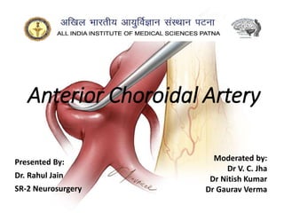
- 1. Anterior Choroidal Artery Presented By: Dr. Rahul Jain SR-2 Neurosurgery Moderated by: Dr V. C. Jha Dr Nitish Kumar Dr Gaurav Verma
- 2. Introduction • Usually arises from C4 as single artery, majority of the time arising near origin of PComA than near bifurcation. • It measures ~1 mm in diameter. • Variant anatomy occasionally it may originate from • internal carotid artery bifurcation • middle cerebral artery • posterior communicating artery duplication of AChA is reported in ~5% of cases
- 3. Embryology • AchA may be most prominent in its vascular supply to the brain during the choroidal stage of development, around five weeks. • During this time, the vertebrobasilar system has yet to develop, and the internal carotid arteries almost exclusively supply the brain. • Thus, the AchA provides cortical branches to the temporal, parietal, and occipital lobes during this early embryonic stage, which is later dominated by the posterior cerebral artery in the adult human.
- 4. • Due to its prominence in early embryonic development, the AchA does have various anatomic variants that may exist as discussed. • In such cases, collateral circulation may diminish, and thus it is essential to keep the singular vessel supplying that area patent.
- 5. Course AChA arises from the posterior wall of the ICA above the origin of the PComA. Initial segment of the AChA is directed posteromedial behind the internal carotid artery and passes backward below the optic tract and lateral to the PCA and cerebral peduncle At the anterior margin of the lateral geniculate body, the AChA again crosses the optic tract from medial to lateral and passes posterolateral through the crural cistern, located between the cerebral peduncle and uncus.
- 6. • The average length that the artery follows the optic tract is 12 mm (range, 5-25 mm). • The AChA passes above the posterior uncal segment and enters the temporal horn by passing through the choroidal fissure located between the thalamus above and fimbria of the fornix below. (Plexal Point) • enter the choroid plexus, courses along the medial border of the choroid plexus in close relation to the lateral posterior choroidal branches of the PCA. • pass dorsally along the medial border of the plexus, reaching the foramen of Monro
- 7. AChA pursues an angulated course, descending along the anterior segment of the uncus, but at the uncal apex it turns sharply upward, reaching the upper part of the posterior uncal segment before entering the temporal horn. The AChA can be divided into two segments: • Cisternal segment: extends from its origin until the choroidal fissure; measures ~2.5 cm (range 1.5-3.5 cm) in length. It passes through carotid cistern, crural cistern and ambient cistern before reaching the choroidal fissure 6,7. • Intraventricular or plexal segment: one or more branches that pass through the choroidal fissure to branch and enter the choroid plexus of the temporal horn.
- 8. 7T MRI - Anterior Choroidal (light blue) can be traced perfectly well towards the choroid plexus (dark red) anterior choroidal perforators (purple) coming off the main trunk (red) prior to the plexal point (light blue). A branch to the choroid distal to the plexal point may be present also (pink). The PComA is orange (with a large thalamic branch of its own). MHT (dark blue) and ILT (green) branches are also visible.
- 9. Vascular territory • cisternal segment • deep brain structures • posterior limb and retrolenticular part of the internal capsule including optic radiations • lateral thalamus including lateral geniculate nucleus • optic tract • lateral cerebral peduncle • globus pallidus internus • tail of caudate nucleus • mesial temporal structures (uncus) • head of hippocampus • amygdala • Intraventricular segment - choroid plexus of the anterior part of the temporal horns
- 10. • Nearly half of hemispheres have anastomoses between the PCA and AChA. The richest anastomoses are those located on the surface of the choroid plexus with the lateral posterior choroidal branches of the PCA. • Anastomoses between the AChA and PCA are also found on the lateral surface of the lateral geniculate body and on the temporal lobe near the uncus. • These complex and variable anastomoses make it difficult to predict the effects of occlusion of a single AChA, but explain some of the inconsistent results of AChA occlusion.
- 11. • There is a marked interchangeability of the field of supply of the AChA and the nearby branches of the C4, PCA, PComA, and MCA. • Within the internal capsule. If the PComA is small, the anterior choroidal artery may take over its normal area of supply to the genu and the anterior third of the internal capsule, or if the AChA is small, the field of supply of the PComA may enlarge to supply the greater part of the posterior limb of the internal capsule.
- 12. Surgical Considerations Intradural exposure of the C4 • In exposing the C4 beyond the origin of the ophthalmic artery, the surgeon often sees the AChA before the PComA, although the AChA arises distal to the PComA. • Three sets of anatomic circumstances • the C4 passes upward in a posterolateral direction, placing the origin of the AChA further lateral to the midline than the origin of the PComA • AChA commonly arises further laterally on the posterior wall of the C4 portion than the PComA. • AChA pursues a more lateral course than the PComA, atter is most commonly directed in its initial course in a posteromedial direction
- 13. Aneurysm • Aneurysms at the junction of the AChA and ICA account for 2%- 4% of all intracranial aneurysms however, distal AChA aneurysms are rare. • point posteriorly or posterolaterally, usually well above the oculomotor nerve. • Most distal AChA aneurysms are located in the choroidal segment beyond the plexal point and are associated with MMD, whereas AChA aneurysms in the cisternal segment are extremely rare.
- 14. • Present with subarachnoid hemorrhage, however, distal AChA aneurysms often present with isolated medial temporal intracerebral hematoma with intraventricular extension. • Because the AChA is vulnerable, inadvertent damage to and occlusion of the AChA during clipping and embolization may have deleterious clinical consequences. • Compared with clipping, coiling AChA aneurysms had a significantly lower incidence of AChA infarction , in a study performed by Bohnstedt et al. in 2013, the ischemic complication rate following surgical treatment of AChA aneurysms was 12%, whereas coiling AChA aneurysms as an alternative to clipping was associated with a 5.5% risk of ischemia.
- 15. • Treatments for distal AChA aneurysms are different from those for proximal AChA aneurysms because of their lack of accessibility and small size the aneurysm location and the preservation of the parent artery are two major prognostic factors. • When distal AChA aneurysms are beyond the plexal point, the AChA can be sacrificed or preserved, if the treatment requires AChA occlusion, preoperative provocative testing should be considered.
- 16. Ach Artery Infarct • triad of contralateral hemiparesis, hemianesthesia, and hemianopia, in AChA syndrome. • contralateral hemiplegia and hemianesthesia (to all sensory modalities) results from infarction in the posterior two-thirds of the posterior limb of the internal capsule and the middle third of the cerebral peduncle. • homonomous hemianopsia of varying degrees results from interruption of the supply to the origin of the optic radiations, the optic tract, and part of the lateral geniculate body. • AChA infarcts can be divided into small vessel and large vessel infarcts, and thrombolytic therapy may be effective for large vessel infarcts.
- 17. Moyamoya disease • chronic occlusive cerebrovascular disease characterized by bilateral stenosis or occlusion at the terminal portion of the ICA and the eponymous vessels at the base of the brain. • site of occlusion or stenosis can occur in one of the following four sites: 1 the top of the ICA or A1 ACA or M1 MCA 2 distal to the AChA 3 between the AChA and posterior communicating artery 4 proximal to the posterior communicating artery
- 18. • collaterals are more likely to arise from the choroidal arteries; therefore, the AChA may act as a major collateral route because it is frequently dilated and exhibits abnormal extension. • The angiographic findings of the AChA can be considered grade 2 according to Suzuki's classification, and the AChA shows dilation and extension beyond the choroidal fissure. • when it functions as a collateral vessel to increase blood flow, the hemodynamic load in the vessels supplying the walls of the posterior areas of the ventricles and the periventricular region is increased. • Under such a hemodynamically stressed state, the dilated branches of the AChA may be more fragile, and the choroidal arteries and their anastomotic channels may rupture. In addition, an aneurysm could develop from an outpouching of the vessel wall in some fragile portions.
- 19. • Distal AChA aneurysms in MMD can be treated endovascularly with embolization. However, disturbing the MMD collaterals should be avoided when using a liquid embolization material. • AChA is a “double-edged sword” in MMD. On one hand, the dilated AChA acts as collateral vessels to prevent brain ischemia; on the other hand, due to the resulting hemodynamic stress, the branches of the AChA may rupture and even form an aneurysm in the AChA. • Following intracranial bleeding, both direct and indirect revascularization may be effective for preventing recurrent bleeding.
- 20. Brain tumors • blood supply to particularly those located in the lateral ventricle; include meningiomas, choroid papilloma and gliomas. • hypervascular nature of the lesions in the lateral ventricle imposes challenges for surgical treatment; therefore, obliterating the feeders from the AChA before surgery could reduce hemorrhage and facilitate the surgery • Since AChA feeds critical regions of the brain, migration of embolic agents through the AChA might cause serious neurological deficits. • Successful embolization requires the catheter to enter the plexal segment beyond the plexal point and during the injection, the speed should be extremely slow to avoid excessive reflux of the embolic agent.
- 21. Arteriovenous malformations • AVMs fed by the AChA are difficult to treat because surgical treatment can cause a high incidence of neurological deficits. • AVM embolization via the AChA may be an appropriate treatment option prior to surgery and radiation therapy or serve as a curative procedure. • Microcatheter tip should be advanced distally beyond the plexal point to avoid serious ischemic complications. • Approximately 38% of capsulo-thalamic arteries arising from the AChA originate from the first part of the plexal segment, and this variation could be an important risk factor
- 22. • In contrast, embolization from the cisternal segment of the AChA does not always result in ischemic complications, suggesting a potential collateral circulation. • To minimize ischemic complications, some authors recommend superselective provocative testing with propofol using motor-evoked potential monitoring to manage AVMs fed by the AChA.
- 23. Parkinson disease • characterized by lesions in the basal ganglia (predominantly in the substantia nigra), and symptoms include tremor, bradykinesia, rigidity, and postural instability. • AChA may play a role in PD because, in rare cases, PD may arise due to AChA territorial infarcts affecting the basal ganglial structures and the striatal pre-synaptic dopaminergic pathways resulting in Vascular Parkinsonism • in 1953, Cooper et al. reported dramatic amelioration of parkinsonism after ligation of the AChA in 8 patients with severely advanced disease and concluded that the procedure had been invariably followed by the disappearance of most of the rigidity and cogwheelism; additionally, neither hemiplegia nor hemianesthesia occurred.
- 24. Pial arteriovenous fistula • cerebral pial arteriovenous fistula (PAVF) is a direct connection between the intracranial artery and vein without a nidus, and the vein often develops venous pouches of different sizes. • Cerebral PAVF should be considered congenital. • Cerebral PAVF involving the AChA is extremely rare. • Currently, endovascular management is the first- line treatment of choice for PAVFs, and the goal is to close feeders at the entry point to the vein. • Coiling is a better choice than glue because the AChA is too short to prevent dangerous reflux during glue embolization.
- 25. Conclusion • Although the AChA is a small thin artery, it supplies an extremely critical region of the brain. • The AChA can be involved in many diseases, including aneurysm, brain infarct, MMD, brain tumor, AVM and traumatic cerebral hemorrhage. • During treatment for aneurysms, tumors, AVM or AVF, the AChA cisternal segments should be preserved as a pathway to prevent the infarction of the critical regions of the brain that receive their blood supply from the AChA.