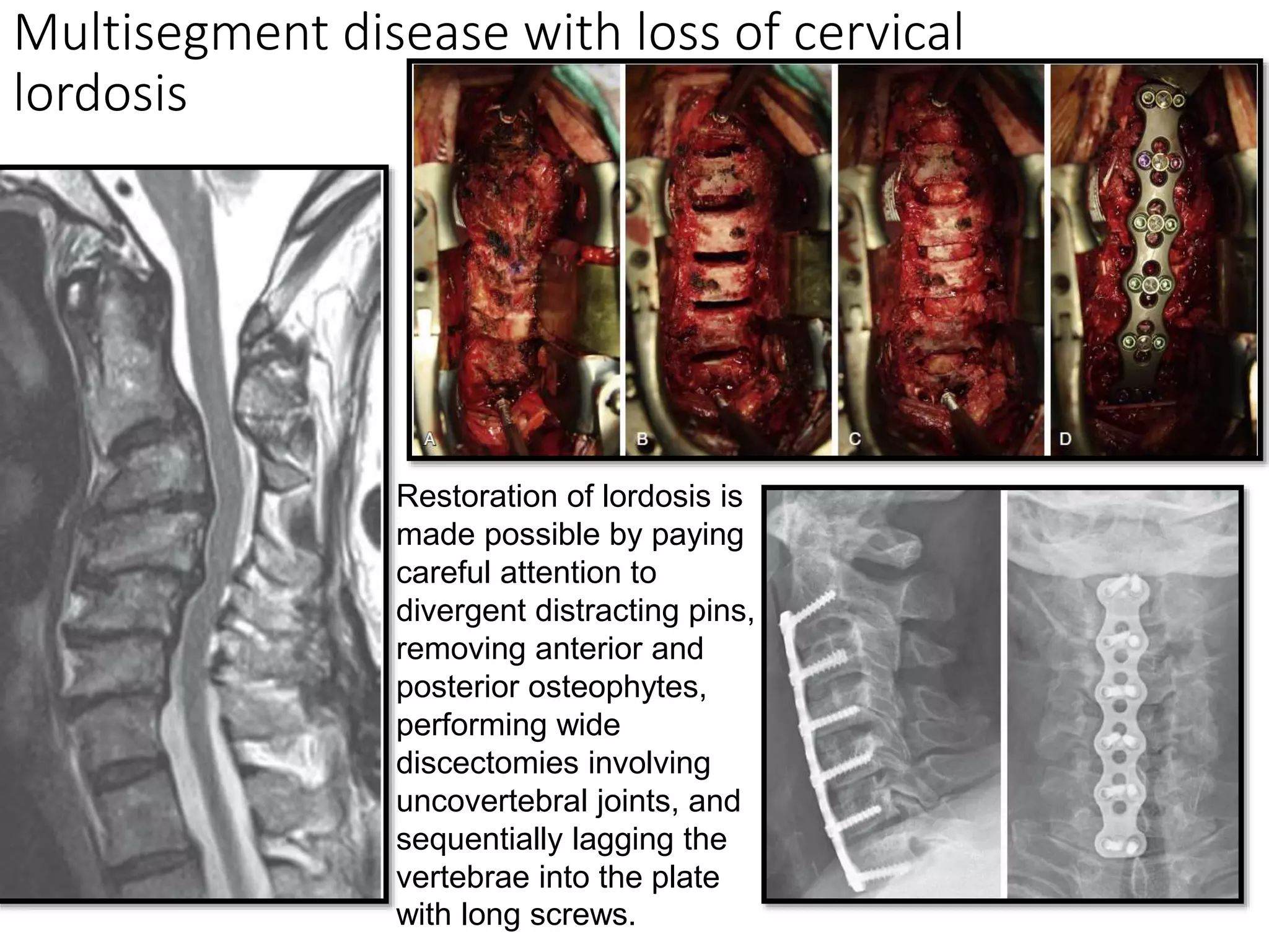
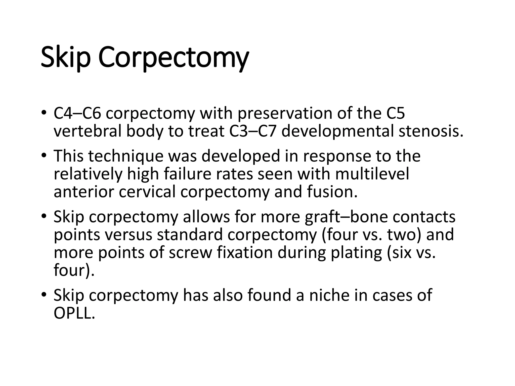
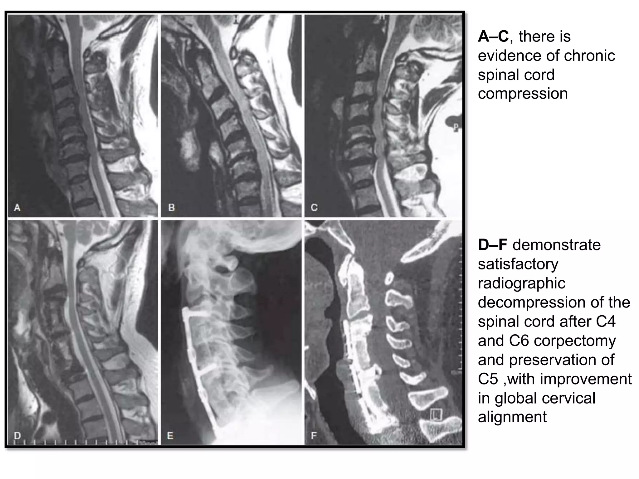
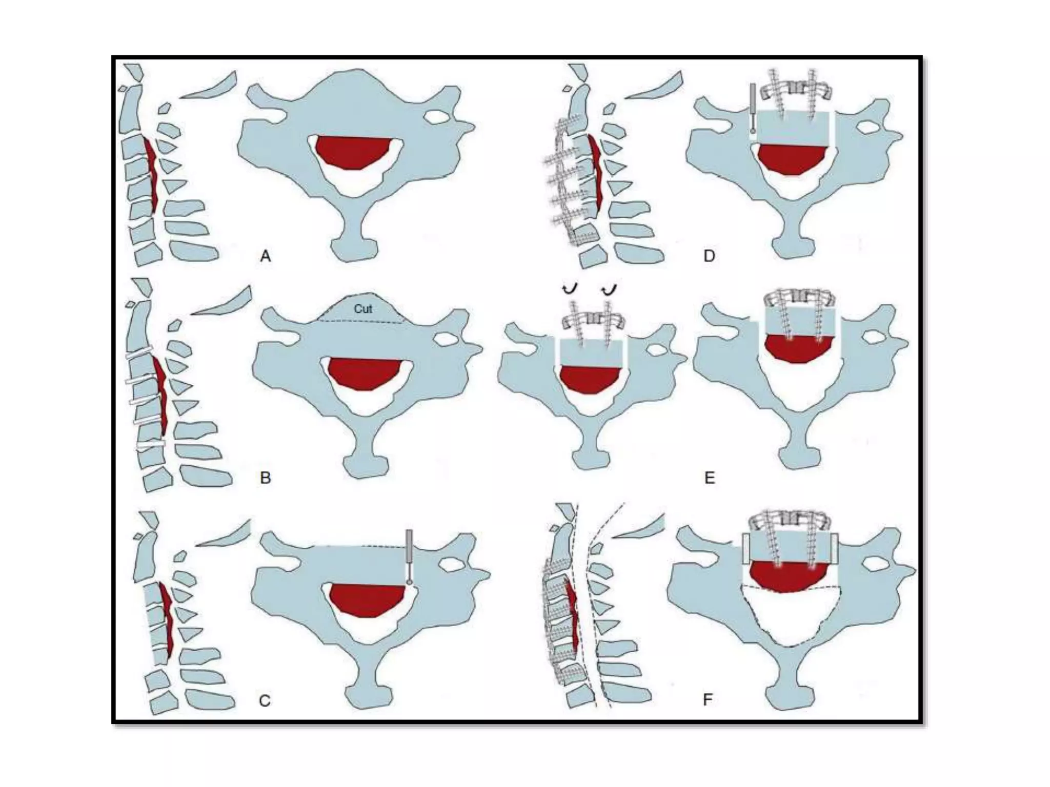
SlideShare a Scribd company logo
1 of 40
Fusion techniques in Subaxial cervical spine
Presented By:
Dr. Rahul Jain
SR-2 Neurosurgery
Moderated by:
Dr V. C. Jha
Dr Nitish Kumar
Dr Gaurav Verma
Anatomy
• more conventional spinal motion segments compared to
C1 and C2
• Superior articulating facets transition from a posteromedial
orientation at C3 to a posterolateral orientation at C7.
• Uncovertebral (Luschka) joints in which the lateral aspects
of the vertebral body superior end plates project upward
as uncinate processes articulating with the unci of the
inferior end plates above, serving to restrain lateral motion
Surgical Planning
• Core principles of spine surgery—
• decompression of the neural elements,
• restoration of normal alignment,
• stabilization of pathologic segments, and
• prevention of further deformity
• Factors such as anterior versus posterior approach,
number of levels, the need for fusion, consideration of
cervical disk arthroplasty (CDA), type of grafting
material, and implant design must all be considered
when developing a surgical strategy.
• In addition, the cost-effectiveness of not only implants
but also the type of procedure itself must be weighed
into the surgical design.
Anterior v/s Posterior
• Anterior approach works well for a central and
posterolateral disk herniation resulting in spinal
cord compression.
• Posterior approaches often result in indirect
decompression of the spinal cord that can shift
away from anterior pathology, whereas an anterior
approach addresses the pathology directly.
• Risks unique to anterior approach are dysphagia,
hoarseness, vocal cord paralysis, anterior vascular
injury, injury to trachea or esophagus.
• Anterior approach is best suited to
address pathology when cervical kyphosis
is present, for restoration of lordosis and
improved sagittal balance.
• Historically more than three levels of
disease were considered a good
indication for posterior approach but
recently there has been increased
interest in three or even four level ACDF.
• In general a simple discectomy is
indicated when the disease is confined to
disk space but when there’s significant
disease posterior to vertebral body then
a corpectomy should be considered.
Anterior fixation techniques
• In 1955, Robinson and Smith – 1st attempt for anterior
approach - operation for ventral removal of cervical
disk material and replacement with a rectangular bone
graft obtained from the iliac crest.
• In 1958, Cloward described his technique, in which a
discectomy was performed and a cylindrical bone
dowel was inserted.
• in 1964 first reported use of screw-plate fixation in the
anterior cervical spine by Bohler and Gaudernak.
• In 1982, Caspar described a novel technique and
instrumentation system (retractors, distractors, and
screw plates) that popularized the practice of anterior
cervical plate application.
Indications
• Degenerative, neoplastic, infectious or inflammatory,
traumatic, and iatrogenic (postsurgical) causes of
vertebral column instability, with or without
concomitant neural compression.
• The two most widely accepted clinical indications for
anterior approaches to the cervical spine are
myelopathy and medically-refractory radiculopathy.
• Whereas structural compromise from discitis or
osteomyelitis and trauma can also necessitate
operative intervention, axial neck pain and cervicogenic
headache with or without kyphotic deformity are not
widely accepted indications for surgery.
Technique
The superior border of the thyroid
cartilage can be used to plan an
exposure of C3‒C4, whereas an
incision based in a skin crease near
the inferior border will allow access
to C4‒C5 and C5‒C6. C6‒C7 about 2
fingerbreadths above the clavicle.
Discectomy Versus Corpectomy
• When circumstances permit, multilevel discectomies
are superior to corpectomies.
• When possible, generous discectomies are
substituted for a corpectomy if adequate neural
decompression can be achieved through wide and
deep undercutting of the offending posterior
vertebral body surfaces.
• The benefits of avoiding a complete corpectomy
include preserving additional sites for screw fixation
along the plate and circumventing the higher risk of
non-union or hardware failure that accompanies
fusion constructs involving multiple corpectomy
segments.
• The primary indications of
corpectomy over discectomy are:
(1) loss of structural integrity of the
vertebral body (anterior column
failure); and
(2) canal compromise behind the
vertebral body, not accessible
through the disc space
• End plate contouring above and
below the decompression zone can
be performed either after
discectomy or after corpectomy
but should be completed before
PLL removal.
• Options for cervical interbodies
include the use of ICBG, structural
allograft, titanium cages, or
polyetheretherketone (PEEK).
Multisegment disease with loss of cervical
lordosis
Restoration of lordosis is
made possible by paying
careful attention to
divergent distracting pins,
removing anterior and
posterior osteophytes,
performing wide
discectomies involving
uncovertebral joints, and
sequentially lagging the
vertebrae into the plate
with long screws.
Skip Corpectomy
• C4–C6 corpectomy with preservation of the C5
vertebral body to treat C3–C7 developmental stenosis.
• This technique was developed in response to the
relatively high failure rates seen with multilevel
anterior cervical corpectomy and fusion.
• Skip corpectomy allows for more graft–bone contacts
points versus standard corpectomy (four vs. two) and
more points of screw fixation during plating (six vs.
four).
• Skip corpectomy has also found a niche in cases of
OPLL.
A–C, there is
evidence of chronic
spinal cord
compression
D–F demonstrate
satisfactory
radiographic
decompression of the
spinal cord after C4
and C6 corpectomy
and preservation of
C5 ,with improvement
in global cervical
alignment
ANTERIOR CONTROLLABLE
ANTEDISPLACEMENT FUSION
• method in treating OPLL, Yang and colleagues described the
anterior controllable antedisplacement fusion (ACAF)
method in 2018.
• Expose the anterior vertebral bodies and intervertebral
discs spanning the vertical length of the OPLL.
• Discectomies are performed above and below the OPLL, and
a partial corpectomy is completed, leaving approximately
50% of the vertebral body intact.
• Vertical troughs are drilled medial to the transverse foramen
and lateral to the OPLL down to the dura. An anterior plate
is affixed to the vertebral bodies above and below the OPLL.
• Finally, screws are placed into the remnant vertebral bodies
spanning the OPLL in an effort to pull the vertebral bodies
and OPLL anteriorly towards the plate.
• Fusions are then performed at the levels of
discectomy/corpectomy.
• The goal of this procedure is to increase the anterior-
posterior diameter of the spinal canal and avoid
potential complications of CSF leak by displacing the
remnant vertebral bodies and OPLL en bloc as done in
less preferred floating method.
• Yang and colleagues compared their results of ACAF
versus ACCF for OPLL and found slightly better mJOA
scores at 6 months (15.4 vs. 14.5). Overall
postoperative complication rate in the ACAF group was
significantly lower compared with the ACCF group.
• Authors reported significantly lower CSF leak rates in
the ACAF versus ACCF (3.6% vs. 22.6%, respectively).
• Contraindications to an anterior-only approach in
traumatic sine injury or degenerative disease
include
1. posterior lesions, which compromise the spinal
cord or nerve roots and need to be addressed,
2. clinically significant posterior dural leaks,
3. locked facet joints that are irreducible by traction
or anterior open surgery, especially in instances
where surgery is delayed
Posterior fixation techniques
• 1891 when Berthold E. Hadra wrapped loops of
silver wire around the spinous processes of the
cervical spine for stabilization.
• 1911 Hibbs’ method of spinal fusion surgery: solid
bony arthrodesis by stripping the periosteum from
spinous processes.
• Goal of posterior cervical spine instrumentation is
to restore anatomic alignment, provide immediate
stability, pro-mote fusion, prevent neurological
compromise, and allow early mobilization of the
patient.
• Cervical instability is the primary indication for
posterior instrumentation of the subaxial cervical
spine; defined as loss of the ability of the spine to
maintain its displacement pattern and to prevent
increased deformity or neurological deficit under
physiologic loading.
• Injury to or destruction of the bony structures or
ligaments may induce instability.
• In general, the anterior and middle columns must
be capable of weight bearing for posterior
instrumentation to be used alone; an anterior or
combined anterior-posterior approach may be
needed if this is not the case.
• Trauma, degenerative disease, deformity,
congenital anomalies, neoplasm, infection, or
inflammatory conditions.
TECHNIQUES
• Bony posterior elements that are available to anchor
instrumentation include the spinous processes,
laminae, facets, and lateral masses.
• The pedicles are also available for instrumentation,
although the small diameters of the C3, C4, and C5
pedicles frequently preclude safe screw placement.
• The individual patient’s disease process, the suitability
of the bony structures to accept instrumentation, the
biomechanics of each construct, and the surgeon’s
experience should be considered when the method of
instrumentation is selected.
Interspinous Wiring
• Technically simple to perform and generally safe,
with minimal risk to the neural elements
• Historically has played a major role in stabilizing the
cervical spine.
• Although wiring restores the posterior tension
band construct, it does not stabilize against
extension, rotation, or lateral bending.
• Since the introduction of fusion and
instrumentation with plates and screws, it has very
limited role usually functioning as a salvage
procedure or as an adjunct to other fixation
constructs.
Indications
• Requires that the
posterior bony ring is
preserved (i.e., lamina,
facet, or spinous
process).
• As it stabilizes flexion
motion, it can be utilized
in a flexion-distraction
injury with facet
subluxation or
dislocation.
• Augment anterior
cervical instrumentation.
• Pathology involving just
one cervical level
Contraindications and limitations
• Any traumatic or pathologic process that compromises
the integrity of the posterior bony elements is a
contraindication.
• Tension band wiring is at risk for failing in osteoporotic
bone.
• In general, stand-alone posterior fixation is relatively
contraindicated when there is instability of the anterior
or middle column.
• wiring only offers resistance against flexion, so
additional fixation technique(s) must be employed to
provide extension, rotation, and lateral bending
stability.
Bohlman Interspinous Wiring Technique
Rogers Interspinous Wiring Technique
Laminar screw fixation
• Traditionally performed in the atlantoaxial and
thoracolumbar spine.
• in the subaxial spine remains an uncommon
practice.
• The feasibility of translaminar screw placement at
C7 is primarily due to its larger laminar size.
Clinically, laminar screw fixation has had promising
results with low complication rates; the main
complication reported was dorsal laminar breach.
• Requires intact posterior elements, specifically
intact laminae, at the levels to be instrumented.
Lateral mass screw fixation
• mainstay technique to achieve posterior fixation of the
subaxial spine.
• First in 1964 by Roy-Camille and later modified by Louis,
Magerl, Anderson, Ebraheim.
• Anatomically, the lateral mass, or articular mass, consists of
the superior and inferior articular facets. The lateral mass
lies anterolateral to the lamina.
• Lateral mass screws ranging in diameter from 2.7 to 4.5 mm
may be used. Screws smaller than 3.2 mm in diameter or
larger than 4.5 mm in diameter have lower pullout
resistance than do screws with diameters within this range.
• Screw length may be 10 to 16 mm; 14-mm screws achieve
bicortical purchase in approximately 92% of lateral masses,
as reported by Sekhon
Entry point is identified in the
center of the exposed dorsal
surface of the lateral mass.
trajectory that aims for the
ventrolateral corner of the lateral
mass
• Fusions supplemented with lateral mass screws and
rods or plates are associated with an overall fusion rate
of 80% to 97%.
• Failure of lateral mass screw–based instrumentation
occurs most commonly at the bone-screw interface.
Pullout resistance is highest at C4, with strength
decreasing about 30% at C7 because of anatomic
variability of the lateral masses.
• Disadvantages
1. including potential nerve root and vertebral artery
injuries.
2. Furthermore, this technique cannot correct kyphotic
deformities, significant translation, or subluxation,
3. Adequate spinal alignment must be achieved before
instrumentation.
Transpedicular Screws
• Not routinely used in the cervical spine because they
are technically difficult to place due to small pedicle
diameter and high medial angulation.
• Pedicles get smaller caudal to C2, reaching a nadir
around C3-C4, and 75% of C3-C4 pedicles have an
average diameter less than 4 mm.
• Furthermore, the lateral wall is the thinnest structure in
the pedicle making screw perforation into the vertebral
artery a significant risk.
• The medial angulation of pedicles increases in the
subaxial cervical spine. Therefore, in order to match the
pedicle trajectory, a far lateral exposure is required.
Indications
• Certain trauma cases, spondyloarthropathies,
osteoporosis, metastatic disease, and revision
surgery, the posterior elements may be
comminuted or deficient, such that lateral mass
fixation is not possible and this is where pedicle
screws can be useful.
• relative indication for its use is posterior correction
of kyphosis and deformity, for which transpedicular
screws offer enhanced biomechanical stability and
resistance to pullout.
Technique
• The starting point is 1 mm
lateral to the center of the
articular mass, near the
cranial end of the superior
articular process.
• high-speed burr is used to
decorticate the starting
point to expose the pedicle
canal. A small pedicle probe
is then inserted into the
canal. The pedicle is tapped
under fluoroscopic guidance
• suggested trajectory is 25 to
45 degrees medially in the
axial plane and parallel to
the superior end plate in the
sagittal plane.
• Technically less demanding
at C7 than in other subaxial
cervical vertebrae because
the C7 pedicles have a
larger mean diameter (5.4
to 9.1 mm) and are
relatively remote from the
vertebral arteries.
• A lateral pedicle
perforation would lead to
violation of the transverse
foramen with potential
vertebral artery injury.
• medial perforation would
violate the spinal canal and
risk dural tear or spinal
cord injury
• Five studies in the literature have analyzed
complications from pedicle screw fixation in the
subaxial cervical spine.
• rates of screw perforation ranged from 6.7% to
30%, and most cases breached the lateral wall
• Despite the relatively high rate of pedicle
perforation with screw placement, the incidence of
neurovascular injury is relatively low. Out of the
350 patients across the five studies, only two
patients had a vertebral artery injury, five patients
had a nerve root injury, and zero patients had a
spinal cord injury.
• high incidence of pedicle perforation, yet the low
incidence of neurovascular injury, can be explained
on an anatomic basis.
• vertebral artery occupies only 35% of its foramen
and distance from the vertebral artery to the lateral
pedicle wall increases from C2 to C7.
• In the cervical spine, nerves occupy the inferior half
of the neural foramen, lie nearly opposed to the
superior part of the caudal pedicle and lie 1.1–1.7
mm from the inferior aspect of the cranial pedicle -
Therefore, a superiorly placed pedicle screw is
more likely to cause nerve damage.
• The medial wall of the pedicle is thickest and the
dural sac is 2.4–3.1 mm away.
360 degree fixation
• Provides very strong construct in severe spine
instability.
• Posterior column + Anterior column Disruption - An
anterior standalone bone graft will not be sufficient for
fixation because
1. Graft extrusion
2. Kyphotic deformity
3. Significant risk of neural injury
• To avoid dislocation and graft extrusion :
1. Anterior plating
2. Supplemental posterior fixation,
3. Rigid external orthosis (halo vest)
Conclusion
• Continued improvements in techniques and technology
havenexpanded the capabilities of both anterior and
posterior cervical spine instrumentation.
• The immediate stability afforded by contemporary
constructs facilitates postoperative patient mobility.
Improved techniques enable the treatment of
increasingly complex spinal disease with greater safety
and efficacy, and potentially with less tissue damage.
• The spinal surgeon, however, must have familiarity with
a wide variety of instrumentation techniques as the
optimal technique for any given patient is determined
by the patient’s unique combination of anatomic and
pathologic variations.
References
1. Youmann’s and winn neurological surgery 8th ed
2. Benzel’s spine surgery 5th ed
3. Posterior Fixation Techniques in the Subaxial
Cervical Spine. A Ghori et al. Cureus. 2015 Oct;
7(10): e338.
4. Mikhail CM, Dowdell JE 3rd, Hecht AC. Posterior
Fusion for the Subaxial Cervical Spine: A Review
of the Major Techniques. HSS J. 2020;16(2):188-
194.

More Related Content

What's hot

Cervical Disc Replacement
Cervical Disc ReplacementCervical Disc Replacement
Cervical Disc Replacementfathi neana
 
Embryology of the Craniovertebral Junction
Embryology of the Craniovertebral JunctionEmbryology of the Craniovertebral Junction
Embryology of the Craniovertebral JunctionShashank Gandhi
 
Frontotemporal FTOZ craniotomy
Frontotemporal FTOZ craniotomyFrontotemporal FTOZ craniotomy
Frontotemporal FTOZ craniotomyDr. Shahnawaz Alam
 
The spine anatomy
The spine anatomyThe spine anatomy
The spine anatomyRakesh Sankpal
 
Cervical trauma
Cervical traumaCervical trauma
Cervical traumaAli Jiwani
 
026 positioning for cranial surgery
026 positioning for cranial surgery026 positioning for cranial surgery
026 positioning for cranial surgeryNeurosurgery Vajira
 
Approaches to Brainstem
Approaches to BrainstemApproaches to Brainstem
Approaches to Brainstemrushabhshah295
 
Anatomy Lect 5 Trunk & Spine
Anatomy Lect 5 Trunk & SpineAnatomy Lect 5 Trunk & Spine
Anatomy Lect 5 Trunk & SpineMiami Dade
 
Craniovertebral junction
Craniovertebral junctionCraniovertebral junction
Craniovertebral junctionNeurologyKota
 
CRANIOSYNOSTOSIS CRANIO SYNOSTOSIS
CRANIOSYNOSTOSIS CRANIO SYNOSTOSIS CRANIOSYNOSTOSIS CRANIO SYNOSTOSIS
CRANIOSYNOSTOSIS CRANIO SYNOSTOSIS Basavaraj Mundaganur
 
Hydrocephalous, shunting & shunt systems
Hydrocephalous, shunting & shunt systemsHydrocephalous, shunting & shunt systems
Hydrocephalous, shunting & shunt systemsMukhtar Khan
 
Punctate midline myelotomy
Punctate midline myelotomy Punctate midline myelotomy
Punctate midline myelotomy Mohamed E Elsebaey
 
Lecture dysphagia following acdf surgery
Lecture dysphagia following acdf surgeryLecture dysphagia following acdf surgery
Lecture dysphagia following acdf surgerySpiro Antoniades
 
Entrapment neuropathies 28.2.12
Entrapment neuropathies 28.2.12Entrapment neuropathies 28.2.12
Entrapment neuropathies 28.2.12nagarjunmn
 

What's hot (20)

Cervical Disc Replacement
Cervical Disc ReplacementCervical Disc Replacement
Cervical Disc Replacement
 
Neuroanatomy
NeuroanatomyNeuroanatomy
Neuroanatomy
 
Embryology of the Craniovertebral Junction
Embryology of the Craniovertebral JunctionEmbryology of the Craniovertebral Junction
Embryology of the Craniovertebral Junction
 
Lateral mass screws
Lateral mass screwsLateral mass screws
Lateral mass screws
 
Frontotemporal FTOZ craniotomy
Frontotemporal FTOZ craniotomyFrontotemporal FTOZ craniotomy
Frontotemporal FTOZ craniotomy
 
Ossified Posterior Longitudinal Ligament (OPLL)
Ossified Posterior Longitudinal Ligament (OPLL)Ossified Posterior Longitudinal Ligament (OPLL)
Ossified Posterior Longitudinal Ligament (OPLL)
 
The spine anatomy
The spine anatomyThe spine anatomy
The spine anatomy
 
Cervical trauma
Cervical traumaCervical trauma
Cervical trauma
 
026 positioning for cranial surgery
026 positioning for cranial surgery026 positioning for cranial surgery
026 positioning for cranial surgery
 
Approaches to Brainstem
Approaches to BrainstemApproaches to Brainstem
Approaches to Brainstem
 
Anatomy Lect 5 Trunk & Spine
Anatomy Lect 5 Trunk & SpineAnatomy Lect 5 Trunk & Spine
Anatomy Lect 5 Trunk & Spine
 
Craniovertebral junction
Craniovertebral junctionCraniovertebral junction
Craniovertebral junction
 
Spine Instrumentation.pptx
Spine Instrumentation.pptxSpine Instrumentation.pptx
Spine Instrumentation.pptx
 
CRANIOSYNOSTOSIS CRANIO SYNOSTOSIS
CRANIOSYNOSTOSIS CRANIO SYNOSTOSIS CRANIOSYNOSTOSIS CRANIO SYNOSTOSIS
CRANIOSYNOSTOSIS CRANIO SYNOSTOSIS
 
Hydrocephalous, shunting & shunt systems
Hydrocephalous, shunting & shunt systemsHydrocephalous, shunting & shunt systems
Hydrocephalous, shunting & shunt systems
 
Dbs final
Dbs finalDbs final
Dbs final
 
Punctate midline myelotomy
Punctate midline myelotomy Punctate midline myelotomy
Punctate midline myelotomy
 
Lecture dysphagia following acdf surgery
Lecture dysphagia following acdf surgeryLecture dysphagia following acdf surgery
Lecture dysphagia following acdf surgery
 
Thalamus
ThalamusThalamus
Thalamus
 
Entrapment neuropathies 28.2.12
Entrapment neuropathies 28.2.12Entrapment neuropathies 28.2.12
Entrapment neuropathies 28.2.12
 

Similar to Subaxial Cervical spine fusion.pptx

thoracolumbar fractures -anterior column reconstruction
 thoracolumbar fractures -anterior column reconstruction  thoracolumbar fractures -anterior column reconstruction
thoracolumbar fractures -anterior column reconstruction Khaled Abdeen
 
Intertrochanteric Fractures of Femur
Intertrochanteric Fractures of FemurIntertrochanteric Fractures of Femur
Intertrochanteric Fractures of FemurPulasthi Kanchana
 
Non pedicular fixation techniques for the treatment of spine
Non pedicular fixation techniques for the treatment of spineNon pedicular fixation techniques for the treatment of spine
Non pedicular fixation techniques for the treatment of spinesabique mp
 
A study of core decompression & free fibular strut grafting in the management...
A study of core decompression & free fibular strut grafting in the management...A study of core decompression & free fibular strut grafting in the management...
A study of core decompression & free fibular strut grafting in the management...Vltech Knr
 
Spondylolisthesis
SpondylolisthesisSpondylolisthesis
SpondylolisthesisAnkit Jain
 
Evaluation of pediatric spinal deformities
Evaluation of pediatric spinal deformitiesEvaluation of pediatric spinal deformities
Evaluation of pediatric spinal deformitiesdrshreyash7987
 
Cervical spine fractures dnbid 2020
Cervical spine fractures dnbid 2020Cervical spine fractures dnbid 2020
Cervical spine fractures dnbid 2020Dibyendunarayan Bid
 
Elbow instability
Elbow instabilityElbow instability
Elbow instabilityAyush Arora
 
Posterior Spine Fixation
Posterior Spine FixationPosterior Spine Fixation
Posterior Spine FixationGhazwan Bayaty
 
management of neck of femur fracture
management of neck of femur fracturemanagement of neck of femur fracture
management of neck of femur fracturePhilson Mensah
 
1PROXIMAL_HUMERUS_FRACTURES_SURGICAL_MANAGEMENT_PHILOS_PLATING_FINAL.pptx
1PROXIMAL_HUMERUS_FRACTURES_SURGICAL_MANAGEMENT_PHILOS_PLATING_FINAL.pptx1PROXIMAL_HUMERUS_FRACTURES_SURGICAL_MANAGEMENT_PHILOS_PLATING_FINAL.pptx
1PROXIMAL_HUMERUS_FRACTURES_SURGICAL_MANAGEMENT_PHILOS_PLATING_FINAL.pptxVigneshwarArumugam1
 
parvathamma potts spine.pptx tgfaTfikvhg
parvathamma potts spine.pptx tgfaTfikvhgparvathamma potts spine.pptx tgfaTfikvhg
parvathamma potts spine.pptx tgfaTfikvhgRAKESH KUMAR
 
200406 Complications of elbow fractures in children
200406 Complications of elbow fractures in children200406 Complications of elbow fractures in children
200406 Complications of elbow fractures in childrenDr MADAN MOHAN
 
Total elbow arthroplasty
Total elbow arthroplastyTotal elbow arthroplasty
Total elbow arthroplastyAnil Kumar Prakash
 
Clowards Anterior approach with Interbody fusion in a patient with AO Spine C...
Clowards Anterior approach with Interbody fusion in a patient with AO Spine C...Clowards Anterior approach with Interbody fusion in a patient with AO Spine C...
Clowards Anterior approach with Interbody fusion in a patient with AO Spine C...Dr. Damian Lastra Copello
 
Osteochondritis dessicans
Osteochondritis dessicansOsteochondritis dessicans
Osteochondritis dessicansPratikDhabalia
 
Adult Orthopedic Imaging Series: Presentation #2 Native Hip Dislocations
Adult Orthopedic Imaging Series: Presentation #2 Native Hip DislocationsAdult Orthopedic Imaging Series: Presentation #2 Native Hip Dislocations
Adult Orthopedic Imaging Series: Presentation #2 Native Hip DislocationsSean M. Fox
 

Similar to Subaxial Cervical spine fusion.pptx (20)

thoracolumbar fractures -anterior column reconstruction
 thoracolumbar fractures -anterior column reconstruction  thoracolumbar fractures -anterior column reconstruction
thoracolumbar fractures -anterior column reconstruction
 
Intertrochanteric Fractures of Femur
Intertrochanteric Fractures of FemurIntertrochanteric Fractures of Femur
Intertrochanteric Fractures of Femur
 
Lumbar interbody fusion.pptx
Lumbar interbody fusion.pptxLumbar interbody fusion.pptx
Lumbar interbody fusion.pptx
 
Non pedicular fixation techniques for the treatment of spine
Non pedicular fixation techniques for the treatment of spineNon pedicular fixation techniques for the treatment of spine
Non pedicular fixation techniques for the treatment of spine
 
A study of core decompression & free fibular strut grafting in the management...
A study of core decompression & free fibular strut grafting in the management...A study of core decompression & free fibular strut grafting in the management...
A study of core decompression & free fibular strut grafting in the management...
 
Spondylolisthesis
SpondylolisthesisSpondylolisthesis
Spondylolisthesis
 
Evaluation of pediatric spinal deformities
Evaluation of pediatric spinal deformitiesEvaluation of pediatric spinal deformities
Evaluation of pediatric spinal deformities
 
Cervical spine fractures dnbid 2020
Cervical spine fractures dnbid 2020Cervical spine fractures dnbid 2020
Cervical spine fractures dnbid 2020
 
Elbow instability
Elbow instabilityElbow instability
Elbow instability
 
Posterior Spine Fixation
Posterior Spine FixationPosterior Spine Fixation
Posterior Spine Fixation
 
Pelvic fractures
Pelvic fracturesPelvic fractures
Pelvic fractures
 
Subaxial spine
Subaxial spineSubaxial spine
Subaxial spine
 
management of neck of femur fracture
management of neck of femur fracturemanagement of neck of femur fracture
management of neck of femur fracture
 
1PROXIMAL_HUMERUS_FRACTURES_SURGICAL_MANAGEMENT_PHILOS_PLATING_FINAL.pptx
1PROXIMAL_HUMERUS_FRACTURES_SURGICAL_MANAGEMENT_PHILOS_PLATING_FINAL.pptx1PROXIMAL_HUMERUS_FRACTURES_SURGICAL_MANAGEMENT_PHILOS_PLATING_FINAL.pptx
1PROXIMAL_HUMERUS_FRACTURES_SURGICAL_MANAGEMENT_PHILOS_PLATING_FINAL.pptx
 
parvathamma potts spine.pptx tgfaTfikvhg
parvathamma potts spine.pptx tgfaTfikvhgparvathamma potts spine.pptx tgfaTfikvhg
parvathamma potts spine.pptx tgfaTfikvhg
 
200406 Complications of elbow fractures in children
200406 Complications of elbow fractures in children200406 Complications of elbow fractures in children
200406 Complications of elbow fractures in children
 
Total elbow arthroplasty
Total elbow arthroplastyTotal elbow arthroplasty
Total elbow arthroplasty
 
Clowards Anterior approach with Interbody fusion in a patient with AO Spine C...
Clowards Anterior approach with Interbody fusion in a patient with AO Spine C...Clowards Anterior approach with Interbody fusion in a patient with AO Spine C...
Clowards Anterior approach with Interbody fusion in a patient with AO Spine C...
 
Osteochondritis dessicans
Osteochondritis dessicansOsteochondritis dessicans
Osteochondritis dessicans
 
Adult Orthopedic Imaging Series: Presentation #2 Native Hip Dislocations
Adult Orthopedic Imaging Series: Presentation #2 Native Hip DislocationsAdult Orthopedic Imaging Series: Presentation #2 Native Hip Dislocations
Adult Orthopedic Imaging Series: Presentation #2 Native Hip Dislocations
 

More from Dr. Rahul Jain

Moya Moya disease (vasculopathy/angiopathy)
Moya Moya disease (vasculopathy/angiopathy)Moya Moya disease (vasculopathy/angiopathy)
Moya Moya disease (vasculopathy/angiopathy)Dr. Rahul Jain
 
Cerebrovascular Vasospasm - Etiopathogenesis and Management
Cerebrovascular Vasospasm - Etiopathogenesis and ManagementCerebrovascular Vasospasm - Etiopathogenesis and Management
Cerebrovascular Vasospasm - Etiopathogenesis and ManagementDr. Rahul Jain
 
Chiari Malformations.pptx
Chiari Malformations.pptxChiari Malformations.pptx
Chiari Malformations.pptxDr. Rahul Jain
 
Journal Club - Extra axial Endoscopic Third Ventriculostomy.pptx
Journal Club - Extra axial Endoscopic Third Ventriculostomy.pptxJournal Club - Extra axial Endoscopic Third Ventriculostomy.pptx
Journal Club - Extra axial Endoscopic Third Ventriculostomy.pptxDr. Rahul Jain
 
Diffuse Midline Gliomas/ Diffuse Pontine Gliomas.pptx
Diffuse Midline Gliomas/ Diffuse Pontine Gliomas.pptxDiffuse Midline Gliomas/ Diffuse Pontine Gliomas.pptx
Diffuse Midline Gliomas/ Diffuse Pontine Gliomas.pptxDr. Rahul Jain
 
Trigeminal Neuralgia.pptx
Trigeminal Neuralgia.pptxTrigeminal Neuralgia.pptx
Trigeminal Neuralgia.pptxDr. Rahul Jain
 
Microscopes and Endoscopes in Neurosurgery.pptx
Microscopes and Endoscopes in Neurosurgery.pptxMicroscopes and Endoscopes in Neurosurgery.pptx
Microscopes and Endoscopes in Neurosurgery.pptxDr. Rahul Jain
 
Carotid Occlusive Disease.pptx
Carotid Occlusive Disease.pptxCarotid Occlusive Disease.pptx
Carotid Occlusive Disease.pptxDr. Rahul Jain
 
Brain AVM (ArterioVenous Malformation) Managment.pptx
Brain AVM (ArterioVenous Malformation) Managment.pptxBrain AVM (ArterioVenous Malformation) Managment.pptx
Brain AVM (ArterioVenous Malformation) Managment.pptxDr. Rahul Jain
 
Intracranial Vascular Bypass.pptx
Intracranial Vascular Bypass.pptxIntracranial Vascular Bypass.pptx
Intracranial Vascular Bypass.pptxDr. Rahul Jain
 
Anterior Choroidal Artery.pptx
Anterior Choroidal Artery.pptxAnterior Choroidal Artery.pptx
Anterior Choroidal Artery.pptxDr. Rahul Jain
 
Decompressive Craniectomy.pptx
Decompressive Craniectomy.pptxDecompressive Craniectomy.pptx
Decompressive Craniectomy.pptxDr. Rahul Jain
 
Audilogical Assessment.pptx
Audilogical Assessment.pptxAudilogical Assessment.pptx
Audilogical Assessment.pptxDr. Rahul Jain
 
HYDROCEPHALUS.pptx
HYDROCEPHALUS.pptxHYDROCEPHALUS.pptx
HYDROCEPHALUS.pptxDr. Rahul Jain
 
CNS WHO 2021 tumor classification.pptx
CNS WHO 2021 tumor classification.pptxCNS WHO 2021 tumor classification.pptx
CNS WHO 2021 tumor classification.pptxDr. Rahul Jain
 
Cerebral Venous thrombosis.pptx
Cerebral Venous thrombosis.pptxCerebral Venous thrombosis.pptx
Cerebral Venous thrombosis.pptxDr. Rahul Jain
 
Endoscopic Endonasal Anatomy.pptx
Endoscopic Endonasal Anatomy.pptxEndoscopic Endonasal Anatomy.pptx
Endoscopic Endonasal Anatomy.pptxDr. Rahul Jain
 
Vertebral Artery Anatomy with Endovascular.pptx
Vertebral Artery Anatomy with Endovascular.pptxVertebral Artery Anatomy with Endovascular.pptx
Vertebral Artery Anatomy with Endovascular.pptxDr. Rahul Jain
 
Minimal Invasive Surgery in CA Rectum
Minimal Invasive Surgery in CA RectumMinimal Invasive Surgery in CA Rectum
Minimal Invasive Surgery in CA RectumDr. Rahul Jain
 

More from Dr. Rahul Jain (20)

Moya Moya disease (vasculopathy/angiopathy)
Moya Moya disease (vasculopathy/angiopathy)Moya Moya disease (vasculopathy/angiopathy)
Moya Moya disease (vasculopathy/angiopathy)
 
Cerebrovascular Vasospasm - Etiopathogenesis and Management
Cerebrovascular Vasospasm - Etiopathogenesis and ManagementCerebrovascular Vasospasm - Etiopathogenesis and Management
Cerebrovascular Vasospasm - Etiopathogenesis and Management
 
Chiari Malformations.pptx
Chiari Malformations.pptxChiari Malformations.pptx
Chiari Malformations.pptx
 
Journal Club - Extra axial Endoscopic Third Ventriculostomy.pptx
Journal Club - Extra axial Endoscopic Third Ventriculostomy.pptxJournal Club - Extra axial Endoscopic Third Ventriculostomy.pptx
Journal Club - Extra axial Endoscopic Third Ventriculostomy.pptx
 
Diffuse Midline Gliomas/ Diffuse Pontine Gliomas.pptx
Diffuse Midline Gliomas/ Diffuse Pontine Gliomas.pptxDiffuse Midline Gliomas/ Diffuse Pontine Gliomas.pptx
Diffuse Midline Gliomas/ Diffuse Pontine Gliomas.pptx
 
Trigeminal Neuralgia.pptx
Trigeminal Neuralgia.pptxTrigeminal Neuralgia.pptx
Trigeminal Neuralgia.pptx
 
Microscopes and Endoscopes in Neurosurgery.pptx
Microscopes and Endoscopes in Neurosurgery.pptxMicroscopes and Endoscopes in Neurosurgery.pptx
Microscopes and Endoscopes in Neurosurgery.pptx
 
Carotid Occlusive Disease.pptx
Carotid Occlusive Disease.pptxCarotid Occlusive Disease.pptx
Carotid Occlusive Disease.pptx
 
Brain AVM (ArterioVenous Malformation) Managment.pptx
Brain AVM (ArterioVenous Malformation) Managment.pptxBrain AVM (ArterioVenous Malformation) Managment.pptx
Brain AVM (ArterioVenous Malformation) Managment.pptx
 
Intracranial Vascular Bypass.pptx
Intracranial Vascular Bypass.pptxIntracranial Vascular Bypass.pptx
Intracranial Vascular Bypass.pptx
 
Anterior Choroidal Artery.pptx
Anterior Choroidal Artery.pptxAnterior Choroidal Artery.pptx
Anterior Choroidal Artery.pptx
 
Decompressive Craniectomy.pptx
Decompressive Craniectomy.pptxDecompressive Craniectomy.pptx
Decompressive Craniectomy.pptx
 
Audilogical Assessment.pptx
Audilogical Assessment.pptxAudilogical Assessment.pptx
Audilogical Assessment.pptx
 
HYDROCEPHALUS.pptx
HYDROCEPHALUS.pptxHYDROCEPHALUS.pptx
HYDROCEPHALUS.pptx
 
CNS WHO 2021 tumor classification.pptx
CNS WHO 2021 tumor classification.pptxCNS WHO 2021 tumor classification.pptx
CNS WHO 2021 tumor classification.pptx
 
Cerebral Venous thrombosis.pptx
Cerebral Venous thrombosis.pptxCerebral Venous thrombosis.pptx
Cerebral Venous thrombosis.pptx
 
Endoscopic Endonasal Anatomy.pptx
Endoscopic Endonasal Anatomy.pptxEndoscopic Endonasal Anatomy.pptx
Endoscopic Endonasal Anatomy.pptx
 
Vertebral Artery Anatomy with Endovascular.pptx
Vertebral Artery Anatomy with Endovascular.pptxVertebral Artery Anatomy with Endovascular.pptx
Vertebral Artery Anatomy with Endovascular.pptx
 
Minimal Invasive Surgery in CA Rectum
Minimal Invasive Surgery in CA RectumMinimal Invasive Surgery in CA Rectum
Minimal Invasive Surgery in CA Rectum
 
Liver anatomy
Liver anatomyLiver anatomy
Liver anatomy
 

Recently uploaded

Call Girl Gurgaon Saloni 9711199012 Independent Escort Service Gurgaon
Call Girl Gurgaon Saloni 9711199012 Independent Escort Service GurgaonCall Girl Gurgaon Saloni 9711199012 Independent Escort Service Gurgaon
Call Girl Gurgaon Saloni 9711199012 Independent Escort Service GurgaonCall Girls Service Gurgaon
 
Call Girls Hyderabad Kirti 9907093804 Independent Escort Service Hyderabad
Call Girls Hyderabad Kirti 9907093804 Independent Escort Service HyderabadCall Girls Hyderabad Kirti 9907093804 Independent Escort Service Hyderabad
Call Girls Hyderabad Kirti 9907093804 Independent Escort Service Hyderabaddelhimodelshub1
 
Hi,Fi Call Girl In Marathahalli - 7001305949 with real photos and phone numbers
Hi,Fi Call Girl In Marathahalli - 7001305949 with real photos and phone numbersHi,Fi Call Girl In Marathahalli - 7001305949 with real photos and phone numbers
Hi,Fi Call Girl In Marathahalli - 7001305949 with real photos and phone numbersnarwatsonia7
 
Call Girls Madhapur 7001305949 all area service COD available Any Time
Call Girls Madhapur 7001305949 all area service COD available Any TimeCall Girls Madhapur 7001305949 all area service COD available Any Time
Call Girls Madhapur 7001305949 all area service COD available Any Timedelhimodelshub1
 
Russian Call Girls in Goa Samaira 7001305949 Independent Escort Service Goa
Russian Call Girls in Goa Samaira 7001305949 Independent Escort Service GoaRussian Call Girls in Goa Samaira 7001305949 Independent Escort Service Goa
Russian Call Girls in Goa Samaira 7001305949 Independent Escort Service Goanarwatsonia7
 
Call Girls Hyderabad Krisha 9907093804 Independent Escort Service Hyderabad
Call Girls Hyderabad Krisha 9907093804 Independent Escort Service HyderabadCall Girls Hyderabad Krisha 9907093804 Independent Escort Service Hyderabad
Call Girls Hyderabad Krisha 9907093804 Independent Escort Service Hyderabaddelhimodelshub1
 
Low Rate Call Girls In Bommanahalli Just Call 7001305949
Low Rate Call Girls In Bommanahalli Just Call 7001305949Low Rate Call Girls In Bommanahalli Just Call 7001305949
Low Rate Call Girls In Bommanahalli Just Call 7001305949ps5894268
 
Russian Call Girls Hyderabad Indira 9907093804 Independent Escort Service Hyd...
Russian Call Girls Hyderabad Indira 9907093804 Independent Escort Service Hyd...Russian Call Girls Hyderabad Indira 9907093804 Independent Escort Service Hyd...
Russian Call Girls Hyderabad Indira 9907093804 Independent Escort Service Hyd...delhimodelshub1
 
Russian Call Girls in Chandigarh Ojaswi ❤️🍑 9907093804 👄🫦 Independent Escort ...
Russian Call Girls in Chandigarh Ojaswi ❤️🍑 9907093804 👄🫦 Independent Escort ...Russian Call Girls in Chandigarh Ojaswi ❤️🍑 9907093804 👄🫦 Independent Escort ...
Russian Call Girls in Chandigarh Ojaswi ❤️🍑 9907093804 👄🫦 Independent Escort ...High Profile Call Girls Chandigarh Aarushi
 
Call Girls Dilsukhnagar 7001305949 all area service COD available Any Time
Call Girls Dilsukhnagar 7001305949 all area service COD available Any TimeCall Girls Dilsukhnagar 7001305949 all area service COD available Any Time
Call Girls Dilsukhnagar 7001305949 all area service COD available Any Timedelhimodelshub1
 
VIP Call Girls Hyderabad Megha 9907093804 Independent Escort Service Hyderabad
VIP Call Girls Hyderabad Megha 9907093804 Independent Escort Service HyderabadVIP Call Girls Hyderabad Megha 9907093804 Independent Escort Service Hyderabad
VIP Call Girls Hyderabad Megha 9907093804 Independent Escort Service Hyderabaddelhimodelshub1
 
Call Girls Service Bommasandra - Call 7001305949 Rs-3500 with A/C Room Cash o...
Call Girls Service Bommasandra - Call 7001305949 Rs-3500 with A/C Room Cash o...Call Girls Service Bommasandra - Call 7001305949 Rs-3500 with A/C Room Cash o...
Call Girls Service Bommasandra - Call 7001305949 Rs-3500 with A/C Room Cash o...narwatsonia7
 
2025 Inpatient Prospective Payment System (IPPS) Proposed Rule
2025 Inpatient Prospective Payment System (IPPS) Proposed Rule2025 Inpatient Prospective Payment System (IPPS) Proposed Rule
2025 Inpatient Prospective Payment System (IPPS) Proposed RuleShelby Lewis
 
Call Girls Gurgaon Parul 9711199012 Independent Escort Service Gurgaon
Call Girls Gurgaon Parul 9711199012 Independent Escort Service GurgaonCall Girls Gurgaon Parul 9711199012 Independent Escort Service Gurgaon
Call Girls Gurgaon Parul 9711199012 Independent Escort Service GurgaonCall Girls Service Gurgaon
 
Russian Call Girls in Raipur 9873940964 Book Hot And Sexy Girls
Russian Call Girls in Raipur 9873940964 Book Hot And Sexy GirlsRussian Call Girls in Raipur 9873940964 Book Hot And Sexy Girls
Russian Call Girls in Raipur 9873940964 Book Hot And Sexy Girlsddev2574
 
Russian Escorts Aishbagh Road * 9548273370 Naughty Call Girls Service in Lucknow
Russian Escorts Aishbagh Road * 9548273370 Naughty Call Girls Service in LucknowRussian Escorts Aishbagh Road * 9548273370 Naughty Call Girls Service in Lucknow
Russian Escorts Aishbagh Road * 9548273370 Naughty Call Girls Service in Lucknowgragteena
 
Kukatpally Call Girls Services 9907093804 High Class Babes Here Call Now
Kukatpally Call Girls Services 9907093804 High Class Babes Here Call NowKukatpally Call Girls Services 9907093804 High Class Babes Here Call Now
Kukatpally Call Girls Services 9907093804 High Class Babes Here Call NowHyderabad Call Girls Services
 
Leading transformational change: inner and outer skills
Leading transformational change: inner and outer skillsLeading transformational change: inner and outer skills
Leading transformational change: inner and outer skillsHelenBevan4
 

Recently uploaded (20)

Call Girl Gurgaon Saloni 9711199012 Independent Escort Service Gurgaon
Call Girl Gurgaon Saloni 9711199012 Independent Escort Service GurgaonCall Girl Gurgaon Saloni 9711199012 Independent Escort Service Gurgaon
Call Girl Gurgaon Saloni 9711199012 Independent Escort Service Gurgaon
 
Call Girls Hyderabad Kirti 9907093804 Independent Escort Service Hyderabad
Call Girls Hyderabad Kirti 9907093804 Independent Escort Service HyderabadCall Girls Hyderabad Kirti 9907093804 Independent Escort Service Hyderabad
Call Girls Hyderabad Kirti 9907093804 Independent Escort Service Hyderabad
 
Hi,Fi Call Girl In Marathahalli - 7001305949 with real photos and phone numbers
Hi,Fi Call Girl In Marathahalli - 7001305949 with real photos and phone numbersHi,Fi Call Girl In Marathahalli - 7001305949 with real photos and phone numbers
Hi,Fi Call Girl In Marathahalli - 7001305949 with real photos and phone numbers
 
Call Girl Lucknow Gauri 🔝 8923113531 🔝 🎶 Independent Escort Service Lucknow
Call Girl Lucknow Gauri 🔝 8923113531  🔝 🎶 Independent Escort Service LucknowCall Girl Lucknow Gauri 🔝 8923113531  🔝 🎶 Independent Escort Service Lucknow
Call Girl Lucknow Gauri 🔝 8923113531 🔝 🎶 Independent Escort Service Lucknow
 
Call Girls Madhapur 7001305949 all area service COD available Any Time
Call Girls Madhapur 7001305949 all area service COD available Any TimeCall Girls Madhapur 7001305949 all area service COD available Any Time
Call Girls Madhapur 7001305949 all area service COD available Any Time
 
Russian Call Girls in Goa Samaira 7001305949 Independent Escort Service Goa
Russian Call Girls in Goa Samaira 7001305949 Independent Escort Service GoaRussian Call Girls in Goa Samaira 7001305949 Independent Escort Service Goa
Russian Call Girls in Goa Samaira 7001305949 Independent Escort Service Goa
 
Call Girls Hyderabad Krisha 9907093804 Independent Escort Service Hyderabad
Call Girls Hyderabad Krisha 9907093804 Independent Escort Service HyderabadCall Girls Hyderabad Krisha 9907093804 Independent Escort Service Hyderabad
Call Girls Hyderabad Krisha 9907093804 Independent Escort Service Hyderabad
 
Low Rate Call Girls In Bommanahalli Just Call 7001305949
Low Rate Call Girls In Bommanahalli Just Call 7001305949Low Rate Call Girls In Bommanahalli Just Call 7001305949
Low Rate Call Girls In Bommanahalli Just Call 7001305949
 
Russian Call Girls Hyderabad Indira 9907093804 Independent Escort Service Hyd...
Russian Call Girls Hyderabad Indira 9907093804 Independent Escort Service Hyd...Russian Call Girls Hyderabad Indira 9907093804 Independent Escort Service Hyd...
Russian Call Girls Hyderabad Indira 9907093804 Independent Escort Service Hyd...
 
Russian Call Girls in Chandigarh Ojaswi ❤️🍑 9907093804 👄🫦 Independent Escort ...
Russian Call Girls in Chandigarh Ojaswi ❤️🍑 9907093804 👄🫦 Independent Escort ...Russian Call Girls in Chandigarh Ojaswi ❤️🍑 9907093804 👄🫦 Independent Escort ...
Russian Call Girls in Chandigarh Ojaswi ❤️🍑 9907093804 👄🫦 Independent Escort ...
 
Call Girls Dilsukhnagar 7001305949 all area service COD available Any Time
Call Girls Dilsukhnagar 7001305949 all area service COD available Any TimeCall Girls Dilsukhnagar 7001305949 all area service COD available Any Time
Call Girls Dilsukhnagar 7001305949 all area service COD available Any Time
 
VIP Call Girls Hyderabad Megha 9907093804 Independent Escort Service Hyderabad
VIP Call Girls Hyderabad Megha 9907093804 Independent Escort Service HyderabadVIP Call Girls Hyderabad Megha 9907093804 Independent Escort Service Hyderabad
VIP Call Girls Hyderabad Megha 9907093804 Independent Escort Service Hyderabad
 
Call Girls Service Bommasandra - Call 7001305949 Rs-3500 with A/C Room Cash o...
Call Girls Service Bommasandra - Call 7001305949 Rs-3500 with A/C Room Cash o...Call Girls Service Bommasandra - Call 7001305949 Rs-3500 with A/C Room Cash o...
Call Girls Service Bommasandra - Call 7001305949 Rs-3500 with A/C Room Cash o...
 
2025 Inpatient Prospective Payment System (IPPS) Proposed Rule
2025 Inpatient Prospective Payment System (IPPS) Proposed Rule2025 Inpatient Prospective Payment System (IPPS) Proposed Rule
2025 Inpatient Prospective Payment System (IPPS) Proposed Rule
 
Call Girls Gurgaon Parul 9711199012 Independent Escort Service Gurgaon
Call Girls Gurgaon Parul 9711199012 Independent Escort Service GurgaonCall Girls Gurgaon Parul 9711199012 Independent Escort Service Gurgaon
Call Girls Gurgaon Parul 9711199012 Independent Escort Service Gurgaon
 
College Call Girls Dehradun Kavya 🔝 7001305949 🔝 📍 Independent Escort Service...
College Call Girls Dehradun Kavya 🔝 7001305949 🔝 📍 Independent Escort Service...College Call Girls Dehradun Kavya 🔝 7001305949 🔝 📍 Independent Escort Service...
College Call Girls Dehradun Kavya 🔝 7001305949 🔝 📍 Independent Escort Service...
 
Russian Call Girls in Raipur 9873940964 Book Hot And Sexy Girls
Russian Call Girls in Raipur 9873940964 Book Hot And Sexy GirlsRussian Call Girls in Raipur 9873940964 Book Hot And Sexy Girls
Russian Call Girls in Raipur 9873940964 Book Hot And Sexy Girls
 
Russian Escorts Aishbagh Road * 9548273370 Naughty Call Girls Service in Lucknow
Russian Escorts Aishbagh Road * 9548273370 Naughty Call Girls Service in LucknowRussian Escorts Aishbagh Road * 9548273370 Naughty Call Girls Service in Lucknow
Russian Escorts Aishbagh Road * 9548273370 Naughty Call Girls Service in Lucknow
 
Kukatpally Call Girls Services 9907093804 High Class Babes Here Call Now
Kukatpally Call Girls Services 9907093804 High Class Babes Here Call NowKukatpally Call Girls Services 9907093804 High Class Babes Here Call Now
Kukatpally Call Girls Services 9907093804 High Class Babes Here Call Now
 
Leading transformational change: inner and outer skills
Leading transformational change: inner and outer skillsLeading transformational change: inner and outer skills
Leading transformational change: inner and outer skills
 

Subaxial Cervical spine fusion.pptx

  • 1. Fusion techniques in Subaxial cervical spine Presented By: Dr. Rahul Jain SR-2 Neurosurgery Moderated by: Dr V. C. Jha Dr Nitish Kumar Dr Gaurav Verma
  • 2. Anatomy • more conventional spinal motion segments compared to C1 and C2 • Superior articulating facets transition from a posteromedial orientation at C3 to a posterolateral orientation at C7. • Uncovertebral (Luschka) joints in which the lateral aspects of the vertebral body superior end plates project upward as uncinate processes articulating with the unci of the inferior end plates above, serving to restrain lateral motion
  • 3.
  • 4. Surgical Planning • Core principles of spine surgery— • decompression of the neural elements, • restoration of normal alignment, • stabilization of pathologic segments, and • prevention of further deformity • Factors such as anterior versus posterior approach, number of levels, the need for fusion, consideration of cervical disk arthroplasty (CDA), type of grafting material, and implant design must all be considered when developing a surgical strategy. • In addition, the cost-effectiveness of not only implants but also the type of procedure itself must be weighed into the surgical design.
  • 5. Anterior v/s Posterior • Anterior approach works well for a central and posterolateral disk herniation resulting in spinal cord compression. • Posterior approaches often result in indirect decompression of the spinal cord that can shift away from anterior pathology, whereas an anterior approach addresses the pathology directly. • Risks unique to anterior approach are dysphagia, hoarseness, vocal cord paralysis, anterior vascular injury, injury to trachea or esophagus.
  • 6. • Anterior approach is best suited to address pathology when cervical kyphosis is present, for restoration of lordosis and improved sagittal balance. • Historically more than three levels of disease were considered a good indication for posterior approach but recently there has been increased interest in three or even four level ACDF. • In general a simple discectomy is indicated when the disease is confined to disk space but when there’s significant disease posterior to vertebral body then a corpectomy should be considered.
  • 7. Anterior fixation techniques • In 1955, Robinson and Smith – 1st attempt for anterior approach - operation for ventral removal of cervical disk material and replacement with a rectangular bone graft obtained from the iliac crest. • In 1958, Cloward described his technique, in which a discectomy was performed and a cylindrical bone dowel was inserted. • in 1964 first reported use of screw-plate fixation in the anterior cervical spine by Bohler and Gaudernak. • In 1982, Caspar described a novel technique and instrumentation system (retractors, distractors, and screw plates) that popularized the practice of anterior cervical plate application.
  • 8. Indications • Degenerative, neoplastic, infectious or inflammatory, traumatic, and iatrogenic (postsurgical) causes of vertebral column instability, with or without concomitant neural compression. • The two most widely accepted clinical indications for anterior approaches to the cervical spine are myelopathy and medically-refractory radiculopathy. • Whereas structural compromise from discitis or osteomyelitis and trauma can also necessitate operative intervention, axial neck pain and cervicogenic headache with or without kyphotic deformity are not widely accepted indications for surgery.
  • 9.
  • 10. Technique The superior border of the thyroid cartilage can be used to plan an exposure of C3‒C4, whereas an incision based in a skin crease near the inferior border will allow access to C4‒C5 and C5‒C6. C6‒C7 about 2 fingerbreadths above the clavicle.
  • 11.
  • 12. Discectomy Versus Corpectomy • When circumstances permit, multilevel discectomies are superior to corpectomies. • When possible, generous discectomies are substituted for a corpectomy if adequate neural decompression can be achieved through wide and deep undercutting of the offending posterior vertebral body surfaces. • The benefits of avoiding a complete corpectomy include preserving additional sites for screw fixation along the plate and circumventing the higher risk of non-union or hardware failure that accompanies fusion constructs involving multiple corpectomy segments.
  • 13. • The primary indications of corpectomy over discectomy are: (1) loss of structural integrity of the vertebral body (anterior column failure); and (2) canal compromise behind the vertebral body, not accessible through the disc space • End plate contouring above and below the decompression zone can be performed either after discectomy or after corpectomy but should be completed before PLL removal. • Options for cervical interbodies include the use of ICBG, structural allograft, titanium cages, or polyetheretherketone (PEEK).
  • 14. Multisegment disease with loss of cervical lordosis Restoration of lordosis is made possible by paying careful attention to divergent distracting pins, removing anterior and posterior osteophytes, performing wide discectomies involving uncovertebral joints, and sequentially lagging the vertebrae into the plate with long screws.
  • 15. Skip Corpectomy • C4–C6 corpectomy with preservation of the C5 vertebral body to treat C3–C7 developmental stenosis. • This technique was developed in response to the relatively high failure rates seen with multilevel anterior cervical corpectomy and fusion. • Skip corpectomy allows for more graft–bone contacts points versus standard corpectomy (four vs. two) and more points of screw fixation during plating (six vs. four). • Skip corpectomy has also found a niche in cases of OPLL.
  • 16. A–C, there is evidence of chronic spinal cord compression D–F demonstrate satisfactory radiographic decompression of the spinal cord after C4 and C6 corpectomy and preservation of C5 ,with improvement in global cervical alignment
  • 17. ANTERIOR CONTROLLABLE ANTEDISPLACEMENT FUSION • method in treating OPLL, Yang and colleagues described the anterior controllable antedisplacement fusion (ACAF) method in 2018. • Expose the anterior vertebral bodies and intervertebral discs spanning the vertical length of the OPLL. • Discectomies are performed above and below the OPLL, and a partial corpectomy is completed, leaving approximately 50% of the vertebral body intact. • Vertical troughs are drilled medial to the transverse foramen and lateral to the OPLL down to the dura. An anterior plate is affixed to the vertebral bodies above and below the OPLL. • Finally, screws are placed into the remnant vertebral bodies spanning the OPLL in an effort to pull the vertebral bodies and OPLL anteriorly towards the plate. • Fusions are then performed at the levels of discectomy/corpectomy.
  • 18. • The goal of this procedure is to increase the anterior- posterior diameter of the spinal canal and avoid potential complications of CSF leak by displacing the remnant vertebral bodies and OPLL en bloc as done in less preferred floating method. • Yang and colleagues compared their results of ACAF versus ACCF for OPLL and found slightly better mJOA scores at 6 months (15.4 vs. 14.5). Overall postoperative complication rate in the ACAF group was significantly lower compared with the ACCF group. • Authors reported significantly lower CSF leak rates in the ACAF versus ACCF (3.6% vs. 22.6%, respectively).
  • 19.
  • 20. • Contraindications to an anterior-only approach in traumatic sine injury or degenerative disease include 1. posterior lesions, which compromise the spinal cord or nerve roots and need to be addressed, 2. clinically significant posterior dural leaks, 3. locked facet joints that are irreducible by traction or anterior open surgery, especially in instances where surgery is delayed
  • 21. Posterior fixation techniques • 1891 when Berthold E. Hadra wrapped loops of silver wire around the spinous processes of the cervical spine for stabilization. • 1911 Hibbs’ method of spinal fusion surgery: solid bony arthrodesis by stripping the periosteum from spinous processes. • Goal of posterior cervical spine instrumentation is to restore anatomic alignment, provide immediate stability, pro-mote fusion, prevent neurological compromise, and allow early mobilization of the patient.
  • 22. • Cervical instability is the primary indication for posterior instrumentation of the subaxial cervical spine; defined as loss of the ability of the spine to maintain its displacement pattern and to prevent increased deformity or neurological deficit under physiologic loading. • Injury to or destruction of the bony structures or ligaments may induce instability. • In general, the anterior and middle columns must be capable of weight bearing for posterior instrumentation to be used alone; an anterior or combined anterior-posterior approach may be needed if this is not the case. • Trauma, degenerative disease, deformity, congenital anomalies, neoplasm, infection, or inflammatory conditions.
  • 23. TECHNIQUES • Bony posterior elements that are available to anchor instrumentation include the spinous processes, laminae, facets, and lateral masses. • The pedicles are also available for instrumentation, although the small diameters of the C3, C4, and C5 pedicles frequently preclude safe screw placement. • The individual patient’s disease process, the suitability of the bony structures to accept instrumentation, the biomechanics of each construct, and the surgeon’s experience should be considered when the method of instrumentation is selected.
  • 24. Interspinous Wiring • Technically simple to perform and generally safe, with minimal risk to the neural elements • Historically has played a major role in stabilizing the cervical spine. • Although wiring restores the posterior tension band construct, it does not stabilize against extension, rotation, or lateral bending. • Since the introduction of fusion and instrumentation with plates and screws, it has very limited role usually functioning as a salvage procedure or as an adjunct to other fixation constructs.
  • 25. Indications • Requires that the posterior bony ring is preserved (i.e., lamina, facet, or spinous process). • As it stabilizes flexion motion, it can be utilized in a flexion-distraction injury with facet subluxation or dislocation. • Augment anterior cervical instrumentation. • Pathology involving just one cervical level
  • 26. Contraindications and limitations • Any traumatic or pathologic process that compromises the integrity of the posterior bony elements is a contraindication. • Tension band wiring is at risk for failing in osteoporotic bone. • In general, stand-alone posterior fixation is relatively contraindicated when there is instability of the anterior or middle column. • wiring only offers resistance against flexion, so additional fixation technique(s) must be employed to provide extension, rotation, and lateral bending stability.
  • 27. Bohlman Interspinous Wiring Technique Rogers Interspinous Wiring Technique
  • 28. Laminar screw fixation • Traditionally performed in the atlantoaxial and thoracolumbar spine. • in the subaxial spine remains an uncommon practice. • The feasibility of translaminar screw placement at C7 is primarily due to its larger laminar size. Clinically, laminar screw fixation has had promising results with low complication rates; the main complication reported was dorsal laminar breach. • Requires intact posterior elements, specifically intact laminae, at the levels to be instrumented.
  • 29. Lateral mass screw fixation • mainstay technique to achieve posterior fixation of the subaxial spine. • First in 1964 by Roy-Camille and later modified by Louis, Magerl, Anderson, Ebraheim. • Anatomically, the lateral mass, or articular mass, consists of the superior and inferior articular facets. The lateral mass lies anterolateral to the lamina. • Lateral mass screws ranging in diameter from 2.7 to 4.5 mm may be used. Screws smaller than 3.2 mm in diameter or larger than 4.5 mm in diameter have lower pullout resistance than do screws with diameters within this range. • Screw length may be 10 to 16 mm; 14-mm screws achieve bicortical purchase in approximately 92% of lateral masses, as reported by Sekhon
  • 30. Entry point is identified in the center of the exposed dorsal surface of the lateral mass. trajectory that aims for the ventrolateral corner of the lateral mass
  • 31. • Fusions supplemented with lateral mass screws and rods or plates are associated with an overall fusion rate of 80% to 97%. • Failure of lateral mass screw–based instrumentation occurs most commonly at the bone-screw interface. Pullout resistance is highest at C4, with strength decreasing about 30% at C7 because of anatomic variability of the lateral masses. • Disadvantages 1. including potential nerve root and vertebral artery injuries. 2. Furthermore, this technique cannot correct kyphotic deformities, significant translation, or subluxation, 3. Adequate spinal alignment must be achieved before instrumentation.
  • 32. Transpedicular Screws • Not routinely used in the cervical spine because they are technically difficult to place due to small pedicle diameter and high medial angulation. • Pedicles get smaller caudal to C2, reaching a nadir around C3-C4, and 75% of C3-C4 pedicles have an average diameter less than 4 mm. • Furthermore, the lateral wall is the thinnest structure in the pedicle making screw perforation into the vertebral artery a significant risk. • The medial angulation of pedicles increases in the subaxial cervical spine. Therefore, in order to match the pedicle trajectory, a far lateral exposure is required.
  • 33. Indications • Certain trauma cases, spondyloarthropathies, osteoporosis, metastatic disease, and revision surgery, the posterior elements may be comminuted or deficient, such that lateral mass fixation is not possible and this is where pedicle screws can be useful. • relative indication for its use is posterior correction of kyphosis and deformity, for which transpedicular screws offer enhanced biomechanical stability and resistance to pullout.
  • 34. Technique • The starting point is 1 mm lateral to the center of the articular mass, near the cranial end of the superior articular process. • high-speed burr is used to decorticate the starting point to expose the pedicle canal. A small pedicle probe is then inserted into the canal. The pedicle is tapped under fluoroscopic guidance • suggested trajectory is 25 to 45 degrees medially in the axial plane and parallel to the superior end plate in the sagittal plane.
  • 35. • Technically less demanding at C7 than in other subaxial cervical vertebrae because the C7 pedicles have a larger mean diameter (5.4 to 9.1 mm) and are relatively remote from the vertebral arteries. • A lateral pedicle perforation would lead to violation of the transverse foramen with potential vertebral artery injury. • medial perforation would violate the spinal canal and risk dural tear or spinal cord injury
  • 36. • Five studies in the literature have analyzed complications from pedicle screw fixation in the subaxial cervical spine. • rates of screw perforation ranged from 6.7% to 30%, and most cases breached the lateral wall • Despite the relatively high rate of pedicle perforation with screw placement, the incidence of neurovascular injury is relatively low. Out of the 350 patients across the five studies, only two patients had a vertebral artery injury, five patients had a nerve root injury, and zero patients had a spinal cord injury. • high incidence of pedicle perforation, yet the low incidence of neurovascular injury, can be explained on an anatomic basis.
  • 37. • vertebral artery occupies only 35% of its foramen and distance from the vertebral artery to the lateral pedicle wall increases from C2 to C7. • In the cervical spine, nerves occupy the inferior half of the neural foramen, lie nearly opposed to the superior part of the caudal pedicle and lie 1.1–1.7 mm from the inferior aspect of the cranial pedicle - Therefore, a superiorly placed pedicle screw is more likely to cause nerve damage. • The medial wall of the pedicle is thickest and the dural sac is 2.4–3.1 mm away.
  • 38. 360 degree fixation • Provides very strong construct in severe spine instability. • Posterior column + Anterior column Disruption - An anterior standalone bone graft will not be sufficient for fixation because 1. Graft extrusion 2. Kyphotic deformity 3. Significant risk of neural injury • To avoid dislocation and graft extrusion : 1. Anterior plating 2. Supplemental posterior fixation, 3. Rigid external orthosis (halo vest)
  • 39. Conclusion • Continued improvements in techniques and technology havenexpanded the capabilities of both anterior and posterior cervical spine instrumentation. • The immediate stability afforded by contemporary constructs facilitates postoperative patient mobility. Improved techniques enable the treatment of increasingly complex spinal disease with greater safety and efficacy, and potentially with less tissue damage. • The spinal surgeon, however, must have familiarity with a wide variety of instrumentation techniques as the optimal technique for any given patient is determined by the patient’s unique combination of anatomic and pathologic variations.
  • 40. References 1. Youmann’s and winn neurological surgery 8th ed 2. Benzel’s spine surgery 5th ed 3. Posterior Fixation Techniques in the Subaxial Cervical Spine. A Ghori et al. Cureus. 2015 Oct; 7(10): e338. 4. Mikhail CM, Dowdell JE 3rd, Hecht AC. Posterior Fusion for the Subaxial Cervical Spine: A Review of the Major Techniques. HSS J. 2020;16(2):188- 194.

Editor's Notes

  1. Incision, platysma divided, undermining platysma caudally nd rostrally for easy exposure, sharply dividing deep cervical fascia over SCM, identifying carotid pulsation over anterior border of SCM and retract laterally, trachea esophagus retracted medially and avascular plane entered.
  2. Disc removal should extend laterally into the uncovertebral joints Resection of the posterior longitudinal ligament begins near the midline and progresses laterally, simultaneously undercutting the superior and inferior endplates for osteophyte removal.