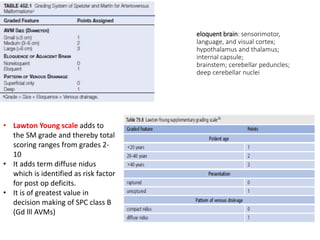
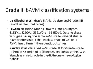
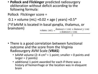
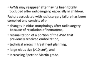
Therapeutic Decision Making in
the Management of
bAVMs
Presented By:
Dr. Rahul Jain
SR-2 Neurosurgery
Moderated by:
Dr V. C. Jha
Dr Nitish Kumar
Dr Gaurav Verma
INTRODUCTION
• AVMs are fistulous connections of cerebral arteries
and veins without a normal intervening capillary
bed, which creates high-flow shunting of arterial
blood into the venous system.
• Due to the direct transmission of arterial pressure
to the venous structures, dilation, tortuosity, and
arterialization of the draining vein(s) occur and
venous hypertension may result.
• Incidence – 1.5 per 100,000 persons, Prevalence –
0.2% to 1.0%
• Among patients aged 15–45 years presenting with
intracerebral hemorrhage, 38% of cases are due to
AVM.
• Patients are typically diagnosed in the third to
fourth decade.
• Most AVMs are supratentorial and solitary. Many
AVMs are situated at the border zone areas of the
anterior, middle, and posterior cerebral arteries.
• They are often pyramidal shaped, with the base
parallel to the cortex and the apex pointing inward
toward the ventricle.
• Up to 40% of patients with AVM present with
symptoms unrelated to the AVM.
• Symptoms related to the AVM, including focal
neurological deficit, headache, and seizure. These
clinical symptoms may be associated with hemorrhage
or simply be due to mechanical compression or
irritation of the surrounding brain.
• Up to 50% (range: 30%–70%) of patients with AVM
present to medical attention due to a ruptured AVM
with hemorrhage with Intraparenchymal hemorrhage
being the most common type of hemorrhage.
• Factors leading to rupture – presence of aneurysms,
venous outflow stenosis, deep location or exclusive
deep drainage and age. Size not considered risk factor.
• Risk of hemorrhage range from 2%-4% per year
• Strongest and most consistent predictor of re-
hemorrhage is prior hemorrhage and highest risk
of rehemorrhage is 20%-40% is within first year.
• Other risk factors for predicting hemorrhage
include age and sex, deep location with exclusive
deep drainage, evidence of microhemorrhage and
large size
• The Spetzler-Martin grading system, the Spetzler-
Ponce classification (SPC), and the Lawton-Young
grading system are three related grading systems of
importance for predicting the risk of surgery for
arteriovenous malformation of the brain (bAVM).
• Three-tiered Spetzler-Ponce Classification (SPC):
SPC class A is Spetzler-Martin grades I and II bAVM,
SPC class B is Spetzler-Martin grade III bAVM, and
SPC class C is Spetzler-Martin grades IV and V
bAVM
• Radiosurgical grading systems are the Pollock-
Flickinger score (and its modification) and the
Virginia Radiosurgery AVM Scale (VRAS).
eloquent brain: sensorimotor,
language, and visual cortex;
hypothalamus and thalamus;
internal capsule;
brainstem; cerebellar peduncles;
deep cerebellar nuclei
• Lawton Young scale adds to
the SM grade and thereby total
scoring ranges from grades 2-
10
• It adds term diffuse nidus
which is identified as risk factor
for post op deficits.
• It is of greatest value in
decision making of SPC class B
(Gd lll AVMs)
Grade III bAVM classification systems
• de Oliveira et al.: Grade IIIA (large size) and Grade IIIB
(small, in eloquent areas)
• Lawton classified Grade III bAVMs into 4 subtypes:
S1E1V1, S2E0V1, S2E1V0, and S3E0V0. Despite these
subtypes having the same S–M Grade, several studies
have demonstrated that each subtype of Grade III
AVMs has different therapeutic outcomes.
• Pandey et al. classified S–M Grade III AVMs into Grade
III (small: <3 cm) and III (large: ≥3 cm) because the AVM
size plays a major role in predicting new neurological
deficits
MANAGEMENT PATHWAY
General consensus on treatment is as follows.
• Small and medium-sized superficially located AVMs
(Spetzler-Martin grades I and II) should be surgically
removed.
• Grade III AVMs fall into a gray zone in terms of
treatment options because of the heterogeneity of
these lesions in terms of location, size, and
angioarchitecture features. Grade III AVMs frequently
require a combined endovascular/surgical approach.
• When Grade IV and V AVMs are incurable, partial
palliative embolization may have a role in reducing flow
demand of the AVM, with improvement of the patient’s
symptoms.
• A Randomised trial of Unruptured Brain Arteriovenous
malformations (ARUBA), study was a comparison of
the risk of death and symptomatic stroke in patients
with unruptured AVMs who were randomly assigned to
receive either only medical management or medical
management with interventional therapy.
• 226 patients between 2007 and 2013 were enrolled,
study concluded that the risk of stroke or death was
lower in the patients who received only medical
management (10.1% versus 30.7%).
• heavily criticized- three main points: (1) only 223 (13%)
of 1740 screened patients were studied; (2) although
76 (68%) of the 112 patients randomly assigned to
receive intervention had low-grade AVMs, only 18
patients underwent surgery; and (3) the short follow-
up period of 33 months inherently favors the outcome
of medical management
A. Conservative Management
• For the conservative management pathway, the
risks are the same as for the natural history of
bAVM.
• Spetzler-Martin grade III bAVMs can be successfully
treated by focused irradiation with an obliteration
rate of around 50% at 3 years and 70% at 5 years.
• Gross and Du*, in a meta-analysis of risk of future
hemorrhage, concluded that the annual rate of
future hemorrhage was 2.2% (95% CI, 1.7%– 2.7%)
for unruptured bAVM and 4.5% (95% CI, 3.7%–
5.5%) for ruptured bAVM.
*Gross B, Du R. Natural history of cerebral arteriovenous malformations: a meta-analysis. J Neurosurg. 2013;118:437–443.
B. Surgery
• three tiers in the SPC have proven to be popular
and reliable in predicting the surgical risk.
• significant separation of SPC class by adverse
outcomes from surgery, with 10% for SPC class A,
18% for SPC class B, and more than 30% for SPC
class C.
• The Lawton-Young grading system has a significant
impact on predicted risk for SPC class B, with an
approximately 10%, 20%, 25%, 40%, and 65%
adverse outcome risk for Lawton-Young grades of 4,
5, 6, 7, and 8, respectively.
• In cases of ruptured AVMs, operate in a delayed fashion
(4–6 weeks) if no neurological deficits.
• During this time period, the risk for early rebleeding is
relatively low, yet the delay allows time for the
hematoma to liquefy, the associated surrounding
edema to resolve, and the dys-autoregulated brain
tissue to recover.
• Spetzler and colleagues reported a significantly higher
incidence of hemorrhagic presentation with small (<3
cm) AVMs compared with large (>6 cm) AVMs (82%
versus 21%).
• These authors compared intraoperative feeding artery
pressure measurements in small and large AVMs. They
found significantly higher feeding artery pressures in
small AVMs and suggested that differences in arterial
feeding pressures may be responsible for the observed
relationship between size of AVMs and the frequency
and severity of hemorrhage.
C. Endovascular Treatment
• results of ARUBA, the only RCT of the management
of unruptured bAVM, suggest that some caution
needs to be exercised with use of embolization as a
management strategy on its own or in combination
with radiosurgery.
• Goals of endovascular embolization include (1) as
adjunctive treatment before surgery or
radiosurgical ablation; (2) as partial, palliative
embolization targeting weak areas of the
angioarchiecture, to decrease the risk of
hemorrhage or improve symptoms related to the
AVM, and (3) to produce curative occlusion.
Angioarchitectural features that are not specified by
the Spetzler-Martin grading scale may have bearing
on the safety and efficacy of AVM treatment are –
• Nidus characteristics (diffuse versus compact),
• Details of the arterial supply (number, location,
size),
• Presence of deep perforator supply, details of the
draining veins (number, size),
• Flow rate (high versus low),
• perinidal angiogenesis, and
• specific pattern of arterial shunting
• Although the transarterial route is routinely used for
AVM embolization, not all AVMs have accessible
arterial feeders.
Transvenous Embolization
• Deep location, small nidus, single draining vein, and
absence of arterial access.
• Using the transvenous approach on select patients who
meet criteria, there is a reported 91.6% immediate
angiographic occlusion of the AVM without any
procedural or clinical complications.
Morbidity associated with AVM embolization is low,
ranging from 2.4% to 2.7%.
Many of the immediate postoperative morbidities are
often transient rather than permanent.
Certain angiographic characteristics of an AVM can
be associated with complete endovascular
obliteration.
These are
• AVM nidus size <30 mm,
• location in noneloquent cortex,
• deep location,
• superficial or large arterial feeders with a single
arterial pedicle,
• anatomy allowing for 2 to 3 cm of embolic material
reflux,
• and unobstructed views of proximal draining veins.
Key technical details that Song and colleagues noted in
achieving successful embolization for cure were that
(1) if multiple feeding arteries are present, the largest
and least tortuous must be embolized;
(2) the tip of the microcatheter must be in the nidus;
(3) Onyx must be injected slowly to increase penetration
while minimizing reflux;
(4) Onyx should be used to cast the inside of the vessel
and then injected within the cast to fill the nidus; and
(5) catheter withdrawal should be quick in straight and
large feeding arteries, whereas slow withdrawal is better
in smaller, more tortuous arteries.
Song D, Leng B, Gu Y, et al. Clinical analysis of 50 cases of BAVM
embolization with Onyx, a novel liquid embolic agent. Interv
Neuroradiol. 2005;11(suppl 1):179–184.
• Embolization as an intent-to-cure treatment
appears to be most successful for treating Spetzler-
Martin grades I and II bAVMs.
• The annual complication rate presented in the
“intent to cure” series reported by Strauss and
associates21 decreased each year throughout the
4-year study.
• single-center case series from 2014 in which
patients were treated with intent to cure
demonstrated a complication rate of 11% in 827
procedures.
• Approximately half of all hemorrhages occurring at
the time of catheterization are caused by arterial
puncture and usually result in subarachnoid
hemorrhage
three major causes of acute hemorrhage
during treatment endovascular
management for cure
• First, the dimethyl sulfoxide (DMSO)–
compatible catheters necessary for Onyx
use are stiffer than those used with
NBCA and more frequently cause arterial
puncture.
• Second, arterial rupture can occur at the
time that the microcatheter is retrieved
from the Onyx cast. This complication is
mitigated by detachable-tip
microcatheters, such as the Apollo
(Medtronic Neurovascular).
• Third cause, is venous compromise that
results from premature advancement of
the embolization material into the
draining veins, which causes pressure in
the AVM nidus .to increase before
embolization
• The most significant long-term complication after
embolization is full or partial recanalization of the
AVM despite initial obliteration, which may occur in
3% to 13% of patients within 1 to 2 years.
• Embolization may also lead to AVM recurrence by
inducing perilesional angiogenesis. Re-formation of
the AVM, in turn, increases the risk of a potentially
fatal intracranial hemorrhage.
EMBOLIC AGENTS
A. Liquid
• nBCA (Trufill) or “glue” and ethylene vinyl alcohol
copolymer (Onyx 18 or 34) are the most commonly
used agents for embolization of brain AVMs.
• Although these agents are considered permanent
embolic agents, AVM recanalization after “complete”
angiographic occlusion has been reported for both.
• NBCA polymerizes to a pastelike consistency on contact
with alkaline solutions such as blood. It may be mixed
with varying amounts of ethiodized oil or glacial acetic
acid to control the rate of solidification.
• The ratio between cyanoacrylate and iodized oil
usually varies between 1:1 (50% cyanoacrylate,
50% iodized oil) and 1:4 (20% cyanoacrylate, 80%
iodized oil) for the embolization of cerebral vascular
malformations
• Ratio of mixture depend on several factors, such as
the position of the microcatheter, the diameter of
the embolized vessel, the velocity of blood flow, the
intended duration of injection and consequently
also the amount of embolic agent to be injected.
• Premature polymerization either within the
delivery microcatheter or proximally in an AVM
feeding artery can be a drawback to the use of
NBCA.
• Prolonged duration while
injecting nBCA can lead to
sticking of catheter tip to vessel
wall.
• Onyx is available in three
premixed concentrations of
ethylene vinyl alcohol copolymer
and tantalum dissolved in
dimethyl sulfoxide(DMSO).
• Compared to the nBCA, Onyx has
(1) slower cast formation, (2)
blood flow-independent delivery,
(3) cohesiveness, and (4) allows
for penetration into a smaller
vessel size. These characteristics
enable the plug-and-push
method and embolization of the
brain AVM microvasculature
• During Onyx injection, the forward advancement of the
material should be observed closely to ensure no
reflux.
• After 30-60 seconds, another slow injection can be
started with a new roadmap, provided there is forward
extension, no reflux and no venous penetration.
• This process is repeated until a compact plug is formed.
• The microcatheter is generally withdrawn if Onyx
material flows back into 1.5 to 2.0 cm of the feeding
artery.
• Once the draining vein has been embolized, it is
important to “commit” to embolizing the remaining
nidus and arterial veins to decrease the chance of
pressure buildup and hemorrhage within the nidus.
• The major advantages of Onyx over nBCA are its low
viscosity and delayed precipitation, thus providing greater
embolization control and repeated injections through the
same microcatheter with deeper nidal penetration.
• The disadvantage of Onyx is its longer precipitation process
leading to longer fluoroscopy and procedure times,
impaired visibility of the microcatheter tip during
embolization, and artifacts in imaging for radiation planning.
• More recently the novel agent, precipitating
hydrophobic injectable liquid (PHIL) was approved
in 2015 for embolization of brain AVMs.
• PHIL is composed of a nonadhesive biocompatible
copolymer (polylactide-co-glycolide and
polyhydroxyethylmethacrylate) dissolved in
dimethyl sulfoxide with an iodine component for
increased radiopacity and visualization.
• In contrast to Onyx, PHIL is ready to use in prefilled
syringes of 1 mL and does not require vigorous
shaking before use.
• PHIL also allows for faster plug formation, more
consistent visibility, lower volumes of PHIL required
for the same extent of embolization compared with
Onyx, and fewer artifacts for postinterventional
radiation planning.
B. Nonliquid
• particulate embolic agent polyvinyl alcohol (PVA) and
platinum coils.
• Appropriately sized PVA particles create mechanical
occlusion in the AVM nidus, promoting stasis and
thrombosis.
• The occluding effects of PVA are transient, however,
and shunting to the lungs is not uncommon with use of
smaller particles.
• Platinum coils may be useful in the slowing of highflow
fistulous AVM compartments, allowing for more
controlled use of liquid embolic agents.
• their main use remains in the treatment of saccular
AVM-associated aneurysms when parent artery
sacrifice is not desired
D. Radiosurgery
• aim of radiosurgery is gradual obliteration of the
AVM by progressive intimal hyperplasia and
progressive nidal thrombosis. Such obliteration
typically takes place over a 2- to 3-year period.
• delivered using a cobalt x-ray source (Gamma Knife;
Elekta AB), with a linear accelerator, or by taking
advantage of the Bragg peak effect of heavy
radioactive particles produced by a cyclotron
(proton or helium beam therapy).
• desired delivery of at least 20 Gy to the bAVM to
induce thrombosis but less than 10 Gy to critical
brain to avoid damage when delivered in a single
fraction (radiosurgery).
• SRS is ideal for small (<3 cm) AVMs located in
eloquent or deep-seated areas of the brain where
the morbidity of surgical excision would be
considered unacceptable.
• It is also a good treatment choice for patients
whose age or comorbidities make the risk of
general anesthesia unacceptable.
• The best obliteration rate is in small bAVMs (<3 cm)
with no prior embolization.
• Radiosurgery has been praised in terms of its cost
effectiveness when used as an alternative to
surgical excision of potentially operable AVMs.
• Pollock and Flickinger predicted radiosurgery
obliteration without deficit according to the
following formula:
Pollock Flickinger score =
0.1 × volume (mL) +0.02 × age ( years) +0.5*
(*if bAVM is located In basal ganglia, thalamus, or
brainstem)
• There is a good correlation between functional
outcome and the score from the Virginia
Radiosurgery AVM Scale (VRAS)
• bAVM volume (2–4 cm3 = 1 point; smaller = 0 points and
larger = 2 points)
• additional 1 point awarded for each if there was a
history of hemorrhage or the location was in eloquent
brain
There are three considerations to weigh in the
choice between surgery and radiosurgery.
I. First, the rate of obliteration is higher for larger
AVMs when higher doses are used (25–45 Gy);
however, the risk of radiation-induced
complications increases significantly.
II. Second, the risk of intracranial hemorrhage
persists during the interval between treatment
and complete obliteration.
III. Third, serial radiographic studies, including
cerebral angiography, are necessary to confirm
complete obliteration.
• Reported obliteration rates range from 100% in
patients with AVMs smaller than 4 cm3, to 78% in
patients undergoing a first-time treatment, to less
than 50% in patients within deeply located AVMs.
• The two main disadvantages of radiosurgery are
the latent period until complete obliteration and
the lack of certainty of obliteration.
• During these periods of delay, which may range
from 1 year to several years, the patient remains at
risk for hemorrhage, and the risk is almost the
same as if no treatment had occurred, at least
during early follow-up.
• Increasing AVM size reciprocally affects obliteration rate,
and repeated radiosurgery for incompletely obliterated
AVMs carries a worse rate for subsequent obliteration.
• No definitive evidence suggests a reduction in the rate of
hemorrhage in patients whose lesion is not completely
obliterated.
• Some patients with grade IV and V AVMs require more
than one course of SRS, and this option has been used in
select patients.
• Disadvantages of such an approach include the second
latency period of 1 to 3 years before obliteration occurs;
the possibility that a second radiosurgical treatment may
still not obliterate the AVM; and the risk of radiation-
induced injury, which may be higher with a second
radiosurgical treatment.
• AVMs may reappear after having been totally
occluded after radiosurgery, especially in children.
Factors associated with radiosurgery failure has been
compiled and consists of :-
• changes in nidus morphology after radiosurgery
because of resolution of hematoma,
• recanalization of a portion of the AVM that
previously received embolization,
• technical errors in treatment planning,
• large nidus size (>10 cm3), and
• Increasing Spetzler-Martin grade.
CONCLUSION
• Brain AVMs are complex and potentially devastating lesions
that often require multimodality treatment at specialized
centers.
• Adjuvant endovascular therapy may be employed to
facilitate definitive AVM eradication by either surgical
resection or radiosurgery.
• Rapid advancement in endovascular technology has enabled
interventionalists to specifically target unique
angioarchitectural features that pose the greatest threat
during either surgery or the radiosurgery latency period.
• Coordinated clinical assessment, risk-benefit analysis, and
implementation of therapy to achieve predetermined
treatment goals will help to ensure the best possible
outcomes for patients with AVMs.
References
1. Youmann and Winn’s Neurological Surgery 8th ed
2. Vollherbs et al. Glue, Onyx, Squid or PHIL? Liquid
Embolic Agents for the Embolization of Cerebral
Arteriovenous Malformations and Dural
Arteriovenous Fistulas. Clin Neuroradiol 32, 25–
38 (2022)
3. Ryu B, Ishikawa T, Kawamata T. Multimodal
Treatment Strategy for Spetzler-Martin Grade III
Arteriovenous Malformations of the Brain. Neurol
Med Chir (Tokyo). 2017;57(2):73-81.

Brain AVM (ArterioVenous Malformation) Managment.pptx

  • 1.
    Therapeutic Decision Makingin the Management of bAVMs Presented By: Dr. Rahul Jain SR-2 Neurosurgery Moderated by: Dr V. C. Jha Dr Nitish Kumar Dr Gaurav Verma
  • 2.
    INTRODUCTION • AVMs arefistulous connections of cerebral arteries and veins without a normal intervening capillary bed, which creates high-flow shunting of arterial blood into the venous system. • Due to the direct transmission of arterial pressure to the venous structures, dilation, tortuosity, and arterialization of the draining vein(s) occur and venous hypertension may result. • Incidence – 1.5 per 100,000 persons, Prevalence – 0.2% to 1.0%
  • 3.
    • Among patientsaged 15–45 years presenting with intracerebral hemorrhage, 38% of cases are due to AVM. • Patients are typically diagnosed in the third to fourth decade. • Most AVMs are supratentorial and solitary. Many AVMs are situated at the border zone areas of the anterior, middle, and posterior cerebral arteries. • They are often pyramidal shaped, with the base parallel to the cortex and the apex pointing inward toward the ventricle.
  • 4.
    • Up to40% of patients with AVM present with symptoms unrelated to the AVM. • Symptoms related to the AVM, including focal neurological deficit, headache, and seizure. These clinical symptoms may be associated with hemorrhage or simply be due to mechanical compression or irritation of the surrounding brain. • Up to 50% (range: 30%–70%) of patients with AVM present to medical attention due to a ruptured AVM with hemorrhage with Intraparenchymal hemorrhage being the most common type of hemorrhage. • Factors leading to rupture – presence of aneurysms, venous outflow stenosis, deep location or exclusive deep drainage and age. Size not considered risk factor.
  • 5.
    • Risk ofhemorrhage range from 2%-4% per year • Strongest and most consistent predictor of re- hemorrhage is prior hemorrhage and highest risk of rehemorrhage is 20%-40% is within first year. • Other risk factors for predicting hemorrhage include age and sex, deep location with exclusive deep drainage, evidence of microhemorrhage and large size
  • 6.
    • The Spetzler-Martingrading system, the Spetzler- Ponce classification (SPC), and the Lawton-Young grading system are three related grading systems of importance for predicting the risk of surgery for arteriovenous malformation of the brain (bAVM). • Three-tiered Spetzler-Ponce Classification (SPC): SPC class A is Spetzler-Martin grades I and II bAVM, SPC class B is Spetzler-Martin grade III bAVM, and SPC class C is Spetzler-Martin grades IV and V bAVM • Radiosurgical grading systems are the Pollock- Flickinger score (and its modification) and the Virginia Radiosurgery AVM Scale (VRAS).
  • 7.
    eloquent brain: sensorimotor, language,and visual cortex; hypothalamus and thalamus; internal capsule; brainstem; cerebellar peduncles; deep cerebellar nuclei • Lawton Young scale adds to the SM grade and thereby total scoring ranges from grades 2- 10 • It adds term diffuse nidus which is identified as risk factor for post op deficits. • It is of greatest value in decision making of SPC class B (Gd lll AVMs)
  • 8.
    Grade III bAVMclassification systems • de Oliveira et al.: Grade IIIA (large size) and Grade IIIB (small, in eloquent areas) • Lawton classified Grade III bAVMs into 4 subtypes: S1E1V1, S2E0V1, S2E1V0, and S3E0V0. Despite these subtypes having the same S–M Grade, several studies have demonstrated that each subtype of Grade III AVMs has different therapeutic outcomes. • Pandey et al. classified S–M Grade III AVMs into Grade III (small: <3 cm) and III (large: ≥3 cm) because the AVM size plays a major role in predicting new neurological deficits
  • 9.
    MANAGEMENT PATHWAY General consensuson treatment is as follows. • Small and medium-sized superficially located AVMs (Spetzler-Martin grades I and II) should be surgically removed. • Grade III AVMs fall into a gray zone in terms of treatment options because of the heterogeneity of these lesions in terms of location, size, and angioarchitecture features. Grade III AVMs frequently require a combined endovascular/surgical approach. • When Grade IV and V AVMs are incurable, partial palliative embolization may have a role in reducing flow demand of the AVM, with improvement of the patient’s symptoms.
  • 11.
    • A Randomisedtrial of Unruptured Brain Arteriovenous malformations (ARUBA), study was a comparison of the risk of death and symptomatic stroke in patients with unruptured AVMs who were randomly assigned to receive either only medical management or medical management with interventional therapy. • 226 patients between 2007 and 2013 were enrolled, study concluded that the risk of stroke or death was lower in the patients who received only medical management (10.1% versus 30.7%). • heavily criticized- three main points: (1) only 223 (13%) of 1740 screened patients were studied; (2) although 76 (68%) of the 112 patients randomly assigned to receive intervention had low-grade AVMs, only 18 patients underwent surgery; and (3) the short follow- up period of 33 months inherently favors the outcome of medical management
  • 12.
    A. Conservative Management •For the conservative management pathway, the risks are the same as for the natural history of bAVM. • Spetzler-Martin grade III bAVMs can be successfully treated by focused irradiation with an obliteration rate of around 50% at 3 years and 70% at 5 years. • Gross and Du*, in a meta-analysis of risk of future hemorrhage, concluded that the annual rate of future hemorrhage was 2.2% (95% CI, 1.7%– 2.7%) for unruptured bAVM and 4.5% (95% CI, 3.7%– 5.5%) for ruptured bAVM. *Gross B, Du R. Natural history of cerebral arteriovenous malformations: a meta-analysis. J Neurosurg. 2013;118:437–443.
  • 13.
    B. Surgery • threetiers in the SPC have proven to be popular and reliable in predicting the surgical risk. • significant separation of SPC class by adverse outcomes from surgery, with 10% for SPC class A, 18% for SPC class B, and more than 30% for SPC class C. • The Lawton-Young grading system has a significant impact on predicted risk for SPC class B, with an approximately 10%, 20%, 25%, 40%, and 65% adverse outcome risk for Lawton-Young grades of 4, 5, 6, 7, and 8, respectively.
  • 14.
    • In casesof ruptured AVMs, operate in a delayed fashion (4–6 weeks) if no neurological deficits. • During this time period, the risk for early rebleeding is relatively low, yet the delay allows time for the hematoma to liquefy, the associated surrounding edema to resolve, and the dys-autoregulated brain tissue to recover. • Spetzler and colleagues reported a significantly higher incidence of hemorrhagic presentation with small (<3 cm) AVMs compared with large (>6 cm) AVMs (82% versus 21%). • These authors compared intraoperative feeding artery pressure measurements in small and large AVMs. They found significantly higher feeding artery pressures in small AVMs and suggested that differences in arterial feeding pressures may be responsible for the observed relationship between size of AVMs and the frequency and severity of hemorrhage.
  • 15.
    C. Endovascular Treatment •results of ARUBA, the only RCT of the management of unruptured bAVM, suggest that some caution needs to be exercised with use of embolization as a management strategy on its own or in combination with radiosurgery. • Goals of endovascular embolization include (1) as adjunctive treatment before surgery or radiosurgical ablation; (2) as partial, palliative embolization targeting weak areas of the angioarchiecture, to decrease the risk of hemorrhage or improve symptoms related to the AVM, and (3) to produce curative occlusion.
  • 16.
    Angioarchitectural features thatare not specified by the Spetzler-Martin grading scale may have bearing on the safety and efficacy of AVM treatment are – • Nidus characteristics (diffuse versus compact), • Details of the arterial supply (number, location, size), • Presence of deep perforator supply, details of the draining veins (number, size), • Flow rate (high versus low), • perinidal angiogenesis, and • specific pattern of arterial shunting
  • 17.
    • Although thetransarterial route is routinely used for AVM embolization, not all AVMs have accessible arterial feeders. Transvenous Embolization • Deep location, small nidus, single draining vein, and absence of arterial access. • Using the transvenous approach on select patients who meet criteria, there is a reported 91.6% immediate angiographic occlusion of the AVM without any procedural or clinical complications. Morbidity associated with AVM embolization is low, ranging from 2.4% to 2.7%. Many of the immediate postoperative morbidities are often transient rather than permanent.
  • 18.
    Certain angiographic characteristicsof an AVM can be associated with complete endovascular obliteration. These are • AVM nidus size <30 mm, • location in noneloquent cortex, • deep location, • superficial or large arterial feeders with a single arterial pedicle, • anatomy allowing for 2 to 3 cm of embolic material reflux, • and unobstructed views of proximal draining veins.
  • 19.
    Key technical detailsthat Song and colleagues noted in achieving successful embolization for cure were that (1) if multiple feeding arteries are present, the largest and least tortuous must be embolized; (2) the tip of the microcatheter must be in the nidus; (3) Onyx must be injected slowly to increase penetration while minimizing reflux; (4) Onyx should be used to cast the inside of the vessel and then injected within the cast to fill the nidus; and (5) catheter withdrawal should be quick in straight and large feeding arteries, whereas slow withdrawal is better in smaller, more tortuous arteries. Song D, Leng B, Gu Y, et al. Clinical analysis of 50 cases of BAVM embolization with Onyx, a novel liquid embolic agent. Interv Neuroradiol. 2005;11(suppl 1):179–184.
  • 20.
    • Embolization asan intent-to-cure treatment appears to be most successful for treating Spetzler- Martin grades I and II bAVMs. • The annual complication rate presented in the “intent to cure” series reported by Strauss and associates21 decreased each year throughout the 4-year study. • single-center case series from 2014 in which patients were treated with intent to cure demonstrated a complication rate of 11% in 827 procedures. • Approximately half of all hemorrhages occurring at the time of catheterization are caused by arterial puncture and usually result in subarachnoid hemorrhage
  • 21.
    three major causesof acute hemorrhage during treatment endovascular management for cure • First, the dimethyl sulfoxide (DMSO)– compatible catheters necessary for Onyx use are stiffer than those used with NBCA and more frequently cause arterial puncture. • Second, arterial rupture can occur at the time that the microcatheter is retrieved from the Onyx cast. This complication is mitigated by detachable-tip microcatheters, such as the Apollo (Medtronic Neurovascular). • Third cause, is venous compromise that results from premature advancement of the embolization material into the draining veins, which causes pressure in the AVM nidus .to increase before embolization
  • 22.
    • The mostsignificant long-term complication after embolization is full or partial recanalization of the AVM despite initial obliteration, which may occur in 3% to 13% of patients within 1 to 2 years. • Embolization may also lead to AVM recurrence by inducing perilesional angiogenesis. Re-formation of the AVM, in turn, increases the risk of a potentially fatal intracranial hemorrhage.
  • 23.
    EMBOLIC AGENTS A. Liquid •nBCA (Trufill) or “glue” and ethylene vinyl alcohol copolymer (Onyx 18 or 34) are the most commonly used agents for embolization of brain AVMs. • Although these agents are considered permanent embolic agents, AVM recanalization after “complete” angiographic occlusion has been reported for both. • NBCA polymerizes to a pastelike consistency on contact with alkaline solutions such as blood. It may be mixed with varying amounts of ethiodized oil or glacial acetic acid to control the rate of solidification.
  • 24.
    • The ratiobetween cyanoacrylate and iodized oil usually varies between 1:1 (50% cyanoacrylate, 50% iodized oil) and 1:4 (20% cyanoacrylate, 80% iodized oil) for the embolization of cerebral vascular malformations • Ratio of mixture depend on several factors, such as the position of the microcatheter, the diameter of the embolized vessel, the velocity of blood flow, the intended duration of injection and consequently also the amount of embolic agent to be injected. • Premature polymerization either within the delivery microcatheter or proximally in an AVM feeding artery can be a drawback to the use of NBCA.
  • 25.
    • Prolonged durationwhile injecting nBCA can lead to sticking of catheter tip to vessel wall. • Onyx is available in three premixed concentrations of ethylene vinyl alcohol copolymer and tantalum dissolved in dimethyl sulfoxide(DMSO). • Compared to the nBCA, Onyx has (1) slower cast formation, (2) blood flow-independent delivery, (3) cohesiveness, and (4) allows for penetration into a smaller vessel size. These characteristics enable the plug-and-push method and embolization of the brain AVM microvasculature
  • 26.
    • During Onyxinjection, the forward advancement of the material should be observed closely to ensure no reflux. • After 30-60 seconds, another slow injection can be started with a new roadmap, provided there is forward extension, no reflux and no venous penetration. • This process is repeated until a compact plug is formed. • The microcatheter is generally withdrawn if Onyx material flows back into 1.5 to 2.0 cm of the feeding artery. • Once the draining vein has been embolized, it is important to “commit” to embolizing the remaining nidus and arterial veins to decrease the chance of pressure buildup and hemorrhage within the nidus.
  • 27.
    • The majoradvantages of Onyx over nBCA are its low viscosity and delayed precipitation, thus providing greater embolization control and repeated injections through the same microcatheter with deeper nidal penetration. • The disadvantage of Onyx is its longer precipitation process leading to longer fluoroscopy and procedure times, impaired visibility of the microcatheter tip during embolization, and artifacts in imaging for radiation planning.
  • 28.
    • More recentlythe novel agent, precipitating hydrophobic injectable liquid (PHIL) was approved in 2015 for embolization of brain AVMs. • PHIL is composed of a nonadhesive biocompatible copolymer (polylactide-co-glycolide and polyhydroxyethylmethacrylate) dissolved in dimethyl sulfoxide with an iodine component for increased radiopacity and visualization. • In contrast to Onyx, PHIL is ready to use in prefilled syringes of 1 mL and does not require vigorous shaking before use. • PHIL also allows for faster plug formation, more consistent visibility, lower volumes of PHIL required for the same extent of embolization compared with Onyx, and fewer artifacts for postinterventional radiation planning.
  • 29.
    B. Nonliquid • particulateembolic agent polyvinyl alcohol (PVA) and platinum coils. • Appropriately sized PVA particles create mechanical occlusion in the AVM nidus, promoting stasis and thrombosis. • The occluding effects of PVA are transient, however, and shunting to the lungs is not uncommon with use of smaller particles. • Platinum coils may be useful in the slowing of highflow fistulous AVM compartments, allowing for more controlled use of liquid embolic agents. • their main use remains in the treatment of saccular AVM-associated aneurysms when parent artery sacrifice is not desired
  • 30.
    D. Radiosurgery • aimof radiosurgery is gradual obliteration of the AVM by progressive intimal hyperplasia and progressive nidal thrombosis. Such obliteration typically takes place over a 2- to 3-year period. • delivered using a cobalt x-ray source (Gamma Knife; Elekta AB), with a linear accelerator, or by taking advantage of the Bragg peak effect of heavy radioactive particles produced by a cyclotron (proton or helium beam therapy). • desired delivery of at least 20 Gy to the bAVM to induce thrombosis but less than 10 Gy to critical brain to avoid damage when delivered in a single fraction (radiosurgery).
  • 31.
    • SRS isideal for small (<3 cm) AVMs located in eloquent or deep-seated areas of the brain where the morbidity of surgical excision would be considered unacceptable. • It is also a good treatment choice for patients whose age or comorbidities make the risk of general anesthesia unacceptable. • The best obliteration rate is in small bAVMs (<3 cm) with no prior embolization. • Radiosurgery has been praised in terms of its cost effectiveness when used as an alternative to surgical excision of potentially operable AVMs.
  • 32.
    • Pollock andFlickinger predicted radiosurgery obliteration without deficit according to the following formula: Pollock Flickinger score = 0.1 × volume (mL) +0.02 × age ( years) +0.5* (*if bAVM is located In basal ganglia, thalamus, or brainstem) • There is a good correlation between functional outcome and the score from the Virginia Radiosurgery AVM Scale (VRAS) • bAVM volume (2–4 cm3 = 1 point; smaller = 0 points and larger = 2 points) • additional 1 point awarded for each if there was a history of hemorrhage or the location was in eloquent brain
  • 33.
    There are threeconsiderations to weigh in the choice between surgery and radiosurgery. I. First, the rate of obliteration is higher for larger AVMs when higher doses are used (25–45 Gy); however, the risk of radiation-induced complications increases significantly. II. Second, the risk of intracranial hemorrhage persists during the interval between treatment and complete obliteration. III. Third, serial radiographic studies, including cerebral angiography, are necessary to confirm complete obliteration.
  • 34.
    • Reported obliterationrates range from 100% in patients with AVMs smaller than 4 cm3, to 78% in patients undergoing a first-time treatment, to less than 50% in patients within deeply located AVMs. • The two main disadvantages of radiosurgery are the latent period until complete obliteration and the lack of certainty of obliteration. • During these periods of delay, which may range from 1 year to several years, the patient remains at risk for hemorrhage, and the risk is almost the same as if no treatment had occurred, at least during early follow-up.
  • 35.
    • Increasing AVMsize reciprocally affects obliteration rate, and repeated radiosurgery for incompletely obliterated AVMs carries a worse rate for subsequent obliteration. • No definitive evidence suggests a reduction in the rate of hemorrhage in patients whose lesion is not completely obliterated. • Some patients with grade IV and V AVMs require more than one course of SRS, and this option has been used in select patients. • Disadvantages of such an approach include the second latency period of 1 to 3 years before obliteration occurs; the possibility that a second radiosurgical treatment may still not obliterate the AVM; and the risk of radiation- induced injury, which may be higher with a second radiosurgical treatment.
  • 36.
    • AVMs mayreappear after having been totally occluded after radiosurgery, especially in children. Factors associated with radiosurgery failure has been compiled and consists of :- • changes in nidus morphology after radiosurgery because of resolution of hematoma, • recanalization of a portion of the AVM that previously received embolization, • technical errors in treatment planning, • large nidus size (>10 cm3), and • Increasing Spetzler-Martin grade.
  • 37.
    CONCLUSION • Brain AVMsare complex and potentially devastating lesions that often require multimodality treatment at specialized centers. • Adjuvant endovascular therapy may be employed to facilitate definitive AVM eradication by either surgical resection or radiosurgery. • Rapid advancement in endovascular technology has enabled interventionalists to specifically target unique angioarchitectural features that pose the greatest threat during either surgery or the radiosurgery latency period. • Coordinated clinical assessment, risk-benefit analysis, and implementation of therapy to achieve predetermined treatment goals will help to ensure the best possible outcomes for patients with AVMs.
  • 38.
    References 1. Youmann andWinn’s Neurological Surgery 8th ed 2. Vollherbs et al. Glue, Onyx, Squid or PHIL? Liquid Embolic Agents for the Embolization of Cerebral Arteriovenous Malformations and Dural Arteriovenous Fistulas. Clin Neuroradiol 32, 25– 38 (2022) 3. Ryu B, Ishikawa T, Kawamata T. Multimodal Treatment Strategy for Spetzler-Martin Grade III Arteriovenous Malformations of the Brain. Neurol Med Chir (Tokyo). 2017;57(2):73-81.