SlideShare a Scribd company logo
1 of 38
Download to read offline
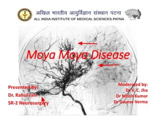
Moya Moya Disease
Presented By:
Dr. Rahul Jain
SR-2 Neurosurgery
Moderated by:
Dr V. C. Jha
Dr Nitish Kumar
Dr Gaurav Verma
INTRODUCTION
• First described in the Japanese medical literature in
1957 by Takeuchi and Shimizu.
• Term moyamoya (Japanese for “puff of smoke”)
was coined by Suzuki and Takaku in 1969.
• Kudo named this disease “spontaneous occlusion of
the circle of Willis” in 1968 from the pathological
point of view, and this name was officially accepted
later by the Research Committee of Ministry of
Welfare and Health, Japan (RCMWHJ), which was
founded in 1977.
EPIDEMIOLOGY
• Presence within non-Asian populations has been
increasingly recognized, though at a lower incidence and
ethnicity seems to play a decisive role in the United States.
• In Japan, an annual incidence of 0.35 per 100,000
population and a prevalence of 3.16 per 100,000
population have been reported.
• Female-to-male ratio is 1.8.
• Overall, there was a bimodal age distribution (with peaks
around age 10 and 40), the peak age of onset in men was
10 to 14 years compared with 20 to 24 years in women.
• In United States, a study between 2002 and 2008 showed
an incidence of 0.57 per 100 000 people/y. The mean age
at diagnosis was 32 years, and the women-to-men ratio
was 2.6:1.
PATHOPHYSIOLOGY AND ETIOLOGY
• The characteristic findings of
intimal thickening and resulting
steno-occlusion at the terminal
portion of the ICA along with
pathologic changes in neighboring
arteries.
• fibrocellular thickening of the
intima, an irregular disruption of
the internal elastic lamina, and
the attenuation of the media are
the main findings.
• observed not only in the carotid
fork but also in cortical branches
of MCA.
• Pluripotent peptides and their receptors, basic FGF,
TGF β, and HGF, detected in increased amounts in the
STA and in the diseased wall of the ICA 
angiogenesis and intimal hyperplasia of intracranial
and extracranial arteries.
• Elevation of serum level of soluble VCAM type 1, ICAM
type 1, and E-selectin and elevation of CSF nitric oxide
metabolites or some specific polypeptides 
endothelial activation cardinal role in inducing MMD.
• Raised Caspase-3 – cardinal for apoptosis  found in
smooth muscle cells of MCA
• Raised HIF-1 alpha subunit endothelial layer MCA.
• The moyamoya collaterals are dilated perforating
arteries believed to be a combination of preexisting
and newly developed vessels.
Familial MMD
• autosomal dominant with
incomplete penetrance.
• Seen in 23%
• Ring Finger Protein 213
(RNF213) is a susceptibility
gene for MMD on Chr 17
locus. (RNF213 p.R4810K)
• Associations between
moyamoya and loci on
chromosomes 3, 6, 8, and 17
(MYMY1, MYMY2, MYMY3).
MMD – primary condition
MM syndrome – aka quasi or secondary moya moya, when moya
moya with other syndromic association.
Moya moya aline refer to arteriopathy independent of etiology
CLINICAL FINDINGS
• Presenting symptoms and
“events” can be grouped
into
• (1) ischemic and
hemorrhagic events,
• (2) other neurological
manifestations, and
• (3) symptoms of asso-
ciated diseases, in the
case of MMS.
• In children TIAs are most common presention (70-80%) and
in adults hemorrhagic events especially in women (upto
66%)are commoner followed by ischemic events.
• A sudden decrease in cognitive performance owing to low
perfusion has been the only clinical manifestation in about
10% of adult patients.
• Infarctions are observed at cortico-subcortical regions
prevalently in watershed territories or posterior cerebral
artery (PCA) territories in about 40% of ischemic cases, but
basal ganglia and thalamus are usually spared.
• The majority of bleeding in adults is intraventricular or
periventricular in location and not subarachnoid.
• Hemorrhages are often repetitive in nature, with an annual rate
of rebleeding of 7%.
• 45% of patients have good neurological recovery, and 7% die.
three main causes of
intracranial bleeding in MMD:
1. rupture of dilated and
stressed perforating
arteries containing
microaneurysms;
2. fibrinoid necrosis of the
arterial wall in the basal
ganglia; and
3. rupture of microaneurysms
in the periventricular
region, especially around
the superolateral wall of
the lateral ventricles
• Recurrent ischemia is more common in adults
with MMD in the United States, whereas
recurrent ischemia is more common in Japanese
adults with MMD with conservative management.
• Patients with angiographic patterns of posterior
circulation involvement (which is usually spared)
have been associated with worse clinical
presentation and higher recurrent
hemorrhages.
• Saccular cerebral aneurysms, a possible cause of
rare subarachnoid hemorrhage in this disease, are
detected in 4% to 14% of patients – 60% around
COW in vertebrobasilar territory; 20% in peripheral
arteries; 20% in abnormal moya moya vasculature.
• Pregnancy and delivery may increase the risk of
ischemic or hemorrhagic stroke in female patients
NEUROIMAGING
• DSA is gold standard.
• Current recommendations of RCMD also
include MRI and MRA for diagnosis (>1 T
desirable).
• MRI and MRA demonstrate the
characteristic findings of steno-occlusive
carotid lesions and basal moyamoya,
especially with the use of high tesla
magnetic resonance.
• Visualization of deep collaterals by 7-T
time-of-flight MRA comparable to cerebral
angiography.
• Xenon enhanced CT, SPECT, PET can be used to measure regional
CBF and metabolic distribution.
• Reduced CBF and cerebrovascular reactivity to carbon dioxide
and/ or acetazolamide are characteristically detected in the ICA
territory.
MRI and CT imaging assessment in an MMA
patient:
Axial FLAIR images show (a) a small lacunar
infarction in the left posterior lenticular region,
which is pointed out by the red arrow;
(b) multiple, non-confluent, white matter
hyperintensities with subcortical distribution in both
hemispheres; yellow arrows indicate the Ivy sign.
(c) MRA with bilateral terminal ICA steno-occlusion
with lack of signal in both M1 MCA.
(d) CTA in coronal MIP showing the same finding as
in (c) with visualization of the distal sylvian segment
of MCA and prominent collateralization in the
lenticulostriatal perforator vessels.
• Reduction in cerebral perfusion pressure (cerebral
blood volume/CBF) is compensated by an increase
in cerebral blood volume and oxygen extraction
fraction.
• These parameters can also be used to confirm the
effectiveness of surgical revascularization.
• Quantitative MRA utilizing the noninvasive optimal
vessel analysis (NOVA) methodology, H2
15O PET for
hemodynamic evaluation of patients with MMD.
• Six-stage classification of Suzuki and Takaku,
angiographic progression. Tend to progress upto
adolescence and stabilise by age of 20 years.
• Most commonly seen stage is 4.
Stage I. Narrowing of the carotid fork.
Stage II. Initiation of the moyamoya
vasculopathy (dilated major cerebral artery
and a slight moyamoya vessel network)
Stage III. Intensification of the moyamoya
vasculopathy (disappearance of the middle
and anterior cerebral arteries, and thick and
distinct moyamoya vessels)
Stage IV. Minimization of the moyamoya
vasculopathy (disappearance of the posterior
cerebral artery and narrowing of individual
moyamoya vessels)
Stage V. Reduction of the moyamoya
vasculopathy (disappearance of all the main
cerebral arteries arising from the internal
carotid artery system, further minimization of
the moyamoya vessels, and an increase in the
collateral pathways from the external carotid
artery system).
Stage VI. Disappearance of the moyamoya
vasculopathy (disappearance of the moyamoya
vessels, with cerebral blood flow derived only
from the external carotid artery and the
vertebrobasilar artery systems).
Limitations of Suzuki classification
• It’s a purely morphological classification focussing
on occlusion and collateralization pattern in DSA
and hence it fails to reflect hemodynamic
compromise and does not correlate with clinical
symptoms or with surgical treatment risks.
• Unclear boundaries between different grades
(almost no difference between 4 and 5) and
inconsistent grading between right and left cerebral
hemispheres.
• No clear quantitative standard for Suzuki
classification, degree of hyperplasia of among
blood vessels and degree of internal carotid artery
branch stenosis cannot be accurately assessed.
Berlin Grading System: Czabanka et al
• 3 Variables: DSA, MRI, and Cerebrovascular Reactivity (CVR).
• CVR determined using cold xenon CT or 123 I-IMP SPECT and judged it as reduced when
CVR < –5% & < 14% respectively.
• A total score (minimum 1 point and maximum 6 points) was determined by summarizing the
numerical values for each variable. Thus, 3 MMD grades were defined: mild (grade I) = 1 to
2 points, moderate (grade II) = 3 to 4 points, and severe (grade III) = 5 to 6 points. The
grading system considered 1 hemisphere only, so it was possible that a patient could have
different grades per hemisphere.
• Houkin et al. established MRA grades for evaluating MMA as an alternative to
conventional angiography, with a scoring system that showed a good correlation with
Suzuki’s stages.
Houkin, K.; Nakayama, N.; Kuroda, S.; Nonaka, T.; Shonai, T.; Yoshimoto, T. Novel magnetic resonance angiography stage grading for
moyamoya disease. Cerebrovasc. Dis. 2005, 20, 347–354
Houkin MRA Score & Grading
TREATMENT
• Because the etiology of MMD is unknown, no medical
treatment is available.
• Acetylsalicylic acid or other antiplatelet drugs are given
because studies have revealed that these may influence
the progression of vascular stenosis.
• Calcium antagonists and steroids have been empirically
administered for headache and involuntary movement
or frequent TIAs, respectively.
• Despite evidence of thromboembolism in some cases,
antithrombotic use has been controversial due to
concerns for increased risk for hemorrhage and lack
of efficacy for prevention of hypoperfusion-related
ischemic events.
• Headaches in moyamoya vasculopathy are
common and often migraine-like, but migraine
therapies that cause vasoconstriction, inhibit
vasodilation, or lower blood pressure should be
avoided.
• Diabetes is an independent predictor of
recurrent ischemic stroke in nonsurgical and
surgically treated patients.
• Increased body mass index and homocysteine
might also be associated with a higher risk of
MMD.
• In a prospective nonrandomized study,
atorvastatin use after surgical revascularization
was shown to improve collateral circulation on
postoperative digital subtraction angiography.
Surgical management
• Surgical revascularization procedures are performed to
augment CBF and to improve impaired hemodynamics that
cannot be resolved by nonsurgical treatment.
• Main categories:
 direct revascularization with microvascular extracranial-to-
intracranial bypass and
 indirect revascularization without microvascular anastomotic
procedures.
 Combined
• Overall evidence for ischemic moyamoya is reflected in the
class 2a, level of evidence C-limited data recommendation
from the AHA/ASA 2021 Guideline which states that
surgical revascularization (both direct or indirect) can be
beneficial for the prevention of ischemic stroke or
transient ischemic attack.
Indication of surgery (2021 RCMD Guidelines)
• Surgical revascularization for MMD patients with
cerebral ischemic attacks has been reported to reduce
the frequency of transient ischemic attacks (TIAs) and
the risk of cerebral infarction and to improve the
postoperative activities of daily living (ADLs) and long-
term prognosis of neurocognitive function.
• In the acute stage, the treatment is the same as for
brain infarction or spontaneous intracerebral
hemorrhage (ICH) due to other etiologies.
• In the event of ventricular hemorrhage, an external
ventricular drainage is performed if the patient
presents in acute evolution with signs of intracranial
hypertension.
• Bypass surgery in the acute stage of the disease is not
indicated in light of the higher risk for perioperative
complications.
• Anastomotic technique requires for its proper
implementation cortical arteries of at least 0.8 to
1.0 mm in diameter.
• It is common to add an indirect bypass more or less
when a direct bypass is scheduled.
• In moyamoya disease, if both cerebral hemispheres
present ischemic symptoms bypass surgery is
required bilaterally. First, one-sided operation for
the hemisphere that is more ischemic is performed,
and then bypass surgery for the opposite side is
scheduled 1 or 2 months later.
• If the patients suffer bilateral ischemic symptoms
equally, the dominant hemisphere should be
operated first.
• Hyperventilation and alpha-adrenergic drugs
should be avoided for their vasoconstrictor effect,
but moderate hypothermia (32°C to 34°C) and
barbiturates, or anesthetics like propofol, are used
for cerebral protection during times of temporary
arterial occlusion.
• Slightly elevated parameters (100 to 130 mm Hg),
and plasma expanders should be used intraop to
prevent any ischemic event.
• Donor vessel (the STA) should be selected with an
external diameter not less than 1 mm because
vessels of smaller diameter have a high percentage
of occlusion, deliver a low blood flow, are not
useful, and are more difficult to anastomose.
• To prevent mechanical vasospasm, it is useful to
apply topical diluted papaverine or nimodipine.
• Avoid incidental injury of occipital
arteries and/or contralateral STA
during head fixation.
• Doppler ultrasound is used over
the donor artery and correlated
with the preoperative
angiography to locate the most
suitable branch of the STA.
• Two branches of the STA, the
frontal and parietal. Both must be
marked during the proceedings.
• The patient is placed on the
operating table with the head
rotated toward the contralateral
side and the temporal bone is
parallel to the floor.
Direct Bypass Surgery (STA-MCA)
-Anastomosis is performed
using 10-0 monofilament
suture.
-Sutures are placed first at the
corners of the linear-shaped
-Incision and then five
interrupted sutures are placed
over the distal wall edges. The
procedure is repeated with five
interrupted sutures over the
nearest wall.
The intima are always included
in the suture to avoid any
Increased tension at the
sutured site.
remove the first distal clip
and then the proximal clip of
the recipient artery.
temporary clipping of the
recipient artery should be no
more than 30 minutes
Indirect Bypass Surgery
(1) Encephalo-Duro-Arterio-Synangiosis(EDAS)
• Alternative to the STA-MCA
bypass.
• EDAS is an indirect way
to increase collateral blood flow to
the ischemic brain.
• Revascularization between 6 and 12
months following intervention.
Encephalo-Myo-Synangiosis
• Flap of the temporalis muscle is sutured to the edges of
the dural surgical opening so that the muscle is
positioned closer to the brain surface.
• several series have shown
that EMS improves the
clinical condition of
patients and promotes
revascularization in the
region of the MCA in 70%
to 80% of all patients
• Performed with direct
bypass for closure.
EDAS Plus Encephalo-Galeo-
Synangiosis
• two stages, initially on
the more
hemodynamically
affected cerebral
hemisphere, with an
average time between
the first and second
procedure being 6 to 8
months.
• To further increase
collateral circulation in
the territory of the
anterior cerebral arteries.
Omental Transplantation
• In the indirect anastomosis, the periosteum, dura
mater, or a slice of the temporal muscle is placed over
the brain surface in anticipation of the development of
new spontaneous anastomoses between extra and
intracranial circulation.
• During the surgical technique (synangiosis), these
arteries must be preserved. In some cases, and to
ensure close contact between the STA and galea
surrounding the cerebral cortex, the extirpation of the
pia mater is done in zones or “windows,” suturing the
edges of the galea to the piamater.
• While using these indirect techniques, a high increase
of CBF does not develop immediately; early
revascularization is frequently observed between 3 and
6 months following the intervention, especially in cases
that course with cerebral ischemia.
Perioperative Management
• selection of timing of surgery at the period of
relatively stable clinical condition without frequent
ischemic episodes;
• optimization of hydration status;
• normocapnia during surgery and judicious selection
of anesthetic agents; and
• Preoperative evaluation of hemodynamic
dysfunction with acetazolamide (Diamox) loading
carried out with caution with surgery carried out
thereafter (usually after 48 hours) when the
hemodynamic and metabolic situation has
stabilized.
PROGNOSIS
• 75% to 80% are thought to have a benign course with regard
to life expectancy with or without surgical treatment.
• However, limited adaptability to social and school life or
impairment of soft neurological signs has been reported.
• After revascularization procedures, the majority of adult
patients with MMD have been reported to be free from TIAs
and ischemic strokes.
• Rebleeding has been reported to occur in about 30% to 65%
of patients during follow-up periods.
• Although a reduction of 12.5% to 20% of rebleeding risk14,44
has been reported, the results of the Japan Adult Moyamoya
Trial in 2014 revealed only a marginal statistical difference
between surgical and nonsurgical groups when testing for
preventive effect of direct bypass against rebleeding.
However the follow-up period was only 5 years.
• Patients with unilateral MMD should be carefully
followed, as 7% to 27% of these patients including
children were reported to progress to bilateral
MMD within a few years.
CONCLUSION
• MMD is a rare disease of unknown etiology. More
recent studies in Japan show some increase of annual
incidence and prevalence. The clinical presentation
seems to have not changed: mostly ischemia in patients
in the first age peak and bleeding in patients in the
second age peak.
• To augment CBF for treatment of the ischemic type of
MMD, use of surgical direct revascularization utilizing
microsurgical techniques is done and its effectiveness is
established in ischemia but reduction of rebleeding in
hemorrhagic types by diminishing hemodynamic stress
of the abnormal moyamoya vasculatures remains
controversial.
References
1. Greenberg 10th ed
2. Youman and winn 8th ed
3. AHA 2023 guidelines of Adult moya moya.
4. 2021 RCMD Japanese guidelines on MMD
management by Fujimura et al.
5. Schmidek and Sweet's Operative Neurosurgical
Techniques 7th Ed

More Related Content

Similar to Moya Moya disease (vasculopathy/angiopathy)

CVST central venous sinus thrombosis.pptx
CVST central venous sinus thrombosis.pptxCVST central venous sinus thrombosis.pptx
CVST central venous sinus thrombosis.pptxajitjagtap13
 
Marantic Endocarditis.pptx
Marantic Endocarditis.pptxMarantic Endocarditis.pptx
Marantic Endocarditis.pptxMouhammad1
 
Noncompaction cardiomyopathy
Noncompaction cardiomyopathyNoncompaction cardiomyopathy
Noncompaction cardiomyopathyKunal Mahajan
 
Acute myeloid leukemia
Acute myeloid leukemiaAcute myeloid leukemia
Acute myeloid leukemiaajayyadav753
 
Carotid endarterectomy
Carotid endarterectomyCarotid endarterectomy
Carotid endarterectomyDheeraj Sharma
 
Blood Supply of Brain
Blood Supply of BrainBlood Supply of Brain
Blood Supply of BrainAditya D
 
Cerebrovascular Vasospasm - Etiopathogenesis and Management
Cerebrovascular Vasospasm - Etiopathogenesis and ManagementCerebrovascular Vasospasm - Etiopathogenesis and Management
Cerebrovascular Vasospasm - Etiopathogenesis and ManagementDr. Rahul Jain
 
Posterior reversible encephalopathy syndrome
Posterior reversible encephalopathy syndromePosterior reversible encephalopathy syndrome
Posterior reversible encephalopathy syndromeNeurologyKota
 
Small cell lung carcinoma anatomy to management
Small cell lung carcinoma anatomy to managementSmall cell lung carcinoma anatomy to management
Small cell lung carcinoma anatomy to managementBrijesh Maheshwari
 
Epidural haematoma extradural haemorrhage
Epidural haematoma extradural haemorrhageEpidural haematoma extradural haemorrhage
Epidural haematoma extradural haemorrhageKaran Rawat
 
Cardiac CT Angiography to detect Myocardial Bridging
Cardiac CT Angiography to detect Myocardial Bridging Cardiac CT Angiography to detect Myocardial Bridging
Cardiac CT Angiography to detect Myocardial Bridging Han Naung Tun
 
Neuroimaging in dementia
Neuroimaging in dementiaNeuroimaging in dementia
Neuroimaging in dementiaNeurologyKota
 
Coronary Artery Aneurysms and Ectasia
Coronary Artery Aneurysms and Ectasia Coronary Artery Aneurysms and Ectasia
Coronary Artery Aneurysms and Ectasia Michael Katz
 

Similar to Moya Moya disease (vasculopathy/angiopathy) (20)

Brain tumors
Brain tumorsBrain tumors
Brain tumors
 
Vasculitis for undergraduates
Vasculitis   for undergraduatesVasculitis   for undergraduates
Vasculitis for undergraduates
 
aSAH Case Study.pptx
aSAH Case Study.pptxaSAH Case Study.pptx
aSAH Case Study.pptx
 
CVST central venous sinus thrombosis.pptx
CVST central venous sinus thrombosis.pptxCVST central venous sinus thrombosis.pptx
CVST central venous sinus thrombosis.pptx
 
Marantic Endocarditis.pptx
Marantic Endocarditis.pptxMarantic Endocarditis.pptx
Marantic Endocarditis.pptx
 
CEREBRAL VENOUS THROMBOSIS
CEREBRAL VENOUS THROMBOSISCEREBRAL VENOUS THROMBOSIS
CEREBRAL VENOUS THROMBOSIS
 
Noncompaction cardiomyopathy
Noncompaction cardiomyopathyNoncompaction cardiomyopathy
Noncompaction cardiomyopathy
 
Acute myeloid leukemia
Acute myeloid leukemiaAcute myeloid leukemia
Acute myeloid leukemia
 
Carotid endarterectomy
Carotid endarterectomyCarotid endarterectomy
Carotid endarterectomy
 
Blood Supply of Brain
Blood Supply of BrainBlood Supply of Brain
Blood Supply of Brain
 
Cerebrovascular Vasospasm - Etiopathogenesis and Management
Cerebrovascular Vasospasm - Etiopathogenesis and ManagementCerebrovascular Vasospasm - Etiopathogenesis and Management
Cerebrovascular Vasospasm - Etiopathogenesis and Management
 
Posterior reversible encephalopathy syndrome
Posterior reversible encephalopathy syndromePosterior reversible encephalopathy syndrome
Posterior reversible encephalopathy syndrome
 
STROKE
STROKESTROKE
STROKE
 
Small cell lung carcinoma anatomy to management
Small cell lung carcinoma anatomy to managementSmall cell lung carcinoma anatomy to management
Small cell lung carcinoma anatomy to management
 
CSVD
CSVDCSVD
CSVD
 
Epidural haematoma extradural haemorrhage
Epidural haematoma extradural haemorrhageEpidural haematoma extradural haemorrhage
Epidural haematoma extradural haemorrhage
 
Cardiac CT Angiography to detect Myocardial Bridging
Cardiac CT Angiography to detect Myocardial Bridging Cardiac CT Angiography to detect Myocardial Bridging
Cardiac CT Angiography to detect Myocardial Bridging
 
Neuroimaging in dementia
Neuroimaging in dementiaNeuroimaging in dementia
Neuroimaging in dementia
 
Coronary Artery Aneurysms and Ectasia
Coronary Artery Aneurysms and Ectasia Coronary Artery Aneurysms and Ectasia
Coronary Artery Aneurysms and Ectasia
 
Stroke
StrokeStroke
Stroke
 

More from Dr. Rahul Jain

Chiari Malformations.pptx
Chiari Malformations.pptxChiari Malformations.pptx
Chiari Malformations.pptxDr. Rahul Jain
 
Journal Club - Extra axial Endoscopic Third Ventriculostomy.pptx
Journal Club - Extra axial Endoscopic Third Ventriculostomy.pptxJournal Club - Extra axial Endoscopic Third Ventriculostomy.pptx
Journal Club - Extra axial Endoscopic Third Ventriculostomy.pptxDr. Rahul Jain
 
Diffuse Midline Gliomas/ Diffuse Pontine Gliomas.pptx
Diffuse Midline Gliomas/ Diffuse Pontine Gliomas.pptxDiffuse Midline Gliomas/ Diffuse Pontine Gliomas.pptx
Diffuse Midline Gliomas/ Diffuse Pontine Gliomas.pptxDr. Rahul Jain
 
Trigeminal Neuralgia.pptx
Trigeminal Neuralgia.pptxTrigeminal Neuralgia.pptx
Trigeminal Neuralgia.pptxDr. Rahul Jain
 
Microscopes and Endoscopes in Neurosurgery.pptx
Microscopes and Endoscopes in Neurosurgery.pptxMicroscopes and Endoscopes in Neurosurgery.pptx
Microscopes and Endoscopes in Neurosurgery.pptxDr. Rahul Jain
 
Subaxial Cervical spine fusion.pptx
Subaxial Cervical spine fusion.pptxSubaxial Cervical spine fusion.pptx
Subaxial Cervical spine fusion.pptxDr. Rahul Jain
 
Carotid Occlusive Disease.pptx
Carotid Occlusive Disease.pptxCarotid Occlusive Disease.pptx
Carotid Occlusive Disease.pptxDr. Rahul Jain
 
Brain AVM (ArterioVenous Malformation) Managment.pptx
Brain AVM (ArterioVenous Malformation) Managment.pptxBrain AVM (ArterioVenous Malformation) Managment.pptx
Brain AVM (ArterioVenous Malformation) Managment.pptxDr. Rahul Jain
 
Intracranial Vascular Bypass.pptx
Intracranial Vascular Bypass.pptxIntracranial Vascular Bypass.pptx
Intracranial Vascular Bypass.pptxDr. Rahul Jain
 
Anterior Choroidal Artery.pptx
Anterior Choroidal Artery.pptxAnterior Choroidal Artery.pptx
Anterior Choroidal Artery.pptxDr. Rahul Jain
 
Decompressive Craniectomy.pptx
Decompressive Craniectomy.pptxDecompressive Craniectomy.pptx
Decompressive Craniectomy.pptxDr. Rahul Jain
 
Audilogical Assessment.pptx
Audilogical Assessment.pptxAudilogical Assessment.pptx
Audilogical Assessment.pptxDr. Rahul Jain
 
CNS WHO 2021 tumor classification.pptx
CNS WHO 2021 tumor classification.pptxCNS WHO 2021 tumor classification.pptx
CNS WHO 2021 tumor classification.pptxDr. Rahul Jain
 
Cerebral Venous thrombosis.pptx
Cerebral Venous thrombosis.pptxCerebral Venous thrombosis.pptx
Cerebral Venous thrombosis.pptxDr. Rahul Jain
 
Endoscopic Endonasal Anatomy.pptx
Endoscopic Endonasal Anatomy.pptxEndoscopic Endonasal Anatomy.pptx
Endoscopic Endonasal Anatomy.pptxDr. Rahul Jain
 
Vertebral Artery Anatomy with Endovascular.pptx
Vertebral Artery Anatomy with Endovascular.pptxVertebral Artery Anatomy with Endovascular.pptx
Vertebral Artery Anatomy with Endovascular.pptxDr. Rahul Jain
 
Minimal Invasive Surgery in CA Rectum
Minimal Invasive Surgery in CA RectumMinimal Invasive Surgery in CA Rectum
Minimal Invasive Surgery in CA RectumDr. Rahul Jain
 

More from Dr. Rahul Jain (20)

Chiari Malformations.pptx
Chiari Malformations.pptxChiari Malformations.pptx
Chiari Malformations.pptx
 
Journal Club - Extra axial Endoscopic Third Ventriculostomy.pptx
Journal Club - Extra axial Endoscopic Third Ventriculostomy.pptxJournal Club - Extra axial Endoscopic Third Ventriculostomy.pptx
Journal Club - Extra axial Endoscopic Third Ventriculostomy.pptx
 
Diffuse Midline Gliomas/ Diffuse Pontine Gliomas.pptx
Diffuse Midline Gliomas/ Diffuse Pontine Gliomas.pptxDiffuse Midline Gliomas/ Diffuse Pontine Gliomas.pptx
Diffuse Midline Gliomas/ Diffuse Pontine Gliomas.pptx
 
Trigeminal Neuralgia.pptx
Trigeminal Neuralgia.pptxTrigeminal Neuralgia.pptx
Trigeminal Neuralgia.pptx
 
Microscopes and Endoscopes in Neurosurgery.pptx
Microscopes and Endoscopes in Neurosurgery.pptxMicroscopes and Endoscopes in Neurosurgery.pptx
Microscopes and Endoscopes in Neurosurgery.pptx
 
Subaxial Cervical spine fusion.pptx
Subaxial Cervical spine fusion.pptxSubaxial Cervical spine fusion.pptx
Subaxial Cervical spine fusion.pptx
 
Carotid Occlusive Disease.pptx
Carotid Occlusive Disease.pptxCarotid Occlusive Disease.pptx
Carotid Occlusive Disease.pptx
 
Brain AVM (ArterioVenous Malformation) Managment.pptx
Brain AVM (ArterioVenous Malformation) Managment.pptxBrain AVM (ArterioVenous Malformation) Managment.pptx
Brain AVM (ArterioVenous Malformation) Managment.pptx
 
Intracranial Vascular Bypass.pptx
Intracranial Vascular Bypass.pptxIntracranial Vascular Bypass.pptx
Intracranial Vascular Bypass.pptx
 
Anterior Choroidal Artery.pptx
Anterior Choroidal Artery.pptxAnterior Choroidal Artery.pptx
Anterior Choroidal Artery.pptx
 
Decompressive Craniectomy.pptx
Decompressive Craniectomy.pptxDecompressive Craniectomy.pptx
Decompressive Craniectomy.pptx
 
Audilogical Assessment.pptx
Audilogical Assessment.pptxAudilogical Assessment.pptx
Audilogical Assessment.pptx
 
HYDROCEPHALUS.pptx
HYDROCEPHALUS.pptxHYDROCEPHALUS.pptx
HYDROCEPHALUS.pptx
 
CNS WHO 2021 tumor classification.pptx
CNS WHO 2021 tumor classification.pptxCNS WHO 2021 tumor classification.pptx
CNS WHO 2021 tumor classification.pptx
 
Cerebral Venous thrombosis.pptx
Cerebral Venous thrombosis.pptxCerebral Venous thrombosis.pptx
Cerebral Venous thrombosis.pptx
 
Endoscopic Endonasal Anatomy.pptx
Endoscopic Endonasal Anatomy.pptxEndoscopic Endonasal Anatomy.pptx
Endoscopic Endonasal Anatomy.pptx
 
Vertebral Artery Anatomy with Endovascular.pptx
Vertebral Artery Anatomy with Endovascular.pptxVertebral Artery Anatomy with Endovascular.pptx
Vertebral Artery Anatomy with Endovascular.pptx
 
Minimal Invasive Surgery in CA Rectum
Minimal Invasive Surgery in CA RectumMinimal Invasive Surgery in CA Rectum
Minimal Invasive Surgery in CA Rectum
 
Liver anatomy
Liver anatomyLiver anatomy
Liver anatomy
 
Ca stomach
Ca stomachCa stomach
Ca stomach
 

Recently uploaded

Russian Call Girls Gurgaon Swara 9711199012 Independent Escort Service Gurgaon
Russian Call Girls Gurgaon Swara 9711199012 Independent Escort Service GurgaonRussian Call Girls Gurgaon Swara 9711199012 Independent Escort Service Gurgaon
Russian Call Girls Gurgaon Swara 9711199012 Independent Escort Service GurgaonCall Girls Service Gurgaon
 
Call Girls Service Chandigarh Grishma ❤️🍑 9907093804 👄🫦 Independent Escort Se...
Call Girls Service Chandigarh Grishma ❤️🍑 9907093804 👄🫦 Independent Escort Se...Call Girls Service Chandigarh Grishma ❤️🍑 9907093804 👄🫦 Independent Escort Se...
Call Girls Service Chandigarh Grishma ❤️🍑 9907093804 👄🫦 Independent Escort Se...High Profile Call Girls Chandigarh Aarushi
 
Chandigarh Call Girls 👙 7001035870 👙 Genuine WhatsApp Number for Real Meet
Chandigarh Call Girls 👙 7001035870 👙 Genuine WhatsApp Number for Real MeetChandigarh Call Girls 👙 7001035870 👙 Genuine WhatsApp Number for Real Meet
Chandigarh Call Girls 👙 7001035870 👙 Genuine WhatsApp Number for Real Meetpriyashah722354
 
VIP Call Girls Noida Jhanvi 9711199171 Best VIP Call Girls Near Me
VIP Call Girls Noida Jhanvi 9711199171 Best VIP Call Girls Near MeVIP Call Girls Noida Jhanvi 9711199171 Best VIP Call Girls Near Me
VIP Call Girls Noida Jhanvi 9711199171 Best VIP Call Girls Near Memriyagarg453
 
pOOJA sexy Call Girls In Sector 49,9999965857 Young Female Escorts Service In...
pOOJA sexy Call Girls In Sector 49,9999965857 Young Female Escorts Service In...pOOJA sexy Call Girls In Sector 49,9999965857 Young Female Escorts Service In...
pOOJA sexy Call Girls In Sector 49,9999965857 Young Female Escorts Service In...Call Girls Noida
 
Call Girl Price Amritsar ❤️🍑 9053900678 Call Girls in Amritsar Suman
Call Girl Price Amritsar ❤️🍑 9053900678 Call Girls in Amritsar SumanCall Girl Price Amritsar ❤️🍑 9053900678 Call Girls in Amritsar Suman
Call Girl Price Amritsar ❤️🍑 9053900678 Call Girls in Amritsar SumanCall Girls Service Chandigarh Ayushi
 
VIP Call Girls Sector 67 Gurgaon Just Call Me 9711199012
VIP Call Girls Sector 67 Gurgaon Just Call Me 9711199012VIP Call Girls Sector 67 Gurgaon Just Call Me 9711199012
VIP Call Girls Sector 67 Gurgaon Just Call Me 9711199012Call Girls Service Gurgaon
 
indian Call Girl Panchkula ❤️🍑 9907093804 Low Rate Call Girls Ludhiana Tulsi
indian Call Girl Panchkula ❤️🍑 9907093804 Low Rate Call Girls Ludhiana Tulsiindian Call Girl Panchkula ❤️🍑 9907093804 Low Rate Call Girls Ludhiana Tulsi
indian Call Girl Panchkula ❤️🍑 9907093804 Low Rate Call Girls Ludhiana TulsiHigh Profile Call Girls Chandigarh Aarushi
 
Call Girls Service Chandigarh Gori WhatsApp ❤9115573837 VIP Call Girls Chandi...
Call Girls Service Chandigarh Gori WhatsApp ❤9115573837 VIP Call Girls Chandi...Call Girls Service Chandigarh Gori WhatsApp ❤9115573837 VIP Call Girls Chandi...
Call Girls Service Chandigarh Gori WhatsApp ❤9115573837 VIP Call Girls Chandi...Niamh verma
 
Hot Call Girl In Ludhiana 👅🥵 9053'900678 Call Girls Service In Ludhiana
Hot  Call Girl In Ludhiana 👅🥵 9053'900678 Call Girls Service In LudhianaHot  Call Girl In Ludhiana 👅🥵 9053'900678 Call Girls Service In Ludhiana
Hot Call Girl In Ludhiana 👅🥵 9053'900678 Call Girls Service In LudhianaRussian Call Girls in Ludhiana
 
Call Now ☎ 9999965857 !! Call Girls in Hauz Khas Escort Service Delhi N.C.R.
Call Now ☎ 9999965857 !! Call Girls in Hauz Khas Escort Service Delhi N.C.R.Call Now ☎ 9999965857 !! Call Girls in Hauz Khas Escort Service Delhi N.C.R.
Call Now ☎ 9999965857 !! Call Girls in Hauz Khas Escort Service Delhi N.C.R.ktanvi103
 
Vip Kolkata Call Girls Cossipore 👉 8250192130 ❣️💯 Available With Room 24×7
Vip Kolkata Call Girls Cossipore 👉 8250192130 ❣️💯 Available With Room 24×7Vip Kolkata Call Girls Cossipore 👉 8250192130 ❣️💯 Available With Room 24×7
Vip Kolkata Call Girls Cossipore 👉 8250192130 ❣️💯 Available With Room 24×7Miss joya
 
Hot Call Girl In Chandigarh 👅🥵 9053'900678 Call Girls Service In Chandigarh
Hot  Call Girl In Chandigarh 👅🥵 9053'900678 Call Girls Service In ChandigarhHot  Call Girl In Chandigarh 👅🥵 9053'900678 Call Girls Service In Chandigarh
Hot Call Girl In Chandigarh 👅🥵 9053'900678 Call Girls Service In ChandigarhVip call girls In Chandigarh
 
Call Girl In Zirakpur ❤️♀️@ 9988299661 Zirakpur Call Girls Near Me ❤️♀️@ Sexy...
Call Girl In Zirakpur ❤️♀️@ 9988299661 Zirakpur Call Girls Near Me ❤️♀️@ Sexy...Call Girl In Zirakpur ❤️♀️@ 9988299661 Zirakpur Call Girls Near Me ❤️♀️@ Sexy...
Call Girl In Zirakpur ❤️♀️@ 9988299661 Zirakpur Call Girls Near Me ❤️♀️@ Sexy...Sheetaleventcompany
 
Call Girl Raipur 📲 9999965857 ヅ10k NiGhT Call Girls In Raipur
Call Girl Raipur 📲 9999965857 ヅ10k NiGhT Call Girls In RaipurCall Girl Raipur 📲 9999965857 ヅ10k NiGhT Call Girls In Raipur
Call Girl Raipur 📲 9999965857 ヅ10k NiGhT Call Girls In Raipurgragmanisha42
 
Call Girl Gurgaon Saloni 9711199012 Independent Escort Service Gurgaon
Call Girl Gurgaon Saloni 9711199012 Independent Escort Service GurgaonCall Girl Gurgaon Saloni 9711199012 Independent Escort Service Gurgaon
Call Girl Gurgaon Saloni 9711199012 Independent Escort Service GurgaonCall Girls Service Gurgaon
 

Recently uploaded (20)

Russian Call Girls Gurgaon Swara 9711199012 Independent Escort Service Gurgaon
Russian Call Girls Gurgaon Swara 9711199012 Independent Escort Service GurgaonRussian Call Girls Gurgaon Swara 9711199012 Independent Escort Service Gurgaon
Russian Call Girls Gurgaon Swara 9711199012 Independent Escort Service Gurgaon
 
Call Girls Service Chandigarh Grishma ❤️🍑 9907093804 👄🫦 Independent Escort Se...
Call Girls Service Chandigarh Grishma ❤️🍑 9907093804 👄🫦 Independent Escort Se...Call Girls Service Chandigarh Grishma ❤️🍑 9907093804 👄🫦 Independent Escort Se...
Call Girls Service Chandigarh Grishma ❤️🍑 9907093804 👄🫦 Independent Escort Se...
 
Chandigarh Call Girls 👙 7001035870 👙 Genuine WhatsApp Number for Real Meet
Chandigarh Call Girls 👙 7001035870 👙 Genuine WhatsApp Number for Real MeetChandigarh Call Girls 👙 7001035870 👙 Genuine WhatsApp Number for Real Meet
Chandigarh Call Girls 👙 7001035870 👙 Genuine WhatsApp Number for Real Meet
 
VIP Call Girls Noida Jhanvi 9711199171 Best VIP Call Girls Near Me
VIP Call Girls Noida Jhanvi 9711199171 Best VIP Call Girls Near MeVIP Call Girls Noida Jhanvi 9711199171 Best VIP Call Girls Near Me
VIP Call Girls Noida Jhanvi 9711199171 Best VIP Call Girls Near Me
 
pOOJA sexy Call Girls In Sector 49,9999965857 Young Female Escorts Service In...
pOOJA sexy Call Girls In Sector 49,9999965857 Young Female Escorts Service In...pOOJA sexy Call Girls In Sector 49,9999965857 Young Female Escorts Service In...
pOOJA sexy Call Girls In Sector 49,9999965857 Young Female Escorts Service In...
 
#9711199012# African Student Escorts in Delhi 😘 Call Girls Delhi
#9711199012# African Student Escorts in Delhi 😘 Call Girls Delhi#9711199012# African Student Escorts in Delhi 😘 Call Girls Delhi
#9711199012# African Student Escorts in Delhi 😘 Call Girls Delhi
 
Call Girl Price Amritsar ❤️🍑 9053900678 Call Girls in Amritsar Suman
Call Girl Price Amritsar ❤️🍑 9053900678 Call Girls in Amritsar SumanCall Girl Price Amritsar ❤️🍑 9053900678 Call Girls in Amritsar Suman
Call Girl Price Amritsar ❤️🍑 9053900678 Call Girls in Amritsar Suman
 
VIP Call Girls Sector 67 Gurgaon Just Call Me 9711199012
VIP Call Girls Sector 67 Gurgaon Just Call Me 9711199012VIP Call Girls Sector 67 Gurgaon Just Call Me 9711199012
VIP Call Girls Sector 67 Gurgaon Just Call Me 9711199012
 
indian Call Girl Panchkula ❤️🍑 9907093804 Low Rate Call Girls Ludhiana Tulsi
indian Call Girl Panchkula ❤️🍑 9907093804 Low Rate Call Girls Ludhiana Tulsiindian Call Girl Panchkula ❤️🍑 9907093804 Low Rate Call Girls Ludhiana Tulsi
indian Call Girl Panchkula ❤️🍑 9907093804 Low Rate Call Girls Ludhiana Tulsi
 
VIP Call Girls Lucknow Isha 🔝 9719455033 🔝 🎶 Independent Escort Service Lucknow
VIP Call Girls Lucknow Isha 🔝 9719455033 🔝 🎶 Independent Escort Service LucknowVIP Call Girls Lucknow Isha 🔝 9719455033 🔝 🎶 Independent Escort Service Lucknow
VIP Call Girls Lucknow Isha 🔝 9719455033 🔝 🎶 Independent Escort Service Lucknow
 
Call Girls Service Chandigarh Gori WhatsApp ❤9115573837 VIP Call Girls Chandi...
Call Girls Service Chandigarh Gori WhatsApp ❤9115573837 VIP Call Girls Chandi...Call Girls Service Chandigarh Gori WhatsApp ❤9115573837 VIP Call Girls Chandi...
Call Girls Service Chandigarh Gori WhatsApp ❤9115573837 VIP Call Girls Chandi...
 
Hot Call Girl In Ludhiana 👅🥵 9053'900678 Call Girls Service In Ludhiana
Hot  Call Girl In Ludhiana 👅🥵 9053'900678 Call Girls Service In LudhianaHot  Call Girl In Ludhiana 👅🥵 9053'900678 Call Girls Service In Ludhiana
Hot Call Girl In Ludhiana 👅🥵 9053'900678 Call Girls Service In Ludhiana
 
Call Now ☎ 9999965857 !! Call Girls in Hauz Khas Escort Service Delhi N.C.R.
Call Now ☎ 9999965857 !! Call Girls in Hauz Khas Escort Service Delhi N.C.R.Call Now ☎ 9999965857 !! Call Girls in Hauz Khas Escort Service Delhi N.C.R.
Call Now ☎ 9999965857 !! Call Girls in Hauz Khas Escort Service Delhi N.C.R.
 
Vip Kolkata Call Girls Cossipore 👉 8250192130 ❣️💯 Available With Room 24×7
Vip Kolkata Call Girls Cossipore 👉 8250192130 ❣️💯 Available With Room 24×7Vip Kolkata Call Girls Cossipore 👉 8250192130 ❣️💯 Available With Room 24×7
Vip Kolkata Call Girls Cossipore 👉 8250192130 ❣️💯 Available With Room 24×7
 
Call Girl Guwahati Aashi 👉 7001305949 👈 🔝 Independent Escort Service Guwahati
Call Girl Guwahati Aashi 👉 7001305949 👈 🔝 Independent Escort Service GuwahatiCall Girl Guwahati Aashi 👉 7001305949 👈 🔝 Independent Escort Service Guwahati
Call Girl Guwahati Aashi 👉 7001305949 👈 🔝 Independent Escort Service Guwahati
 
Hot Call Girl In Chandigarh 👅🥵 9053'900678 Call Girls Service In Chandigarh
Hot  Call Girl In Chandigarh 👅🥵 9053'900678 Call Girls Service In ChandigarhHot  Call Girl In Chandigarh 👅🥵 9053'900678 Call Girls Service In Chandigarh
Hot Call Girl In Chandigarh 👅🥵 9053'900678 Call Girls Service In Chandigarh
 
Call Girl In Zirakpur ❤️♀️@ 9988299661 Zirakpur Call Girls Near Me ❤️♀️@ Sexy...
Call Girl In Zirakpur ❤️♀️@ 9988299661 Zirakpur Call Girls Near Me ❤️♀️@ Sexy...Call Girl In Zirakpur ❤️♀️@ 9988299661 Zirakpur Call Girls Near Me ❤️♀️@ Sexy...
Call Girl In Zirakpur ❤️♀️@ 9988299661 Zirakpur Call Girls Near Me ❤️♀️@ Sexy...
 
Call Girl Raipur 📲 9999965857 ヅ10k NiGhT Call Girls In Raipur
Call Girl Raipur 📲 9999965857 ヅ10k NiGhT Call Girls In RaipurCall Girl Raipur 📲 9999965857 ヅ10k NiGhT Call Girls In Raipur
Call Girl Raipur 📲 9999965857 ヅ10k NiGhT Call Girls In Raipur
 
Call Girl Gurgaon Saloni 9711199012 Independent Escort Service Gurgaon
Call Girl Gurgaon Saloni 9711199012 Independent Escort Service GurgaonCall Girl Gurgaon Saloni 9711199012 Independent Escort Service Gurgaon
Call Girl Gurgaon Saloni 9711199012 Independent Escort Service Gurgaon
 
Model Call Girl in Subhash Nagar Delhi reach out to us at 🔝9953056974🔝
Model Call Girl in Subhash Nagar Delhi reach out to us at 🔝9953056974🔝Model Call Girl in Subhash Nagar Delhi reach out to us at 🔝9953056974🔝
Model Call Girl in Subhash Nagar Delhi reach out to us at 🔝9953056974🔝
 

Moya Moya disease (vasculopathy/angiopathy)

  • 1. Moya Moya Disease Presented By: Dr. Rahul Jain SR-2 Neurosurgery Moderated by: Dr V. C. Jha Dr Nitish Kumar Dr Gaurav Verma
  • 2. INTRODUCTION • First described in the Japanese medical literature in 1957 by Takeuchi and Shimizu. • Term moyamoya (Japanese for “puff of smoke”) was coined by Suzuki and Takaku in 1969. • Kudo named this disease “spontaneous occlusion of the circle of Willis” in 1968 from the pathological point of view, and this name was officially accepted later by the Research Committee of Ministry of Welfare and Health, Japan (RCMWHJ), which was founded in 1977.
  • 3. EPIDEMIOLOGY • Presence within non-Asian populations has been increasingly recognized, though at a lower incidence and ethnicity seems to play a decisive role in the United States. • In Japan, an annual incidence of 0.35 per 100,000 population and a prevalence of 3.16 per 100,000 population have been reported. • Female-to-male ratio is 1.8. • Overall, there was a bimodal age distribution (with peaks around age 10 and 40), the peak age of onset in men was 10 to 14 years compared with 20 to 24 years in women. • In United States, a study between 2002 and 2008 showed an incidence of 0.57 per 100 000 people/y. The mean age at diagnosis was 32 years, and the women-to-men ratio was 2.6:1.
  • 4. PATHOPHYSIOLOGY AND ETIOLOGY • The characteristic findings of intimal thickening and resulting steno-occlusion at the terminal portion of the ICA along with pathologic changes in neighboring arteries. • fibrocellular thickening of the intima, an irregular disruption of the internal elastic lamina, and the attenuation of the media are the main findings. • observed not only in the carotid fork but also in cortical branches of MCA.
  • 5. • Pluripotent peptides and their receptors, basic FGF, TGF β, and HGF, detected in increased amounts in the STA and in the diseased wall of the ICA  angiogenesis and intimal hyperplasia of intracranial and extracranial arteries. • Elevation of serum level of soluble VCAM type 1, ICAM type 1, and E-selectin and elevation of CSF nitric oxide metabolites or some specific polypeptides  endothelial activation cardinal role in inducing MMD. • Raised Caspase-3 – cardinal for apoptosis  found in smooth muscle cells of MCA • Raised HIF-1 alpha subunit endothelial layer MCA. • The moyamoya collaterals are dilated perforating arteries believed to be a combination of preexisting and newly developed vessels.
  • 6. Familial MMD • autosomal dominant with incomplete penetrance. • Seen in 23% • Ring Finger Protein 213 (RNF213) is a susceptibility gene for MMD on Chr 17 locus. (RNF213 p.R4810K) • Associations between moyamoya and loci on chromosomes 3, 6, 8, and 17 (MYMY1, MYMY2, MYMY3). MMD – primary condition MM syndrome – aka quasi or secondary moya moya, when moya moya with other syndromic association. Moya moya aline refer to arteriopathy independent of etiology
  • 7. CLINICAL FINDINGS • Presenting symptoms and “events” can be grouped into • (1) ischemic and hemorrhagic events, • (2) other neurological manifestations, and • (3) symptoms of asso- ciated diseases, in the case of MMS.
  • 8. • In children TIAs are most common presention (70-80%) and in adults hemorrhagic events especially in women (upto 66%)are commoner followed by ischemic events. • A sudden decrease in cognitive performance owing to low perfusion has been the only clinical manifestation in about 10% of adult patients. • Infarctions are observed at cortico-subcortical regions prevalently in watershed territories or posterior cerebral artery (PCA) territories in about 40% of ischemic cases, but basal ganglia and thalamus are usually spared.
  • 9. • The majority of bleeding in adults is intraventricular or periventricular in location and not subarachnoid. • Hemorrhages are often repetitive in nature, with an annual rate of rebleeding of 7%. • 45% of patients have good neurological recovery, and 7% die. three main causes of intracranial bleeding in MMD: 1. rupture of dilated and stressed perforating arteries containing microaneurysms; 2. fibrinoid necrosis of the arterial wall in the basal ganglia; and 3. rupture of microaneurysms in the periventricular region, especially around the superolateral wall of the lateral ventricles
  • 10. • Recurrent ischemia is more common in adults with MMD in the United States, whereas recurrent ischemia is more common in Japanese adults with MMD with conservative management. • Patients with angiographic patterns of posterior circulation involvement (which is usually spared) have been associated with worse clinical presentation and higher recurrent hemorrhages. • Saccular cerebral aneurysms, a possible cause of rare subarachnoid hemorrhage in this disease, are detected in 4% to 14% of patients – 60% around COW in vertebrobasilar territory; 20% in peripheral arteries; 20% in abnormal moya moya vasculature. • Pregnancy and delivery may increase the risk of ischemic or hemorrhagic stroke in female patients
  • 11. NEUROIMAGING • DSA is gold standard. • Current recommendations of RCMD also include MRI and MRA for diagnosis (>1 T desirable). • MRI and MRA demonstrate the characteristic findings of steno-occlusive carotid lesions and basal moyamoya, especially with the use of high tesla magnetic resonance. • Visualization of deep collaterals by 7-T time-of-flight MRA comparable to cerebral angiography. • Xenon enhanced CT, SPECT, PET can be used to measure regional CBF and metabolic distribution. • Reduced CBF and cerebrovascular reactivity to carbon dioxide and/ or acetazolamide are characteristically detected in the ICA territory.
  • 12. MRI and CT imaging assessment in an MMA patient: Axial FLAIR images show (a) a small lacunar infarction in the left posterior lenticular region, which is pointed out by the red arrow; (b) multiple, non-confluent, white matter hyperintensities with subcortical distribution in both hemispheres; yellow arrows indicate the Ivy sign. (c) MRA with bilateral terminal ICA steno-occlusion with lack of signal in both M1 MCA. (d) CTA in coronal MIP showing the same finding as in (c) with visualization of the distal sylvian segment of MCA and prominent collateralization in the lenticulostriatal perforator vessels.
  • 13. • Reduction in cerebral perfusion pressure (cerebral blood volume/CBF) is compensated by an increase in cerebral blood volume and oxygen extraction fraction. • These parameters can also be used to confirm the effectiveness of surgical revascularization. • Quantitative MRA utilizing the noninvasive optimal vessel analysis (NOVA) methodology, H2 15O PET for hemodynamic evaluation of patients with MMD. • Six-stage classification of Suzuki and Takaku, angiographic progression. Tend to progress upto adolescence and stabilise by age of 20 years. • Most commonly seen stage is 4.
  • 14. Stage I. Narrowing of the carotid fork. Stage II. Initiation of the moyamoya vasculopathy (dilated major cerebral artery and a slight moyamoya vessel network) Stage III. Intensification of the moyamoya vasculopathy (disappearance of the middle and anterior cerebral arteries, and thick and distinct moyamoya vessels)
  • 15. Stage IV. Minimization of the moyamoya vasculopathy (disappearance of the posterior cerebral artery and narrowing of individual moyamoya vessels) Stage V. Reduction of the moyamoya vasculopathy (disappearance of all the main cerebral arteries arising from the internal carotid artery system, further minimization of the moyamoya vessels, and an increase in the collateral pathways from the external carotid artery system). Stage VI. Disappearance of the moyamoya vasculopathy (disappearance of the moyamoya vessels, with cerebral blood flow derived only from the external carotid artery and the vertebrobasilar artery systems).
  • 16. Limitations of Suzuki classification • It’s a purely morphological classification focussing on occlusion and collateralization pattern in DSA and hence it fails to reflect hemodynamic compromise and does not correlate with clinical symptoms or with surgical treatment risks. • Unclear boundaries between different grades (almost no difference between 4 and 5) and inconsistent grading between right and left cerebral hemispheres. • No clear quantitative standard for Suzuki classification, degree of hyperplasia of among blood vessels and degree of internal carotid artery branch stenosis cannot be accurately assessed.
  • 17. Berlin Grading System: Czabanka et al • 3 Variables: DSA, MRI, and Cerebrovascular Reactivity (CVR). • CVR determined using cold xenon CT or 123 I-IMP SPECT and judged it as reduced when CVR < –5% & < 14% respectively. • A total score (minimum 1 point and maximum 6 points) was determined by summarizing the numerical values for each variable. Thus, 3 MMD grades were defined: mild (grade I) = 1 to 2 points, moderate (grade II) = 3 to 4 points, and severe (grade III) = 5 to 6 points. The grading system considered 1 hemisphere only, so it was possible that a patient could have different grades per hemisphere.
  • 18. • Houkin et al. established MRA grades for evaluating MMA as an alternative to conventional angiography, with a scoring system that showed a good correlation with Suzuki’s stages. Houkin, K.; Nakayama, N.; Kuroda, S.; Nonaka, T.; Shonai, T.; Yoshimoto, T. Novel magnetic resonance angiography stage grading for moyamoya disease. Cerebrovasc. Dis. 2005, 20, 347–354 Houkin MRA Score & Grading
  • 19. TREATMENT • Because the etiology of MMD is unknown, no medical treatment is available. • Acetylsalicylic acid or other antiplatelet drugs are given because studies have revealed that these may influence the progression of vascular stenosis. • Calcium antagonists and steroids have been empirically administered for headache and involuntary movement or frequent TIAs, respectively. • Despite evidence of thromboembolism in some cases, antithrombotic use has been controversial due to concerns for increased risk for hemorrhage and lack of efficacy for prevention of hypoperfusion-related ischemic events.
  • 20.
  • 21. • Headaches in moyamoya vasculopathy are common and often migraine-like, but migraine therapies that cause vasoconstriction, inhibit vasodilation, or lower blood pressure should be avoided. • Diabetes is an independent predictor of recurrent ischemic stroke in nonsurgical and surgically treated patients. • Increased body mass index and homocysteine might also be associated with a higher risk of MMD. • In a prospective nonrandomized study, atorvastatin use after surgical revascularization was shown to improve collateral circulation on postoperative digital subtraction angiography.
  • 22. Surgical management • Surgical revascularization procedures are performed to augment CBF and to improve impaired hemodynamics that cannot be resolved by nonsurgical treatment. • Main categories:  direct revascularization with microvascular extracranial-to- intracranial bypass and  indirect revascularization without microvascular anastomotic procedures.  Combined • Overall evidence for ischemic moyamoya is reflected in the class 2a, level of evidence C-limited data recommendation from the AHA/ASA 2021 Guideline which states that surgical revascularization (both direct or indirect) can be beneficial for the prevention of ischemic stroke or transient ischemic attack.
  • 23. Indication of surgery (2021 RCMD Guidelines) • Surgical revascularization for MMD patients with cerebral ischemic attacks has been reported to reduce the frequency of transient ischemic attacks (TIAs) and the risk of cerebral infarction and to improve the postoperative activities of daily living (ADLs) and long- term prognosis of neurocognitive function. • In the acute stage, the treatment is the same as for brain infarction or spontaneous intracerebral hemorrhage (ICH) due to other etiologies. • In the event of ventricular hemorrhage, an external ventricular drainage is performed if the patient presents in acute evolution with signs of intracranial hypertension. • Bypass surgery in the acute stage of the disease is not indicated in light of the higher risk for perioperative complications.
  • 24. • Anastomotic technique requires for its proper implementation cortical arteries of at least 0.8 to 1.0 mm in diameter. • It is common to add an indirect bypass more or less when a direct bypass is scheduled. • In moyamoya disease, if both cerebral hemispheres present ischemic symptoms bypass surgery is required bilaterally. First, one-sided operation for the hemisphere that is more ischemic is performed, and then bypass surgery for the opposite side is scheduled 1 or 2 months later. • If the patients suffer bilateral ischemic symptoms equally, the dominant hemisphere should be operated first.
  • 25. • Hyperventilation and alpha-adrenergic drugs should be avoided for their vasoconstrictor effect, but moderate hypothermia (32°C to 34°C) and barbiturates, or anesthetics like propofol, are used for cerebral protection during times of temporary arterial occlusion. • Slightly elevated parameters (100 to 130 mm Hg), and plasma expanders should be used intraop to prevent any ischemic event. • Donor vessel (the STA) should be selected with an external diameter not less than 1 mm because vessels of smaller diameter have a high percentage of occlusion, deliver a low blood flow, are not useful, and are more difficult to anastomose. • To prevent mechanical vasospasm, it is useful to apply topical diluted papaverine or nimodipine.
  • 26. • Avoid incidental injury of occipital arteries and/or contralateral STA during head fixation. • Doppler ultrasound is used over the donor artery and correlated with the preoperative angiography to locate the most suitable branch of the STA. • Two branches of the STA, the frontal and parietal. Both must be marked during the proceedings. • The patient is placed on the operating table with the head rotated toward the contralateral side and the temporal bone is parallel to the floor.
  • 28. -Anastomosis is performed using 10-0 monofilament suture. -Sutures are placed first at the corners of the linear-shaped -Incision and then five interrupted sutures are placed over the distal wall edges. The procedure is repeated with five interrupted sutures over the nearest wall. The intima are always included in the suture to avoid any Increased tension at the sutured site. remove the first distal clip and then the proximal clip of the recipient artery. temporary clipping of the recipient artery should be no more than 30 minutes
  • 29. Indirect Bypass Surgery (1) Encephalo-Duro-Arterio-Synangiosis(EDAS) • Alternative to the STA-MCA bypass. • EDAS is an indirect way to increase collateral blood flow to the ischemic brain. • Revascularization between 6 and 12 months following intervention.
  • 30. Encephalo-Myo-Synangiosis • Flap of the temporalis muscle is sutured to the edges of the dural surgical opening so that the muscle is positioned closer to the brain surface. • several series have shown that EMS improves the clinical condition of patients and promotes revascularization in the region of the MCA in 70% to 80% of all patients • Performed with direct bypass for closure.
  • 31. EDAS Plus Encephalo-Galeo- Synangiosis • two stages, initially on the more hemodynamically affected cerebral hemisphere, with an average time between the first and second procedure being 6 to 8 months. • To further increase collateral circulation in the territory of the anterior cerebral arteries.
  • 33. • In the indirect anastomosis, the periosteum, dura mater, or a slice of the temporal muscle is placed over the brain surface in anticipation of the development of new spontaneous anastomoses between extra and intracranial circulation. • During the surgical technique (synangiosis), these arteries must be preserved. In some cases, and to ensure close contact between the STA and galea surrounding the cerebral cortex, the extirpation of the pia mater is done in zones or “windows,” suturing the edges of the galea to the piamater. • While using these indirect techniques, a high increase of CBF does not develop immediately; early revascularization is frequently observed between 3 and 6 months following the intervention, especially in cases that course with cerebral ischemia.
  • 34. Perioperative Management • selection of timing of surgery at the period of relatively stable clinical condition without frequent ischemic episodes; • optimization of hydration status; • normocapnia during surgery and judicious selection of anesthetic agents; and • Preoperative evaluation of hemodynamic dysfunction with acetazolamide (Diamox) loading carried out with caution with surgery carried out thereafter (usually after 48 hours) when the hemodynamic and metabolic situation has stabilized.
  • 35. PROGNOSIS • 75% to 80% are thought to have a benign course with regard to life expectancy with or without surgical treatment. • However, limited adaptability to social and school life or impairment of soft neurological signs has been reported. • After revascularization procedures, the majority of adult patients with MMD have been reported to be free from TIAs and ischemic strokes. • Rebleeding has been reported to occur in about 30% to 65% of patients during follow-up periods. • Although a reduction of 12.5% to 20% of rebleeding risk14,44 has been reported, the results of the Japan Adult Moyamoya Trial in 2014 revealed only a marginal statistical difference between surgical and nonsurgical groups when testing for preventive effect of direct bypass against rebleeding. However the follow-up period was only 5 years.
  • 36. • Patients with unilateral MMD should be carefully followed, as 7% to 27% of these patients including children were reported to progress to bilateral MMD within a few years.
  • 37. CONCLUSION • MMD is a rare disease of unknown etiology. More recent studies in Japan show some increase of annual incidence and prevalence. The clinical presentation seems to have not changed: mostly ischemia in patients in the first age peak and bleeding in patients in the second age peak. • To augment CBF for treatment of the ischemic type of MMD, use of surgical direct revascularization utilizing microsurgical techniques is done and its effectiveness is established in ischemia but reduction of rebleeding in hemorrhagic types by diminishing hemodynamic stress of the abnormal moyamoya vasculatures remains controversial.
  • 38. References 1. Greenberg 10th ed 2. Youman and winn 8th ed 3. AHA 2023 guidelines of Adult moya moya. 4. 2021 RCMD Japanese guidelines on MMD management by Fujimura et al. 5. Schmidek and Sweet's Operative Neurosurgical Techniques 7th Ed