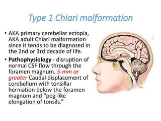
Chiari Malformtions
Presented By:
Dr. Rahul Jain
SR-2 Neurosurgery
Moderated by:
Dr V. C. Jha
Dr Nitish Kumar
Dr Gaurav Verma
Introduction
• Collection of hindbrain abnormalities ranging from
simple herniation of the cerebellar tonsils through
the foramen magnum to complete agenesis of the
cerebellum.
• Focus of treatment for symptomatic patients with
Chiari malformations mainly consists of restoring
normal cerebrospinal fluid (CSF) dynamics across
the craniocervical junction.
• Wide variability in the clinical presentation, imaging
findings, and technical aspects of decompression
for each type of Chiari malformation.
HISTORY
• Observationes Medicae, by the Dutch physician and
anatomist Nicholas Tulp (1593–1674), reference to
hindbrain herniation in a myelodysplastic individual.
• In 1883, John Cleland of Scotland reported a single
myelodysplastic patient with hindbrain herniation and
hydrocephalus
• Early 1890s, Dr. Hans Chiari, professor of pathologic
anatomy at the German University in Prague, used
autopsy specimens to describe four congenital
anomalies later termed the Chiari malformations (types
I–IV).
• Julius Arnold (1835–1915), professor of anatomy at
Heidelberg, described a single myelodysplastic patient
with hindbrain herniation and no hydrocephalus.
• The term “Chiari malformation” is preferred for
type 1 malformations (due to the more significant
contribution of pathologist, Hans Chiari).
• Although the term Arnold-Chiari malformation has
been used specifically in reference to hindbrain
herniation in myelodysplastic patients, it was Chiari
who described and attempted to delineate the
pathophysiology of these posterior fossa
abnormalities. Therefore it is most appropriate to
refer to this abnormality as the Chiari II
malformation (CIIM).
Pathophysiology Theories
• Grouped into the hindbrain dysgenesis and
developmental arrest theory; the caudal traction
theory; the hydrocephalus and hydrodynamic theory of
Gardner; the small posterior fossa/hindbrain
overgrowth theory; and the lack of embryologic
ventricular distention theory.
• Nishikawa and colleagues suggested that the
underdevelopment of occipital somites within the
paraxial mesoderm creates a small posterior fossa and
CIM.
• The association of craniosynostosis and CIM appears
strongest in cases of syndromic, multisuture, and
lambdoid synostosis. Lambdoid suture closure, typical
in Crouzon syndrome, can directly reduce posterior
fossa volume.
• Development of a craniospinal pressure gradient
across the foramen magnum may cause or hasten
the development of CIM.
• The gradient results from impaired CSF flow across
the foramen magnum.
• Negative CSF pressure in the spinal compartment
relative to the intracranial compartment creates a
“sump effect” that forces the tonsils down through
the foramen magnum.
• Once CSF flow is blocked at the foramen magnum,
low intraspinal pressures can be accentuated and
perpetuated by continuous absorption of CSF
through spinal pathways, further worsening the
clinical situation.
• Chiari II Malformation may occur because the
cerebellar vermis develops before the tonsils,
anabnormal pressure differential that develops in
utero would cause abnormal displacement of the
vermis and brainstem structures, without any
tonsillar involvement.
• Such a pressure differential could occur with fluid
leakage from the myelomeningocele.
• McLone and Knepper gave “unified theory” based
on the aforementioned assumption that the neural
tube defect occurs first, and all the other
manifestations, including the Chiari malformation
and hydrocephalus, follow secondarily.
• Leakage of CSF through the spinal defect causes a
lack of distention of the primitive cranial ventricular
system.
• In experimental animals, venting fluid from the
embryonic ventricular system can cause, for
example, disorganization of the developing cerebral
cortex and abnormal development of the pontine
flexure.
Type 1 Chiari malformation
• AKA primary cerebellar ectopia,
AKA adult Chiari malformation
since it tends to be diagnosed in
the 2nd or 3rd decade of life.
• Pathophysiology - disruption of
normal CSF flow through the
foramen magnum. 5-mm or
greater Caudal displacement of
cerebellum with tonsillar
herniation below the foramen
magnum and “peg-like
elongation of tonsils.”
• Prevalence (radiographically): ≈ 0.5%.
• Average age 41 years (12-73 yrs),
slight female preponderance
(female:male = 1.3:1).
• Patients may present due to
compression of brainstem at the level
of the foramen magnum or
hydrocephalus or syringomyelia; 15-
30% are asymptomatic.
• The most common presenting
symptom is pain (60%–70%)
especially headache, usually occipital
and upper cervical in location, and
often induced by Valsalva maneuvers
such as laughing, sneezing, and
coughing (tussive headache).
• Signs of CIM
1. Downbeat nystagmus considered characteristic. 10%
will have a normal neurologic exam with occipital H/A
as their only complaint. Some patients may present
primarily with spasticity.
2. Three main patterns of clustering of signs
Foramen Magnum Compression
Syndrome (22%)
• Ataxia, Corticospinal
and sensory deficits
• cerebellar signs,
lower cranial nerve
palsies
• 37% severe headache
Central Cord Syndrome (65%)
• dissociated sensory loss
(loss of pain &
temperature sensation
with preserved touch &
JPS)
• 11% cranial nerve palsies
Cerebellar Syndrome (11%)
• Truncal and
limb ataxia
• Nystagmus
• dysarthia
Evaluation
• MRI of brain and C-spine
are the diagnostic tests
of choice.
• Cine MRI may
demonstrate blockage of
CSF flow at FM.
• Unenhanced CT is poor
for evaluating the neural
structures, but very
good for HCP.
Management of CIM
• Patients with Chiari malformation and
hydrocephalus – Treatment of the hydrocephalus
with CSF shunting may also resolve the tonsillar
descent and syringomyelia.
Indications for surgical decompression
• Since patients respond best when operated on
within 2 years of the onset of symptoms (see
below), early surgery is recommended for
symptomatic patients.
Operative results
• Pain generally respond well to surgery.
• Weakness is less responsive to surgery, especially
when muscle atrophy is present.
• Sensation may improve when the posterior
columns are unaffected and the deficit is due to
spinothalamic involvement alone.
• Factors that correlate with a worse outcome are
the presence of atrophy, ataxia, scoliosis, and
symptoms lasting longer than 2 years.
• Most favorable results occur in patients with
cerebellar syndrome.
Type 2 (Arnold)–Chiari malformation
• CIIM occurs in most (>95%) patients with
myelomeningocele and is the leading cause of
death in treated myelodysplastic patients today.
• Almost always associated with myelomeningocele,
often accompanied by hydrocephalus.
• Pathophysiology - More likely due to primary
dysgenesis of the brainstem with multiple other
developmental anomalies.
• Findings of Caudally dislocated cervicomedullary
junction, pons, 4th ventricle and medulla.
Cerebellar tonsils located at or below the foramen
magnum.
• Associated neurological
anomalies include tectal
beaking - secondary to
partial or complete fusion of
the colliculi into a single
backward pointed peak
kinking at the level of the
cervicomedullary junction
caused by the caudal
displacement of a portion of
the medulla in conjunction
with a spinal cord that is held
in relative immobility by the
denticulate ligaments
• Other conditions related with CIIM are poorly
myelinated cerebellar folia, hydrocephalus: present
in most, heterotopias, hypoplasia of falx,
microgyria, degeneration of lower cranial nerve
nuclei.
• absence of the septum pellucidum, thought to be
due to necrosis with resorption secondary to
hydrocephalus
• Bony abnormalities associated with CIIM are of
cervicomedullary junction, assimilation of atlas,
platybasia, basilar impression, Klippel–Feil
deformity, craniolacunia of the skull.
Presentation
• About one-third of these patients develop
brainstem symptoms by age of 5 years.
• 20% may present as a neurological emergency -
dysfunction of the 9th and 10th cranial nerves,
affecting respiration, swallowing, and vocal cord
functions; this is often accompanied by stridor,
opisthotonos, and nystagmus.
• Symptomatic deterioration with progressive
brainstem dysfunction may be irreversible and lead
to death. This potentially catastrophic syndrome
occurs most frequently in infants younger than 2
years, particularly younger than 3 months.
• Findings include:
• swallowing difficulties (neurogenic dysphagia) (69%)
• apneic spells (58%)
• stridor (56%)
• aspiration (40%)
• armweakness (27%) that may progress to quadriparesis
Diagnostic evaluation
• Skull films - Craniolacunia (AKA Lückenschädel) in
85% (round defects in the skull with sharp borders,
separated by irregularly branching bands of bone;
not due to increased ICP.
• Cranial and cervical MRI is the diagnostic test of
choice
More
common
in
neonates
Management of CIIM
• Expeditious brainstem decompression should be
carried out when any of the following critical
warning signs develop: neurogenic dysphagia,
stridor, apneic spells.
• Pre-op status and the rapidity of neurologic
deterioration are the most important
prognosticators.
• 68% had complete or near-complete resolution of
symptoms, 20% had no improvement (in general,
neonates fared worse than older children)
• Mortality rate is 71% in infants having
cardiopulmonary arrest, vocal cord paralysis or arm
weakness within 2 weeks of presentation.
• Before surgical decompression is considered,
patients with symptomatic CIIM must have
physiologic intracranial pressure.
• Properly functioning ventricular shunt can often
obviate the need for decompression of hindbrain
herniation.
• Tomita and McLone # concluded that shunt revision
can reverse acute respiratoryarrest. In contrast,
lower cranial nerve findings may not improve after
the shunt revision but rather only after posterior
fossa decompression.
# Tomita T, Mclone DG. Acute respiratory arrest: a Complication of malformation of the
shunt in children with myelomeningocele and arnold-chiari malformation. Am J Dis Child.
1983. https://doi.org/10.1001/archpedi.1983.02140280040011
• The Chiari II anomaly is a challenging surgical entity
with unusual and highly variable anatomy.
• Cerebellar tissue usually extends into the lower cervical
spine; it may be very adherent to the medulla, and
occasionally the two tissues may even seem
indistinguishable or fused.
• The confluence of sinuses can be as low as the rim of
the foramen magnum, and the dura may contain large
venous sinuses.
• Helpful preoperative diagnostic studies include BAEPs,
swallow study, direct vocal cord visualization by an
otolaryngologist, and assessment of pulmonary
function including obstructive and central apnea (sleep
study), as well as hypercapnic ventilatory drive by a
pulmonary specialist.
• Once adequate shunt function is ensured and when the
symptoms are progressive – surgical decompression
Chiari III Malformation
• Rarest and most severe form of the Chiari
malformations; even the existence are
controversial
• Herniation of cerebellum and brainstem into a
posterior occipital or high cervical encephalocele
with other intracranial anomalies that are seen in
CIIM.
• Given the severity of hindbrain herniation, its
management is often problematic from both a
technical and an ethical perspective; poor
prognosis and usually incompatible with life.
Chiari IV Malformation
• Cerebellar hypoplasia or aplasis
without cerebellar herniation.
• Associated with a small posterior
fossa.
• Existence as a distinct clinical
entity is debated.
• Although this was included in
Chiari’s classification of
rhombencephalic
malformations, it is more
appropriate to include this in the
category of posterior fossa cysts
Chiari type 0
• Syringohydromyelia without hindbrain herniation.
• “Chiari-like” pathophysiology may be present in the
absence of tonsillar herniation, which may be
intermittent.
• Crowded foramen magnum, multiple arachnoid
adhesions, fourth ventricular arachnoid veil.
• Abnormal CSF flow at the posterior fossa or
foramen magnum
• Responds to posterior fossa decompression
Chiari type 0.5
• Cerebellar tonsils descend less than 5 mm below
the foramen magnum, but exhibit ventral
herniation defined as unilateral or bilateral crossing
of the cerebellar tonsils anterior to the horizontal
anatomic line bisecting the caudal medulla at the
level of the foramen magnum.
• Symptoms include: dysphagia & sleep apnea in very
young children, and exertion induced H/A
• and paresthesias in older children. Other possible
symptoms: ataxia, behavioral changes.
Chiari 1.5 Malformation
• Severe form of Chiari 1.
• Entire cervicomedullary junction
(and obex) is situated below the
foramen magnum.
• Platybasia
• Clinical manifestations and
response to suboccipital
decompression are similar to
Chiari I with the exception that
syringomyelia persisted in almost
twice as many Chiari 1.5 cases
(13.6%) as Chiari I (6.9%).
Chiari malformation and pregnancy
• Issues during pregnancy
• increased ICP during vaginal labor & delivery elevates
the risk of acute tonsillar herniation
• spinal fluid leak with spinal anesthesia or inadvertent
entry into subarachnoid space during attempted
epidural anesthesia may precipitate tonsillar herniation.
• Recommendations (proposed guidelines)
1. before pregnancy in a patient with known CIM
• MRI of brain and cervical spine to assess degree of
obstruction
• lumbar MRI to evaluate for occult spinal dysraphism
• If symptomatic other than headache or has HCP -
operate
2. patients with CIM considering pregnancy or
already pregnant
• MDT team -fetal medicine, anesthesia, and
neurosurgery
• management at a specialized center since there’s
increased risk of medical and obstetrical
complications including ARDS, stroke, seizures,
sepsis, pre-eclampsia and eclampsia.
3. pregnant patients with CIM who are asymptomatic
or only have H/A
epidural or spinal anesthesia are low risk and should
be made available
4. pregnant CIM patients with more symptoms
• for vaginal delivery, minimizing Valsalva maneuvers
• C-section under regional anesthesia or general
anesthesia
5. pregnant CIM patients with unshunted
hydrocephalus or findings of increased ICP
• considered high-risk for vaginal delivery and
neuraxial anesthesia.
• C-section under general anesthesia should be
considered
• suboccipital decompression may be considered if
the fetus has not reached viability
Surgical technique for suboccipital
decompression
• most frequently performed operation is posterior
fossa decompression of the cerebellar tonsilsusing
a suboccipital craniectomy.
• usually combined with dural patch grafting and
cervical laminectomy, which must be carried down
to the bottom of the tonsillar tip, which usually
includes C1, and sometimes C2 or C3.
• Options for grafts: same incision (pericranium),
separate incision (e.g., fascia lata), and allograft.
• Tonsillar herniation is present in all cases (by
definition), the most common position being at C1
(62%).
• Fibrous adhesions between dura, arachnoid and
tonsils with occlusion of foramina of Luschka and
Magendie in 41%.
• The tonsils separated easily in 40%.
• Posterior rim of the foramen magnum (FM) is the
inferior part of the occipital bone.
• FM is enlarged no higher than ≈ 3cm above the FM
and approximately as wide as the FM (also ≈ 3 cm).
• Compression is at the foramen magnum, not in the
p-fossa, so keep the posterior-fossa exposure small;
the emphasis is to decompress the tonsils by
opening the FM and the upper cervical spine as far
inferiorly as the tonsils extend.
• Thick constricting dural band is usually found
between the C1 arch and foramen magnum and
may be lysed separately.
• Shrinking the tonsils with bipolar cautery.
• dividing adhesions to separate the tonsils from
each other as well as from the underlying medulla.
• Excessive removal of occipital bone – cerebellar
hemispheres to herniate through the FM
(“cerebellar ptosis” AKA “cerebellar sag).
Controversies in management
1. Need for Dural Opening
• opening of the dural covering of the foramen
magnum is thought to be necessary.
• increased interest in bony decompression alone
• The surgeon needs to consider the higher
reoperation rates with bone-only decompression
and share this information with patients.
2. Arachnoid Opening
• Confirming the adequate flow of CSF through the
obex without any veil obstructing the site.
• Some surgeons feel keeping the arachnoid intact is
important to minimize scarring.
3. Dural Closure
• autograft material (periosteum, and ligamentum
nuchae) and allograft material (cadaveric, animal,
or artificial tonsil coagulation or resection).
• no consensus on type of dural closure or even lack
of closure.
• Opponents argue that scarring is induced by such
maneuvers and can lead to redo operations
4. Open Versus Endoscopic Approaches
• benefits of this technique over open surgery
remain to be elucidated.
• Some surgeons raise concerns about added surgical
time, limited bony removal, and reliance on
tonsillar shrinkage to gain adequate
decompression, with increased surgical times and
potential for postoperative scarring.
5. Extent of Bony Decompression
• Most surgeons limit the bony exposure to about 2
cm to prevent cerebellar sag.
• Titanium mesh with or without duraplasty is
typically used to address subsequent cerebellar
ectopia.
6. Chiari Decompression in Ehlers-Danlos Syndrome
• CIM has been reported as a comorbidity with Ehlers-
Danlos syndrome (EDS), may be complicated by
craniocervical instability or basilar invagination.
• The term instability is certainly a misnomer, because
the increased range of motion with ligamentous laxity
is not clinically the same as instability secondary to
trauma or rheumatoid disease.
• some reports showing improvement in symptoms after
craniocervical fixation does not provide adequate or
sufficient scientific data to recommend this as a first-
line treatment.
• Even after more than a decade of treating this
condition, we are still unable to decide which patients
will improve with which surgical intervention.
7. Tethered Cord Release
• tethered spinal cord with CM or syringomyelia –
Incidence ranges from 2.2% to 6%.
• subgroup of Chiari patients with atypical
presentations and thickened filum may benefit
from detethering.
8. Scoliosis
• 25% to 50% of CIM patients.
• role for cilia-driven CSF flow in spine
morphogenesis, with irregularities in CSF flow
being a potential driver for the development of
scoliosis.
• asymmetric anterior horn cell dysfunction caused
by syringomyelia results in unbalanced innervation
of the paraspinal musculature, predisposing the
patient to scoliosis.
• Presence of a syrinx is responsible for the increased
risk of scoliosis in patients with Chiari 1
malformations.
• In patients with Chiari-related scoliosis, a
decompression may lead to stabilization or even
improvement of the scoliosis.
• Nu ber of small case series reported variable rates
of improvement ranging from 0% to 73%, with
similarly variable rates of progression ranging from
18% to 72% following decompression.
• In practice, a Chiari decompression first-line treatment
for Chiari-related scoliosis in younger patients (i.e.,
those <10 years of age) and/or patients with smaller
Cobb angles (20 to 50 degree).
• progressive scoliosis – treatment of spinal curvature.
• Less than 20 degree – observation with 6 mnth
standing xrays for skeletal immature patients.
• Between 20 to 40 degree – spinal bracing
• Cobb angle > 50 degree – deformity correction with
fusion
• Chiari 1 patients with syringomyelia who undergo
scoliosis surgery, lengthening of the spinal column
carries a risk of neurological deficit, and a Chiari
decompression is often recommended first in an effort
to reduce the size of the syrinx.
9. Occipitocervical Fusion
• subset of Chiari patients have a variety of
radiological findings other than tonsillar herniation,
including brainstem herniation through the
foramen magnum; medullary kinking, retroflexion
of the odontoid, an abnormal clival-cervical angle
(CXA), assimilation of the atlas, and/or basilar
invagination - complex Chiari malformations
• key radiological features
1. the degree of odontoid retroflexion – pBC2
distance > 9 mm
2. the CXA < 125 degree
• Combination of odontoid retroflexion (pBC2
distance >9 mm) and a CXA less than 125 degrees is
highly predictive of progressive craniocervical
kyphosis and the need for occipitocervical fusion,
particularly following disruption of the posterior
tension band during a standard Chiari
decompression.
CXA, Clival-cervical
angle
pBC2 line
McRae’s line
10. Syringomyelia
• occurs in approximately 50% to 75% of patients
with Chiari 1 malformations.
• How? obstruction of CSF flow across the
cervicomedullary junction results in a downward,
piston-like movement of the cerebellar tonsils
during the systolic phase of the cardiac cycle, which
in turn produces a pressure wave that forces fluid
into the spinal cord.
• Posterior fossa decompression with duroplasty
successfully improves the symptoms of a syrinx in
over two thirds of patients, with an initial success
rate of 80% in their own series.
• Clinical and radiological improvement is expected
within 6 months of surgery.
• Some surgeons prefer to place syringo-
subarachnoid and syringo-pleural shunts into the
syrinx in addition to performing a decompression in
patients with suboccipital headaches and an 8 mm
syrinx.
Conclusion
Chiari presented his series of hindbrain herniations more
than 100 years ago. Today, we understand the Chiari
malformations to involve an abnormality at the
craniocervical junction resulting in impaired neural
function and CSF hydrodynamics. However, the
pathophysiology of each malformation is likely very
different, and the management is tailored to each
individual but simple tenets in the management of these
patients: (1) patient selection; 2) assurance of normal
intracranial physiology; 3) restoration of normal CSF
dynamic flow from the fourth ventricle to the
subarachnoid space and relief of direct brainstem
compression are the goals of surgery.
References
1. Greenberg 10th ed
2. Youman and winn 8th ed
3. Schmidek and Sweets 7th ed
4. Internet

Chiari Malformations.pptx

  • 1.
    Chiari Malformtions Presented By: Dr.Rahul Jain SR-2 Neurosurgery Moderated by: Dr V. C. Jha Dr Nitish Kumar Dr Gaurav Verma
  • 2.
    Introduction • Collection ofhindbrain abnormalities ranging from simple herniation of the cerebellar tonsils through the foramen magnum to complete agenesis of the cerebellum. • Focus of treatment for symptomatic patients with Chiari malformations mainly consists of restoring normal cerebrospinal fluid (CSF) dynamics across the craniocervical junction. • Wide variability in the clinical presentation, imaging findings, and technical aspects of decompression for each type of Chiari malformation.
  • 3.
    HISTORY • Observationes Medicae,by the Dutch physician and anatomist Nicholas Tulp (1593–1674), reference to hindbrain herniation in a myelodysplastic individual. • In 1883, John Cleland of Scotland reported a single myelodysplastic patient with hindbrain herniation and hydrocephalus • Early 1890s, Dr. Hans Chiari, professor of pathologic anatomy at the German University in Prague, used autopsy specimens to describe four congenital anomalies later termed the Chiari malformations (types I–IV). • Julius Arnold (1835–1915), professor of anatomy at Heidelberg, described a single myelodysplastic patient with hindbrain herniation and no hydrocephalus.
  • 4.
    • The term“Chiari malformation” is preferred for type 1 malformations (due to the more significant contribution of pathologist, Hans Chiari). • Although the term Arnold-Chiari malformation has been used specifically in reference to hindbrain herniation in myelodysplastic patients, it was Chiari who described and attempted to delineate the pathophysiology of these posterior fossa abnormalities. Therefore it is most appropriate to refer to this abnormality as the Chiari II malformation (CIIM).
  • 5.
    Pathophysiology Theories • Groupedinto the hindbrain dysgenesis and developmental arrest theory; the caudal traction theory; the hydrocephalus and hydrodynamic theory of Gardner; the small posterior fossa/hindbrain overgrowth theory; and the lack of embryologic ventricular distention theory. • Nishikawa and colleagues suggested that the underdevelopment of occipital somites within the paraxial mesoderm creates a small posterior fossa and CIM. • The association of craniosynostosis and CIM appears strongest in cases of syndromic, multisuture, and lambdoid synostosis. Lambdoid suture closure, typical in Crouzon syndrome, can directly reduce posterior fossa volume.
  • 6.
    • Development ofa craniospinal pressure gradient across the foramen magnum may cause or hasten the development of CIM. • The gradient results from impaired CSF flow across the foramen magnum. • Negative CSF pressure in the spinal compartment relative to the intracranial compartment creates a “sump effect” that forces the tonsils down through the foramen magnum. • Once CSF flow is blocked at the foramen magnum, low intraspinal pressures can be accentuated and perpetuated by continuous absorption of CSF through spinal pathways, further worsening the clinical situation.
  • 7.
    • Chiari IIMalformation may occur because the cerebellar vermis develops before the tonsils, anabnormal pressure differential that develops in utero would cause abnormal displacement of the vermis and brainstem structures, without any tonsillar involvement. • Such a pressure differential could occur with fluid leakage from the myelomeningocele. • McLone and Knepper gave “unified theory” based on the aforementioned assumption that the neural tube defect occurs first, and all the other manifestations, including the Chiari malformation and hydrocephalus, follow secondarily.
  • 8.
    • Leakage ofCSF through the spinal defect causes a lack of distention of the primitive cranial ventricular system. • In experimental animals, venting fluid from the embryonic ventricular system can cause, for example, disorganization of the developing cerebral cortex and abnormal development of the pontine flexure.
  • 9.
    Type 1 Chiarimalformation • AKA primary cerebellar ectopia, AKA adult Chiari malformation since it tends to be diagnosed in the 2nd or 3rd decade of life. • Pathophysiology - disruption of normal CSF flow through the foramen magnum. 5-mm or greater Caudal displacement of cerebellum with tonsillar herniation below the foramen magnum and “peg-like elongation of tonsils.”
  • 10.
    • Prevalence (radiographically):≈ 0.5%. • Average age 41 years (12-73 yrs), slight female preponderance (female:male = 1.3:1). • Patients may present due to compression of brainstem at the level of the foramen magnum or hydrocephalus or syringomyelia; 15- 30% are asymptomatic. • The most common presenting symptom is pain (60%–70%) especially headache, usually occipital and upper cervical in location, and often induced by Valsalva maneuvers such as laughing, sneezing, and coughing (tussive headache).
  • 11.
    • Signs ofCIM 1. Downbeat nystagmus considered characteristic. 10% will have a normal neurologic exam with occipital H/A as their only complaint. Some patients may present primarily with spasticity. 2. Three main patterns of clustering of signs Foramen Magnum Compression Syndrome (22%) • Ataxia, Corticospinal and sensory deficits • cerebellar signs, lower cranial nerve palsies • 37% severe headache Central Cord Syndrome (65%) • dissociated sensory loss (loss of pain & temperature sensation with preserved touch & JPS) • 11% cranial nerve palsies Cerebellar Syndrome (11%) • Truncal and limb ataxia • Nystagmus • dysarthia
  • 12.
    Evaluation • MRI ofbrain and C-spine are the diagnostic tests of choice. • Cine MRI may demonstrate blockage of CSF flow at FM. • Unenhanced CT is poor for evaluating the neural structures, but very good for HCP.
  • 13.
  • 14.
    • Patients withChiari malformation and hydrocephalus – Treatment of the hydrocephalus with CSF shunting may also resolve the tonsillar descent and syringomyelia. Indications for surgical decompression • Since patients respond best when operated on within 2 years of the onset of symptoms (see below), early surgery is recommended for symptomatic patients.
  • 15.
    Operative results • Paingenerally respond well to surgery. • Weakness is less responsive to surgery, especially when muscle atrophy is present. • Sensation may improve when the posterior columns are unaffected and the deficit is due to spinothalamic involvement alone. • Factors that correlate with a worse outcome are the presence of atrophy, ataxia, scoliosis, and symptoms lasting longer than 2 years. • Most favorable results occur in patients with cerebellar syndrome.
  • 16.
    Type 2 (Arnold)–Chiarimalformation • CIIM occurs in most (>95%) patients with myelomeningocele and is the leading cause of death in treated myelodysplastic patients today. • Almost always associated with myelomeningocele, often accompanied by hydrocephalus. • Pathophysiology - More likely due to primary dysgenesis of the brainstem with multiple other developmental anomalies. • Findings of Caudally dislocated cervicomedullary junction, pons, 4th ventricle and medulla. Cerebellar tonsils located at or below the foramen magnum.
  • 17.
    • Associated neurological anomaliesinclude tectal beaking - secondary to partial or complete fusion of the colliculi into a single backward pointed peak kinking at the level of the cervicomedullary junction caused by the caudal displacement of a portion of the medulla in conjunction with a spinal cord that is held in relative immobility by the denticulate ligaments
  • 18.
    • Other conditionsrelated with CIIM are poorly myelinated cerebellar folia, hydrocephalus: present in most, heterotopias, hypoplasia of falx, microgyria, degeneration of lower cranial nerve nuclei. • absence of the septum pellucidum, thought to be due to necrosis with resorption secondary to hydrocephalus • Bony abnormalities associated with CIIM are of cervicomedullary junction, assimilation of atlas, platybasia, basilar impression, Klippel–Feil deformity, craniolacunia of the skull.
  • 19.
    Presentation • About one-thirdof these patients develop brainstem symptoms by age of 5 years. • 20% may present as a neurological emergency - dysfunction of the 9th and 10th cranial nerves, affecting respiration, swallowing, and vocal cord functions; this is often accompanied by stridor, opisthotonos, and nystagmus. • Symptomatic deterioration with progressive brainstem dysfunction may be irreversible and lead to death. This potentially catastrophic syndrome occurs most frequently in infants younger than 2 years, particularly younger than 3 months.
  • 20.
    • Findings include: •swallowing difficulties (neurogenic dysphagia) (69%) • apneic spells (58%) • stridor (56%) • aspiration (40%) • armweakness (27%) that may progress to quadriparesis Diagnostic evaluation • Skull films - Craniolacunia (AKA Lückenschädel) in 85% (round defects in the skull with sharp borders, separated by irregularly branching bands of bone; not due to increased ICP. • Cranial and cervical MRI is the diagnostic test of choice More common in neonates
  • 21.
  • 22.
    • Expeditious brainstemdecompression should be carried out when any of the following critical warning signs develop: neurogenic dysphagia, stridor, apneic spells. • Pre-op status and the rapidity of neurologic deterioration are the most important prognosticators. • 68% had complete or near-complete resolution of symptoms, 20% had no improvement (in general, neonates fared worse than older children) • Mortality rate is 71% in infants having cardiopulmonary arrest, vocal cord paralysis or arm weakness within 2 weeks of presentation.
  • 23.
    • Before surgicaldecompression is considered, patients with symptomatic CIIM must have physiologic intracranial pressure. • Properly functioning ventricular shunt can often obviate the need for decompression of hindbrain herniation. • Tomita and McLone # concluded that shunt revision can reverse acute respiratoryarrest. In contrast, lower cranial nerve findings may not improve after the shunt revision but rather only after posterior fossa decompression. # Tomita T, Mclone DG. Acute respiratory arrest: a Complication of malformation of the shunt in children with myelomeningocele and arnold-chiari malformation. Am J Dis Child. 1983. https://doi.org/10.1001/archpedi.1983.02140280040011
  • 24.
    • The ChiariII anomaly is a challenging surgical entity with unusual and highly variable anatomy. • Cerebellar tissue usually extends into the lower cervical spine; it may be very adherent to the medulla, and occasionally the two tissues may even seem indistinguishable or fused. • The confluence of sinuses can be as low as the rim of the foramen magnum, and the dura may contain large venous sinuses. • Helpful preoperative diagnostic studies include BAEPs, swallow study, direct vocal cord visualization by an otolaryngologist, and assessment of pulmonary function including obstructive and central apnea (sleep study), as well as hypercapnic ventilatory drive by a pulmonary specialist. • Once adequate shunt function is ensured and when the symptoms are progressive – surgical decompression
  • 25.
    Chiari III Malformation •Rarest and most severe form of the Chiari malformations; even the existence are controversial • Herniation of cerebellum and brainstem into a posterior occipital or high cervical encephalocele with other intracranial anomalies that are seen in CIIM. • Given the severity of hindbrain herniation, its management is often problematic from both a technical and an ethical perspective; poor prognosis and usually incompatible with life.
  • 27.
    Chiari IV Malformation •Cerebellar hypoplasia or aplasis without cerebellar herniation. • Associated with a small posterior fossa. • Existence as a distinct clinical entity is debated. • Although this was included in Chiari’s classification of rhombencephalic malformations, it is more appropriate to include this in the category of posterior fossa cysts
  • 28.
    Chiari type 0 •Syringohydromyelia without hindbrain herniation. • “Chiari-like” pathophysiology may be present in the absence of tonsillar herniation, which may be intermittent. • Crowded foramen magnum, multiple arachnoid adhesions, fourth ventricular arachnoid veil. • Abnormal CSF flow at the posterior fossa or foramen magnum • Responds to posterior fossa decompression
  • 29.
    Chiari type 0.5 •Cerebellar tonsils descend less than 5 mm below the foramen magnum, but exhibit ventral herniation defined as unilateral or bilateral crossing of the cerebellar tonsils anterior to the horizontal anatomic line bisecting the caudal medulla at the level of the foramen magnum. • Symptoms include: dysphagia & sleep apnea in very young children, and exertion induced H/A • and paresthesias in older children. Other possible symptoms: ataxia, behavioral changes.
  • 31.
    Chiari 1.5 Malformation •Severe form of Chiari 1. • Entire cervicomedullary junction (and obex) is situated below the foramen magnum. • Platybasia • Clinical manifestations and response to suboccipital decompression are similar to Chiari I with the exception that syringomyelia persisted in almost twice as many Chiari 1.5 cases (13.6%) as Chiari I (6.9%).
  • 32.
    Chiari malformation andpregnancy • Issues during pregnancy • increased ICP during vaginal labor & delivery elevates the risk of acute tonsillar herniation • spinal fluid leak with spinal anesthesia or inadvertent entry into subarachnoid space during attempted epidural anesthesia may precipitate tonsillar herniation. • Recommendations (proposed guidelines) 1. before pregnancy in a patient with known CIM • MRI of brain and cervical spine to assess degree of obstruction • lumbar MRI to evaluate for occult spinal dysraphism • If symptomatic other than headache or has HCP - operate
  • 33.
    2. patients withCIM considering pregnancy or already pregnant • MDT team -fetal medicine, anesthesia, and neurosurgery • management at a specialized center since there’s increased risk of medical and obstetrical complications including ARDS, stroke, seizures, sepsis, pre-eclampsia and eclampsia. 3. pregnant patients with CIM who are asymptomatic or only have H/A epidural or spinal anesthesia are low risk and should be made available
  • 34.
    4. pregnant CIMpatients with more symptoms • for vaginal delivery, minimizing Valsalva maneuvers • C-section under regional anesthesia or general anesthesia 5. pregnant CIM patients with unshunted hydrocephalus or findings of increased ICP • considered high-risk for vaginal delivery and neuraxial anesthesia. • C-section under general anesthesia should be considered • suboccipital decompression may be considered if the fetus has not reached viability
  • 35.
    Surgical technique forsuboccipital decompression • most frequently performed operation is posterior fossa decompression of the cerebellar tonsilsusing a suboccipital craniectomy. • usually combined with dural patch grafting and cervical laminectomy, which must be carried down to the bottom of the tonsillar tip, which usually includes C1, and sometimes C2 or C3. • Options for grafts: same incision (pericranium), separate incision (e.g., fascia lata), and allograft.
  • 37.
    • Tonsillar herniationis present in all cases (by definition), the most common position being at C1 (62%). • Fibrous adhesions between dura, arachnoid and tonsils with occlusion of foramina of Luschka and Magendie in 41%. • The tonsils separated easily in 40%.
  • 38.
    • Posterior rimof the foramen magnum (FM) is the inferior part of the occipital bone. • FM is enlarged no higher than ≈ 3cm above the FM and approximately as wide as the FM (also ≈ 3 cm). • Compression is at the foramen magnum, not in the p-fossa, so keep the posterior-fossa exposure small; the emphasis is to decompress the tonsils by opening the FM and the upper cervical spine as far inferiorly as the tonsils extend. • Thick constricting dural band is usually found between the C1 arch and foramen magnum and may be lysed separately.
  • 39.
    • Shrinking thetonsils with bipolar cautery. • dividing adhesions to separate the tonsils from each other as well as from the underlying medulla. • Excessive removal of occipital bone – cerebellar hemispheres to herniate through the FM (“cerebellar ptosis” AKA “cerebellar sag).
  • 40.
    Controversies in management 1.Need for Dural Opening • opening of the dural covering of the foramen magnum is thought to be necessary. • increased interest in bony decompression alone • The surgeon needs to consider the higher reoperation rates with bone-only decompression and share this information with patients.
  • 41.
    2. Arachnoid Opening •Confirming the adequate flow of CSF through the obex without any veil obstructing the site. • Some surgeons feel keeping the arachnoid intact is important to minimize scarring. 3. Dural Closure • autograft material (periosteum, and ligamentum nuchae) and allograft material (cadaveric, animal, or artificial tonsil coagulation or resection). • no consensus on type of dural closure or even lack of closure. • Opponents argue that scarring is induced by such maneuvers and can lead to redo operations
  • 42.
    4. Open VersusEndoscopic Approaches • benefits of this technique over open surgery remain to be elucidated. • Some surgeons raise concerns about added surgical time, limited bony removal, and reliance on tonsillar shrinkage to gain adequate decompression, with increased surgical times and potential for postoperative scarring. 5. Extent of Bony Decompression • Most surgeons limit the bony exposure to about 2 cm to prevent cerebellar sag. • Titanium mesh with or without duraplasty is typically used to address subsequent cerebellar ectopia.
  • 43.
    6. Chiari Decompressionin Ehlers-Danlos Syndrome • CIM has been reported as a comorbidity with Ehlers- Danlos syndrome (EDS), may be complicated by craniocervical instability or basilar invagination. • The term instability is certainly a misnomer, because the increased range of motion with ligamentous laxity is not clinically the same as instability secondary to trauma or rheumatoid disease. • some reports showing improvement in symptoms after craniocervical fixation does not provide adequate or sufficient scientific data to recommend this as a first- line treatment. • Even after more than a decade of treating this condition, we are still unable to decide which patients will improve with which surgical intervention.
  • 44.
    7. Tethered CordRelease • tethered spinal cord with CM or syringomyelia – Incidence ranges from 2.2% to 6%. • subgroup of Chiari patients with atypical presentations and thickened filum may benefit from detethering. 8. Scoliosis • 25% to 50% of CIM patients. • role for cilia-driven CSF flow in spine morphogenesis, with irregularities in CSF flow being a potential driver for the development of scoliosis.
  • 45.
    • asymmetric anteriorhorn cell dysfunction caused by syringomyelia results in unbalanced innervation of the paraspinal musculature, predisposing the patient to scoliosis. • Presence of a syrinx is responsible for the increased risk of scoliosis in patients with Chiari 1 malformations. • In patients with Chiari-related scoliosis, a decompression may lead to stabilization or even improvement of the scoliosis. • Nu ber of small case series reported variable rates of improvement ranging from 0% to 73%, with similarly variable rates of progression ranging from 18% to 72% following decompression.
  • 46.
    • In practice,a Chiari decompression first-line treatment for Chiari-related scoliosis in younger patients (i.e., those <10 years of age) and/or patients with smaller Cobb angles (20 to 50 degree). • progressive scoliosis – treatment of spinal curvature. • Less than 20 degree – observation with 6 mnth standing xrays for skeletal immature patients. • Between 20 to 40 degree – spinal bracing • Cobb angle > 50 degree – deformity correction with fusion • Chiari 1 patients with syringomyelia who undergo scoliosis surgery, lengthening of the spinal column carries a risk of neurological deficit, and a Chiari decompression is often recommended first in an effort to reduce the size of the syrinx.
  • 47.
    9. Occipitocervical Fusion •subset of Chiari patients have a variety of radiological findings other than tonsillar herniation, including brainstem herniation through the foramen magnum; medullary kinking, retroflexion of the odontoid, an abnormal clival-cervical angle (CXA), assimilation of the atlas, and/or basilar invagination - complex Chiari malformations • key radiological features 1. the degree of odontoid retroflexion – pBC2 distance > 9 mm 2. the CXA < 125 degree
  • 48.
    • Combination ofodontoid retroflexion (pBC2 distance >9 mm) and a CXA less than 125 degrees is highly predictive of progressive craniocervical kyphosis and the need for occipitocervical fusion, particularly following disruption of the posterior tension band during a standard Chiari decompression.
  • 49.
  • 50.
    10. Syringomyelia • occursin approximately 50% to 75% of patients with Chiari 1 malformations. • How? obstruction of CSF flow across the cervicomedullary junction results in a downward, piston-like movement of the cerebellar tonsils during the systolic phase of the cardiac cycle, which in turn produces a pressure wave that forces fluid into the spinal cord. • Posterior fossa decompression with duroplasty successfully improves the symptoms of a syrinx in over two thirds of patients, with an initial success rate of 80% in their own series.
  • 51.
    • Clinical andradiological improvement is expected within 6 months of surgery. • Some surgeons prefer to place syringo- subarachnoid and syringo-pleural shunts into the syrinx in addition to performing a decompression in patients with suboccipital headaches and an 8 mm syrinx.
  • 52.
    Conclusion Chiari presented hisseries of hindbrain herniations more than 100 years ago. Today, we understand the Chiari malformations to involve an abnormality at the craniocervical junction resulting in impaired neural function and CSF hydrodynamics. However, the pathophysiology of each malformation is likely very different, and the management is tailored to each individual but simple tenets in the management of these patients: (1) patient selection; 2) assurance of normal intracranial physiology; 3) restoration of normal CSF dynamic flow from the fourth ventricle to the subarachnoid space and relief of direct brainstem compression are the goals of surgery.
  • 53.
    References 1. Greenberg 10thed 2. Youman and winn 8th ed 3. Schmidek and Sweets 7th ed 4. Internet

Editor's Notes

  • #11 aclassically: downbeat nystagmus on vertical movement, and rotatory nystagmus on horizontal movement