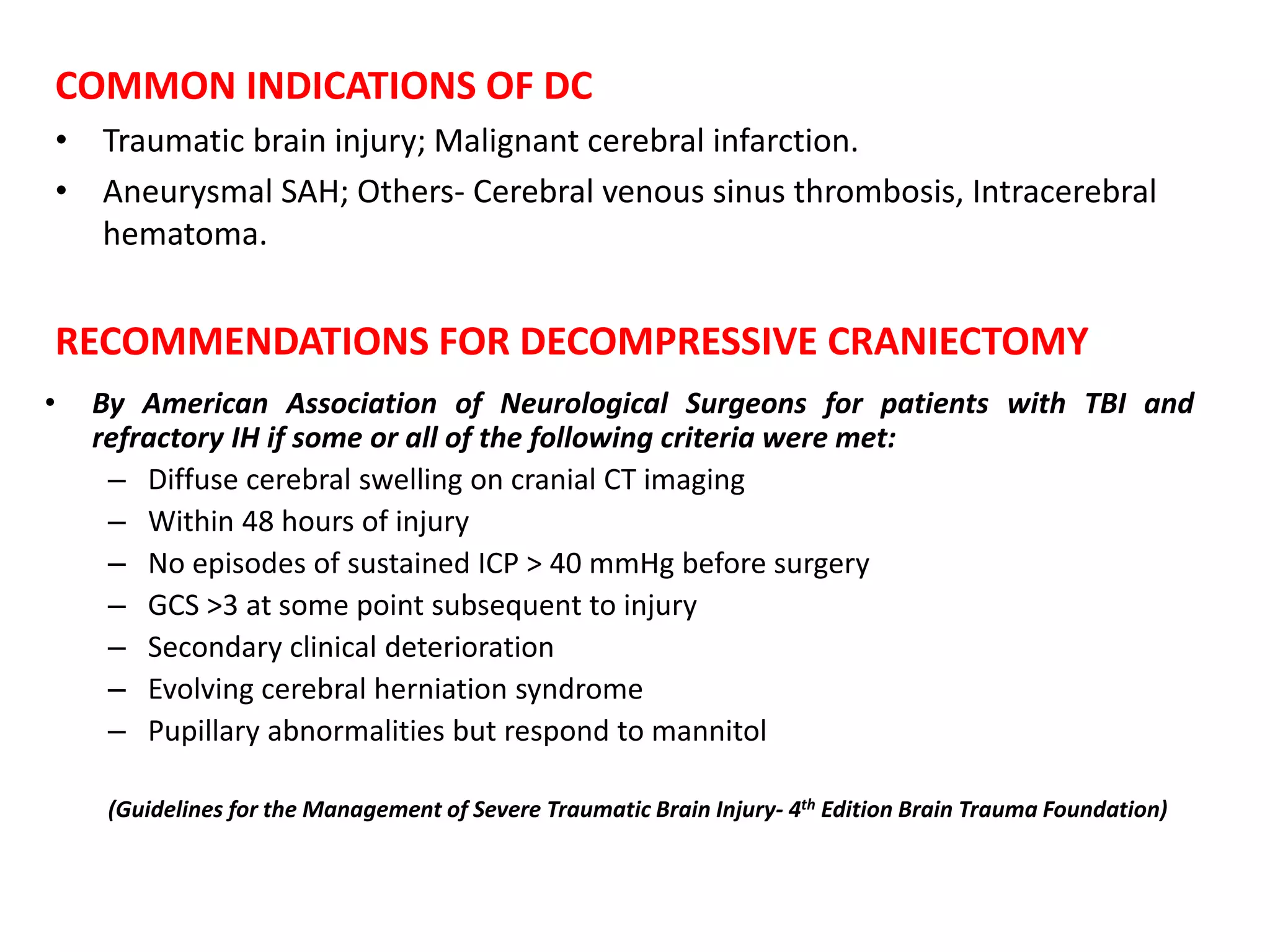
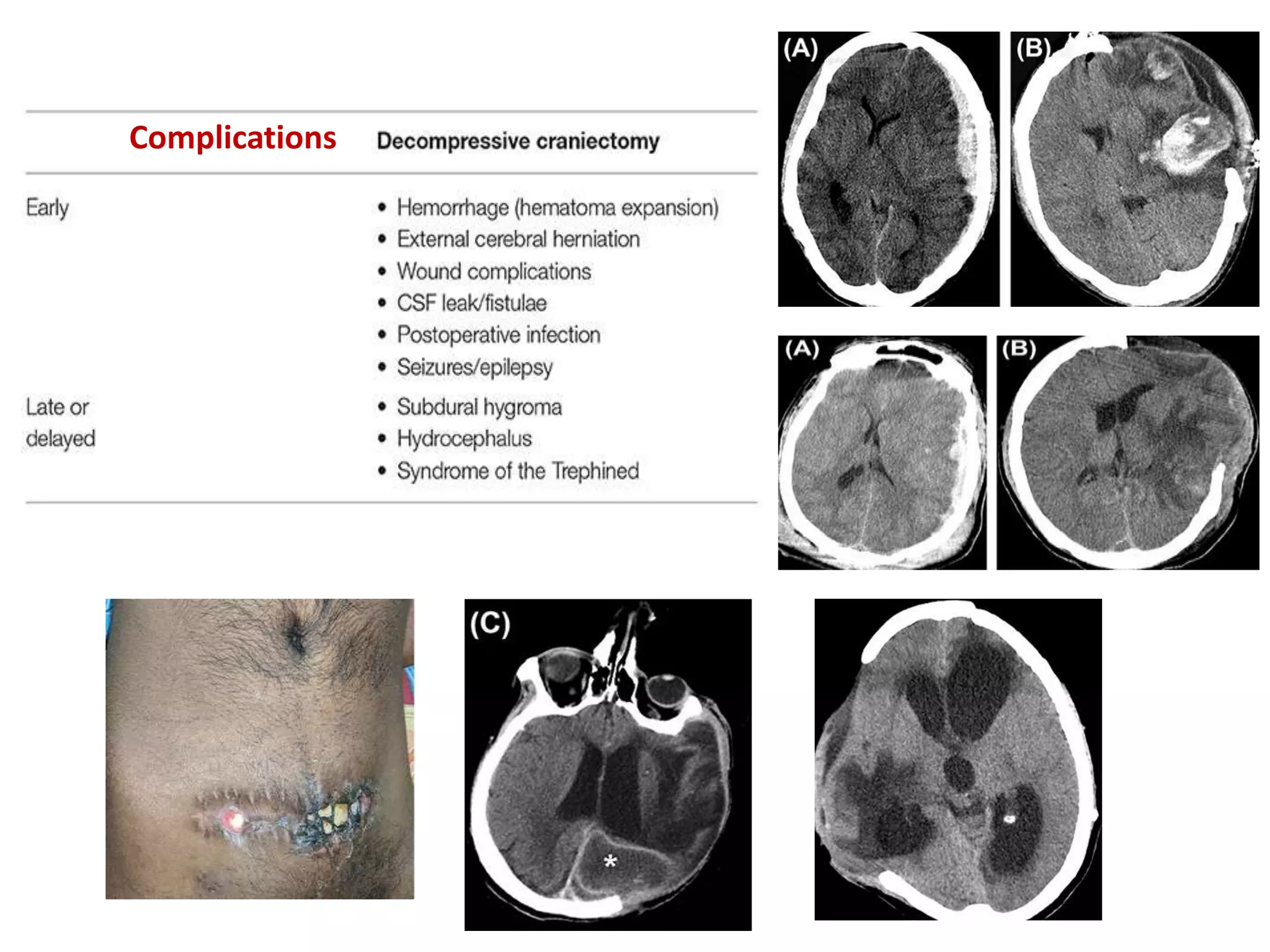
SlideShare a Scribd company logo
Decompressive Craniectomy
Presented by:-
Dr Rahul Jain
SR-2 Neurosurgery
Moderated By:-
Dr V. C. Jha
HoD Neurosurgery
INTRODUCTION
• First described by Ernst von Bergmann first described the technique in 1880
• Annandale in 1894; Performed as palliative procedure for inoperable brain
tumor.
• Kocher(1901) first proposed DC as treatment of raised ICP.
• Cushing: Sub-temporal and suboccipital decompression.
• Decompressive craniectomy (DC) has been used as a final option in
the management of refractory intracranial hypertension/edema.
• Reduction of death and disability from traumatic brain injury (TBI) is
the goal of cranial decompressive surgery.
• Medically refractory cerebral edema is defined as ICP >20 mmHg
for >15 minutes in a given hour despite standard medical
interventions and external ventricular drainage.
• A method of “giving room to the swelling brain” and can be “ a
life-saving procedure.”
PATHOPHYSIOLOGY OF RAISED ICP
• ICP is pressure with in cranial vault, a
rigid structure with fixed volume.
• Contents of cranial vault:
– Brain parenchyma
– CSF
– Blood
Monro-Kellie Doctrine:
• Beyond compensatory mechanisms rise
in any of these, will increase ICP
exponentially
• ICP is one of the factors that govern
cerebral perfusion pressure.
FEED FORWARD CYCLE OF RAISED ICP
• Cranial vault compartmentalized by thick dural folds like falx, tentorium.
• Raised ICP leads to herniation across these compartments.
• Herniation distort vascular anatomy and CSF flow pathways leading to
further increase in ICP.
ROLE OF DC
• Increases buffering capacity of cranium.
• Allows outward herniation, preventing compression of brainstem
structures and reconstruct brain perfusion.
• ICP reduction vary from 15-85% depending on size of bone removed.
• Durotomy further decreases ICP.
AIMS OF DECOMPRESSIVE CRANIECTOMY
• Reduce ICP; Improve blood flow.
• Reduce damage to surrounding brain tissue i.e. reduce secondary brain
injury.
COMMON INDICATIONS OF DC
• Traumatic brain injury; Malignant cerebral infarction.
• Aneurysmal SAH; Others- Cerebral venous sinus thrombosis, Intracerebral
hematoma.
RECOMMENDATIONS FOR DECOMPRESSIVE CRANIECTOMY
• By American Association of Neurological Surgeons for patients with TBI and
refractory IH if some or all of the following criteria were met:
– Diffuse cerebral swelling on cranial CT imaging
– Within 48 hours of injury
– No episodes of sustained ICP > 40 mmHg before surgery
– GCS >3 at some point subsequent to injury
– Secondary clinical deterioration
– Evolving cerebral herniation syndrome
– Pupillary abnormalities but respond to mannitol
(Guidelines for the Management of Severe Traumatic Brain Injury- 4th Edition Brain Trauma Foundation)
TYPES OF DC
Primary/Prophylactic DC
• Any surgical decompression performed, with or without brain tissue
removal, in patients undergoing surgery primarily for the evacuation of any
type of intra-dural lesion.
• Aim: not to control refractory ICP but to avoid increase in ICP.
• Decision taken based on CT, not on ICP.
Secondary/Therapeutic DC
• Continuous ICP monitoring is conducted.
• DC performed when ICP is refractory to medical treatment.
RECOMMENDATION OF BRAIN TRAUMA FOUNDATION
• General maneuvers
– head elevation & neutral position (to avoid venous congestion)
– Normothermia (36.5-37.5 F)
– sedation
– volume resuscitation
• First line therapy: CSF drainage, hypocapnia (PaCO2 30-35mmHg), mannitol
administration.
• Second tier: high dose barbiturates, intense hyperventilation (PaCO2-
<30mmHg), increase in MAP, mild to moderate hypothermia, decompressive
craniectomy.
• Inadequate craniectomy size is associated with parenchymal hemorrhage-
infarction and increased mortality.
(Wagner S et al. Journal of Neurosurgery, May 2001, vol./is. 94/5(693-6)
• TBI: most common indication for DC; Pathophysiology of TBI: Primary injury Vs
Secondary injury.
• Secondary injury is amenable to treatment. Aim of the treatment is to prevent
secondary injury.
APPROACHES OF DC
Fronto-temporo-parietal approach.
Bifrontal decompressive approach.
Temporal approach.
FTP APPROACH
• A large reverse question mark incision is
made starting at the level of the zygoma and
curving posteriorly above the ear, over the
parietooccipital region, then superiorly and
anteriorly, approximately 2 cm lateral to the
midline, and stopping just behind the hairline.
• The posterior extent of the incision should be
more than 15 cm behind the keyhole to allow
for an adequate craniectomy flap.
• Medially, the craniectomy should extend to
only about 2 to 3 cm from the midline to
prevent damage to any draining veins and
arachnoid granulations near the superior sagittal
sinus.
• The temporalis dissection should be carried down to the zygoma to adequately
expose the temporal bone and maximize the temporal decompression.
• Preferences for the location and number of burr holes vary, but typically three
burr holes are made: one at the keyhole, one more inferiorly in the temporal
bone and posterior to the sphenoid bone, and one supero-posteriorly in the
parietal bone.
• Large FTP DC (standard trauma
craniectomy-12x15cm2) significantly
improved the outcome in severe TBI
patients with refractory intracranial
hypertension, compared with routine
temporoparietal craniectomy (6x8cm2),
and had a better effect in terms of
decreasing ICP.
• Small size craniectomies associated
with pericraniectomy hemorrhage and
infarct (diameter < 8 cm).
• A suboptimal DC will lead to exacerbated external brain herniation and shear
forces at the bone edges, which can cause intraparenchymal hemorrhage and
kinking of the cerebral veins.
• Durotomy enhances further decrease in ICP; Durotomy with augmentative
duraplasty recommended.
• Only durotomy without duraplasty; increase risk of herniation of parenchyma;
epilepsy; CSF leak.
In a decompressive hemicraniectomy, the dural opening is just as important as the craniectomy.
Hemostasis should be obtained prior to dural opening (A), and the dura should be opened in a stellate
fashion to maximize cerebral decompression (B and C). The dura can be left open and the dural flaps laid
over a dural substitute prior to skin closure (D).
BIFRONTAL CRANIECTOMY
• In contrast to DHC, the bifrontal
craniectomy used in situations of bifrontal
contusions or diffuse cerebral edema.
• An incision is made beginning at the level of
the zygomatic arch just anterior to the tragus
on one side, coursing back 2 to 3 cm behind
the coronal suture, and down the
contralateral zygomatic arch.
• The myocutaneous flap is then brought
forward over the orbital rim. Care should be
taken to dissect out the supraorbital nerves
from the supraorbital notch on either side.
• A series of symmetric burr holes are then made in the bilateral keyholes,
bilateral squamous portions of the temporal bone, and straddling the
superior sagittal sinus just posterior to the coronal suture. Some authors
advocate drilling burr holes adjacent to the anterior most portion of the superior
sagittal sinus as well.
• The superior sagittal sinus can be dissected away from the overlying bone by
passing a Penfield 3 between the posterior and anterior burr holes. The burr holes
are then connected with a cutting drill bit and footplate, making sure to cross the
superior sagittal sinus last.
• Once the bone flap is elevated, bleeding from the superior sagittal sinus can be
controlled with strips of Gelfoam. The temporal portion of the craniectomy is
then extended downward to the floor of the middle fossa.
• The key to opening the dura with a bifrontal craniectomy is to divide the
anterior portion of the superior sagittal sinus and underlying falx. Without
this step, the brain is not free to expand and can be damaged by herniation
against a tight dural edge.
• As with DHC, the dural opening must be wide. It begins at the floor of the
middle fossa and courses anteriorly along the course of the craniectomy, spanning
the entire anterior width of the craniectomy and crossing the anterior portion of
the superior sagittal suture to reach the contralateral middle fossa floor.
• The dural opening should then also be taken from the middle fossa floor
posteriorly to within approximately 2 cm of the posterior portion of the
superior sagittal sinus on either side.
• To cross the anterior superior sagittal sinus, two heavy sutures are passed around
the sinus and tied to occlude it. The sinus is then divided with scissors. The
underlying falx should then be divided, with care taken to avoid the bilateral
anterior cerebral arteries.
• Polin technique bifrontal craniectomy
– anteriorly roof of orbit; posteriorly
3-5 cm behind coronal suture
– base of temporal bone
– Saggital sinus ligation and division
 Subtemporal decompression: area 26-33 cm3
• decreases ICP; unable to improve blood flow
• temporal lobe herniation and necrosis
Complications
Syndrome of the Trephined
• Overall prevalence of 10%; initially described by Grant and Norcross in 1939.
• The sinking of the scalp due to lack of bony support causes cerebral blood flow
anomaly and dysfunction in the underlying cortex due to impaired CSF flow
dynamics.
• Most common symptoms: motor weakness (61.1%) followed by cognitive
deficits (44.4%), language deficits (29.6%), altered level of consciousness
(27.8%), headache (20.4%), psychosomatic disturbances (18.5%), seizures or
electroencephalographic changes (11.1%), and cranial nerve deficits (5.6%).
• It manifests either as new symptoms causing deterioration of the patient
condition or as failure to retain the early gains. It could manifest as early as 3
days to as late as 7 years (with an average of 5 months).
• These symptoms, as well as, cerebral blood flow abnormalities improve
dramatically after a cranioplasty. Yang has suggested it is safe to do early
cranioplasty within 5-8 weeks to mitigate this risk.
DECRA trail
(Decompressive Craniectomy in Diffuse Traumatic Brain Injury . N Engl J Med. 2011 Apr 21;364(16):1493-502)
• Multicenter, randomized, controlled trial to test the efficacy of bi-fronto-
temporo-parietal DC in adults under the age of 60 years with TBI in whom
first-tier intensive care and neurosurgical therapies had not maintained
intracranial pressure below accepted targets; Dec 2002- April 2010; 15
tertiary care centres in Australia, New Zealand, Saudi Arabia.
• Inclusion criteria: Age 15-59 yrs, Non penetrating traumatic brain injury, GCS
3-8, Marshall class III (moderate diffuse injury on CT); Exclusion: dilated non
reactive pupil, mass lesion, spinal cord injury or cardiac arrest at scene.
• Treatment protocol: Standard treatment Vs surgery + standard treatment;
Within 72 hrs; ICP measurement in all patient (12 before and 36 hrs after
randomization).
• Surgery: BFTP craniectomy with bilateral Dural opening (modified Polin
technique); Sagittal sinus and falx not divided.
• Results compared based on extended Glasgow outcome scale at 6 months
RESULTS
Conclusion
 In adults with severe diffuse
traumatic brain injury and
refractory IH , early bi-fronto-
temporo-parietal DC decreased
ICP and the length of stay in the
ICU but was associated with
more unfavourable outcomes.
RESCUEicp TRIAL
• (Randomized Evaluation of Surgery with Craniectomy for Uncontrollable
Elevation of ICP); 2004-2014; International, multicenter, parallel-group,
randomized trial; 52 centers in 20 countries.
• Aim: Provide class I evidence as to whether decompressive is effective for
the management of patients with raised and refractory ICP following
traumatic brain injury (TBI).
• n=398; Surgery (n=202) vs Control (n=196); With refractory intracranial
hypertension following TBI.
• Primary outcome: Extended Glasgow Outcome Scale (GOS-E) at 6 months
after randomization
• Secondary outcomes:
– GOS-E results at 12 and 24 months after randomization
– Mortality at 6, 12, and 24 months after randomization
– Quality of life at 6, 12, and 24 months after randomization
– Glasgow Coma Scale (GCS) score at discharge from the neurosciences
hospital
– Assessment of intracranial-pressure control
– Time in the ICU
– Time to discharge from the neurosciences hospital
– Economic evaluation.
• Inclusion Criteria
– Age 10 and 65 years
– TBI with an abnormal brain CT
– Intracranial pressure monitor already in place
– Raised ICP (>25 mm Hg for 1 to 12 hours) despite intensive medical
management.
– Patients who had undergone an immediate operation for evacuation
of an intracranial hematoma were included as long as the operation
was not a craniectomy.
– Trial sites were hospitals that provided acute neurosciences care for
patients with severe TBI and that have 24-hour neurosurgical services.
• Exclusion Criteria
– Bilateral fixed and dilated pupils
– Bleeding diathesis
– Injury that was deemed to be unsurvivable
PRIMARY OUTCOMES
• Comparisons are surgery vs. control groups.
• GOS-E distributions at 6 months:
– Death: 26.9% versus 48.9%
– Vegetative state: 8.5% versus 2.1%
– Lower severe disability (dependent on others for care): 21.9% versus
14.4%
– Upper severe disability (independent at home): 15.4% versus 8.0%
– Moderate disability: 23.4% versus 19.7%
– Good recovery: 4.0% versus 6.9%
• Favorable outcomes (upper severe disability or better on the GOS-E) at 6
months: 42.8% versus 34.6% (P = 0.12).
SECONDARY OUTCOMES
• GOS-E distributions at 12 months
– Death:30.4% versus 52.0%
– Vegetative state: 6.2% versus 1.7%
– Lower severe disability: 18.0% versus 14.0%
– Upper severe disability: 13.4% versus 3.9%
– Moderate disability: 22.2% versus 20.1%
– Good recovery: 9.8% versus 8.4%
• Favorable outcomes (upper severe disability or better) at 12 months:
45.4% versus 32.4% of those in the medical group (P=0.01)
• Median time to discharge among survivors: 15.0 days vs. 20.8 days
(P=0.01)
• Control of ICP: Better in surgical group than in the medical group.
ADVERSE EVENTS & CRITICISMS
Adverse event
• 16.3% vs. 9.2% (P=0.03); Most commonly pneumonia, SSI, postoperative
hematoma
Criticism
• Recruitment was slow, taking over 10 years. Half of centers recruited 3 or
fewer patients.
• Clinical teams who cared for the patients were aware of trial group
assignments, although this was mitigated by outcome adjudication.
• A large proportion of patients in the medical group underwent
decompressive craniectomy (37%)
DECRA vs RESCUEicp
• DECRA and RESCUEicp are higher quality studies whose
findings supersede those of lesser quality investigations.
• Though both studied secondary DC for the treatment of
refractory ICP elevation, a key difference in the study
protocols for DECRA and RESCUEicp is that they were
designed to investigate conditions
of early and late refractory ICP elevations, respectively.
• Indeed, DECRA enrolled TBI patients with ICP above 20 mm
Hg for 15 min over a 1-h period despite the optimization of
tier 1 treatments within the first 72 h of care (early), while
RESCUEicp enrolled patients with ICP greater than 25 mm
Hg for 1 to 12 h refractory to 2 tiers of treatment within 10
d of admission (late).
UPDATED RECOMMENDATIONS
(after RERSCUEicp)
PRESENT STATUS OF DC IN TBI
• Pediatric population: DC reduces risk of death and unfavorable outcome.
• For adults: Role of DC in raised ICP due to TBI as second tier treatment is
still controversial.
• Early DC reduces brain edema formation by more than 50% and prevents
secondary brain damage when performed early enough (i.e., during the
first 3 h after trauma).
• Malignant Cerebral Infarction: Large territorial parenchymal infarction
with post ischemic edema and associated with uncal or axial herniation;
Occlusion of proximal MCA, more than 50% of supplied territory involved.
• Treatment: medical management or surgery (DC); DC clearly reduce
mortality however survivors suffered high morbidity .
• Malignant MCA Infarction: An infarction of at least two thirds MCA
territory upward; Present clinically with severe hemispheric stroke
syndrome and progressive deterioration of consciousness within the first 2
days.
• Thereafter, symptoms of transtentorial herniation occur within 2-4 days of
stroke onset; Prognosis is poor and mortality is as high as 80%.
• Very high mortality despite maximal medical treatment: 70% (37/ 53 )
died in NICU (33/37 died within first 5 days); 78% (35/45) died within 1
week
• 3 RCTs conducted and results published:
 DECIMAL (DEcompressive Craniectomy In MALignant middle cerebral artery
infarction)
 DESTINY (DEcompressive Surgery for Treatment of INfarction of malignant
middle cerebral arterY)
 HAMLET ( Hemicraniectomy After Middle cerebral artery infarction with Life -
threatening Edema Trial )
POOLED ANALYSIS
Results:
• Only patient underwent decompression with in 48 hrs were considered for pooled
analysis; n=109, Surgery-58, Medical-51.
• Effect on mortality:
 DECIMAL: absolute reduction in risk 53% at 6 months.
 DESTINY: 12% mortality in surgical and 53% in medical group at 30 days.
 HAMLET: 38% reduction in risk at 1 year.
• Effect on mortality: all 3 trial shows significant reduction in mortality;
Absolute reduction in mortality is 49.9%.
• Effect on severe disability (mRS score 5): absolute reduction in risk of
bad functional outcome was 41.9%.
• Conclusion: surgical decompression with in 48 hrs of onset of stroke
reduced risk of significant morbidity.
• Disability in survivors: while demonstrating an undeniable increase in
the number of survivors among surgical patients, also showed an
increase in the number of survivors with moderately severe disability
(mRS score of 4).
• Pooled analysis of all 3 trials provides Class I evidence for the
performance of early decompressive craniectomy in the setting of large
unilateral infarcts (volume > 145 cc) within 48 hours of the ischemic
event.
Current algorithm for use of
DC
 The only Class I data (recently
published from the DECRA trial)
suggest that primary DC may
increase morbidity with equal
chance of survival compared to
medical management.
 The current standard practice of
performing secondary DC (red
arrow) is up to the discretion of
the treating neurosurgeon
(though the results of the
RESCUEicp trial may provide
Class I evidence for this
management decision in the
future).
Neurological Outcome Scores
References:
• Youmans and Winn neurological surgery 8th edition
• Ramamurthi & Tandon's textbook of neurosurgery 3rd edition
• Schmidek and Sweet: Operative Neurosurgical Techniques 7th edition
• Management of Severe TBI, Brain Trauma foundation Guidwelines.
Neurosurgery 87(3):p 427-434, September 2020.
THANK YOU

More Related Content

Similar to Decompressive Craniectomy.pptx

Case 2
Case   2Case   2
Hydrocephalus
HydrocephalusHydrocephalus
Hydrocephalus
Biswajit Deka
 
Seminar on Hydrocephalus
Seminar on HydrocephalusSeminar on Hydrocephalus
Seminar on Hydrocephalus
Biswajit Deka
 
Perioperative management of Traumatic Brain Injury.pptx
Perioperative management of Traumatic Brain Injury.pptxPerioperative management of Traumatic Brain Injury.pptx
Perioperative management of Traumatic Brain Injury.pptx
Sheikh Muhammad Farhad Sazib
 
How to Read a Head CT.pptx
How to Read a Head CT.pptxHow to Read a Head CT.pptx
How to Read a Head CT.pptx
Maheen Fatima
 
Head injury
Head injuryHead injury
Head injury
HIRANGER
 
basilar artery.pptx
basilar artery.pptxbasilar artery.pptx
basilar artery.pptx
Anas Ahmed
 
Supratentorial masses excision -anaesthetic implication
Supratentorial masses excision -anaesthetic implication Supratentorial masses excision -anaesthetic implication
Supratentorial masses excision -anaesthetic implication
ZIKRULLAH MALLICK
 
BMS2-K13 Pemeriksaan Radiologi pada Sistem Saraf.pptx
BMS2-K13 Pemeriksaan Radiologi pada Sistem Saraf.pptxBMS2-K13 Pemeriksaan Radiologi pada Sistem Saraf.pptx
BMS2-K13 Pemeriksaan Radiologi pada Sistem Saraf.pptx
ssuser144901
 
369 Microsurgery of DACA
369 Microsurgery of DACA369 Microsurgery of DACA
369 Microsurgery of DACA
Neurosurgery Vajira
 
Acute Stroke management
Acute Stroke managementAcute Stroke management
Acute Stroke management
Javed Ahamed
 
Parasagittal meningioma
Parasagittal meningioma Parasagittal meningioma
Parasagittal meningioma
Mohamed E Elsebaey
 
Neuro surgeries (pt)
Neuro surgeries (pt)Neuro surgeries (pt)
Neuro surgeries (pt)
DR NIYATI PATEL
 
Neurotrauma
NeurotraumaNeurotrauma
Neurotrauma
Amit Ghosh
 
Stroke Care of Patient With Post Decompressive Craniectomy
Stroke Care of Patient With Post Decompressive CraniectomyStroke Care of Patient With Post Decompressive Craniectomy
Stroke Care of Patient With Post Decompressive Craniectomy
Pei Yin (Charissa) Wong
 
Vascular diseases of the Central Nervous Sysytem
Vascular diseases of the Central Nervous SysytemVascular diseases of the Central Nervous Sysytem
Vascular diseases of the Central Nervous Sysytem
Kevin Tan
 
HEAD INJURY IN THE ED.pptx
HEAD INJURY IN THE ED.pptxHEAD INJURY IN THE ED.pptx
HEAD INJURY IN THE ED.pptx
masoom parwez
 
Decompressive craniectomy
Decompressive craniectomyDecompressive craniectomy
Decompressive craniectomy
Dr Praveen kumar tripathi
 
Imaginginacutestroke dr anoop.k.r
Imaginginacutestroke dr anoop.k.rImaginginacutestroke dr anoop.k.r
Imaginginacutestroke dr anoop.k.r
anoop k r
 
Digital Subtraction Neuroangiography: What a Resident Should Know
Digital Subtraction Neuroangiography: What a Resident Should Know Digital Subtraction Neuroangiography: What a Resident Should Know
Digital Subtraction Neuroangiography: What a Resident Should Know
Dr. Shahnawaz Alam
 

Similar to Decompressive Craniectomy.pptx (20)

Case 2
Case   2Case   2
Case 2
 
Hydrocephalus
HydrocephalusHydrocephalus
Hydrocephalus
 
Seminar on Hydrocephalus
Seminar on HydrocephalusSeminar on Hydrocephalus
Seminar on Hydrocephalus
 
Perioperative management of Traumatic Brain Injury.pptx
Perioperative management of Traumatic Brain Injury.pptxPerioperative management of Traumatic Brain Injury.pptx
Perioperative management of Traumatic Brain Injury.pptx
 
How to Read a Head CT.pptx
How to Read a Head CT.pptxHow to Read a Head CT.pptx
How to Read a Head CT.pptx
 
Head injury
Head injuryHead injury
Head injury
 
basilar artery.pptx
basilar artery.pptxbasilar artery.pptx
basilar artery.pptx
 
Supratentorial masses excision -anaesthetic implication
Supratentorial masses excision -anaesthetic implication Supratentorial masses excision -anaesthetic implication
Supratentorial masses excision -anaesthetic implication
 
BMS2-K13 Pemeriksaan Radiologi pada Sistem Saraf.pptx
BMS2-K13 Pemeriksaan Radiologi pada Sistem Saraf.pptxBMS2-K13 Pemeriksaan Radiologi pada Sistem Saraf.pptx
BMS2-K13 Pemeriksaan Radiologi pada Sistem Saraf.pptx
 
369 Microsurgery of DACA
369 Microsurgery of DACA369 Microsurgery of DACA
369 Microsurgery of DACA
 
Acute Stroke management
Acute Stroke managementAcute Stroke management
Acute Stroke management
 
Parasagittal meningioma
Parasagittal meningioma Parasagittal meningioma
Parasagittal meningioma
 
Neuro surgeries (pt)
Neuro surgeries (pt)Neuro surgeries (pt)
Neuro surgeries (pt)
 
Neurotrauma
NeurotraumaNeurotrauma
Neurotrauma
 
Stroke Care of Patient With Post Decompressive Craniectomy
Stroke Care of Patient With Post Decompressive CraniectomyStroke Care of Patient With Post Decompressive Craniectomy
Stroke Care of Patient With Post Decompressive Craniectomy
 
Vascular diseases of the Central Nervous Sysytem
Vascular diseases of the Central Nervous SysytemVascular diseases of the Central Nervous Sysytem
Vascular diseases of the Central Nervous Sysytem
 
HEAD INJURY IN THE ED.pptx
HEAD INJURY IN THE ED.pptxHEAD INJURY IN THE ED.pptx
HEAD INJURY IN THE ED.pptx
 
Decompressive craniectomy
Decompressive craniectomyDecompressive craniectomy
Decompressive craniectomy
 
Imaginginacutestroke dr anoop.k.r
Imaginginacutestroke dr anoop.k.rImaginginacutestroke dr anoop.k.r
Imaginginacutestroke dr anoop.k.r
 
Digital Subtraction Neuroangiography: What a Resident Should Know
Digital Subtraction Neuroangiography: What a Resident Should Know Digital Subtraction Neuroangiography: What a Resident Should Know
Digital Subtraction Neuroangiography: What a Resident Should Know
 

More from Dr. Rahul Jain

Moya Moya disease (vasculopathy/angiopathy)
Moya Moya disease (vasculopathy/angiopathy)Moya Moya disease (vasculopathy/angiopathy)
Moya Moya disease (vasculopathy/angiopathy)
Dr. Rahul Jain
 
Cerebrovascular Vasospasm - Etiopathogenesis and Management
Cerebrovascular Vasospasm - Etiopathogenesis and ManagementCerebrovascular Vasospasm - Etiopathogenesis and Management
Cerebrovascular Vasospasm - Etiopathogenesis and Management
Dr. Rahul Jain
 
Chiari Malformations.pptx
Chiari Malformations.pptxChiari Malformations.pptx
Chiari Malformations.pptx
Dr. Rahul Jain
 
Journal Club - Extra axial Endoscopic Third Ventriculostomy.pptx
Journal Club - Extra axial Endoscopic Third Ventriculostomy.pptxJournal Club - Extra axial Endoscopic Third Ventriculostomy.pptx
Journal Club - Extra axial Endoscopic Third Ventriculostomy.pptx
Dr. Rahul Jain
 
Diffuse Midline Gliomas/ Diffuse Pontine Gliomas.pptx
Diffuse Midline Gliomas/ Diffuse Pontine Gliomas.pptxDiffuse Midline Gliomas/ Diffuse Pontine Gliomas.pptx
Diffuse Midline Gliomas/ Diffuse Pontine Gliomas.pptx
Dr. Rahul Jain
 
Trigeminal Neuralgia.pptx
Trigeminal Neuralgia.pptxTrigeminal Neuralgia.pptx
Trigeminal Neuralgia.pptx
Dr. Rahul Jain
 
Microscopes and Endoscopes in Neurosurgery.pptx
Microscopes and Endoscopes in Neurosurgery.pptxMicroscopes and Endoscopes in Neurosurgery.pptx
Microscopes and Endoscopes in Neurosurgery.pptx
Dr. Rahul Jain
 
Subaxial Cervical spine fusion.pptx
Subaxial Cervical spine fusion.pptxSubaxial Cervical spine fusion.pptx
Subaxial Cervical spine fusion.pptx
Dr. Rahul Jain
 
Carotid Occlusive Disease.pptx
Carotid Occlusive Disease.pptxCarotid Occlusive Disease.pptx
Carotid Occlusive Disease.pptx
Dr. Rahul Jain
 
Brain AVM (ArterioVenous Malformation) Managment.pptx
Brain AVM (ArterioVenous Malformation) Managment.pptxBrain AVM (ArterioVenous Malformation) Managment.pptx
Brain AVM (ArterioVenous Malformation) Managment.pptx
Dr. Rahul Jain
 
Intracranial Vascular Bypass.pptx
Intracranial Vascular Bypass.pptxIntracranial Vascular Bypass.pptx
Intracranial Vascular Bypass.pptx
Dr. Rahul Jain
 
Anterior Choroidal Artery.pptx
Anterior Choroidal Artery.pptxAnterior Choroidal Artery.pptx
Anterior Choroidal Artery.pptx
Dr. Rahul Jain
 
Audilogical Assessment.pptx
Audilogical Assessment.pptxAudilogical Assessment.pptx
Audilogical Assessment.pptx
Dr. Rahul Jain
 
HYDROCEPHALUS.pptx
HYDROCEPHALUS.pptxHYDROCEPHALUS.pptx
HYDROCEPHALUS.pptx
Dr. Rahul Jain
 
CNS WHO 2021 tumor classification.pptx
CNS WHO 2021 tumor classification.pptxCNS WHO 2021 tumor classification.pptx
CNS WHO 2021 tumor classification.pptx
Dr. Rahul Jain
 
Cerebral Venous thrombosis.pptx
Cerebral Venous thrombosis.pptxCerebral Venous thrombosis.pptx
Cerebral Venous thrombosis.pptx
Dr. Rahul Jain
 
Endoscopic Endonasal Anatomy.pptx
Endoscopic Endonasal Anatomy.pptxEndoscopic Endonasal Anatomy.pptx
Endoscopic Endonasal Anatomy.pptx
Dr. Rahul Jain
 
Vertebral Artery Anatomy with Endovascular.pptx
Vertebral Artery Anatomy with Endovascular.pptxVertebral Artery Anatomy with Endovascular.pptx
Vertebral Artery Anatomy with Endovascular.pptx
Dr. Rahul Jain
 
Minimal Invasive Surgery in CA Rectum
Minimal Invasive Surgery in CA RectumMinimal Invasive Surgery in CA Rectum
Minimal Invasive Surgery in CA Rectum
Dr. Rahul Jain
 
Liver anatomy
Liver anatomyLiver anatomy
Liver anatomy
Dr. Rahul Jain
 

More from Dr. Rahul Jain (20)

Moya Moya disease (vasculopathy/angiopathy)
Moya Moya disease (vasculopathy/angiopathy)Moya Moya disease (vasculopathy/angiopathy)
Moya Moya disease (vasculopathy/angiopathy)
 
Cerebrovascular Vasospasm - Etiopathogenesis and Management
Cerebrovascular Vasospasm - Etiopathogenesis and ManagementCerebrovascular Vasospasm - Etiopathogenesis and Management
Cerebrovascular Vasospasm - Etiopathogenesis and Management
 
Chiari Malformations.pptx
Chiari Malformations.pptxChiari Malformations.pptx
Chiari Malformations.pptx
 
Journal Club - Extra axial Endoscopic Third Ventriculostomy.pptx
Journal Club - Extra axial Endoscopic Third Ventriculostomy.pptxJournal Club - Extra axial Endoscopic Third Ventriculostomy.pptx
Journal Club - Extra axial Endoscopic Third Ventriculostomy.pptx
 
Diffuse Midline Gliomas/ Diffuse Pontine Gliomas.pptx
Diffuse Midline Gliomas/ Diffuse Pontine Gliomas.pptxDiffuse Midline Gliomas/ Diffuse Pontine Gliomas.pptx
Diffuse Midline Gliomas/ Diffuse Pontine Gliomas.pptx
 
Trigeminal Neuralgia.pptx
Trigeminal Neuralgia.pptxTrigeminal Neuralgia.pptx
Trigeminal Neuralgia.pptx
 
Microscopes and Endoscopes in Neurosurgery.pptx
Microscopes and Endoscopes in Neurosurgery.pptxMicroscopes and Endoscopes in Neurosurgery.pptx
Microscopes and Endoscopes in Neurosurgery.pptx
 
Subaxial Cervical spine fusion.pptx
Subaxial Cervical spine fusion.pptxSubaxial Cervical spine fusion.pptx
Subaxial Cervical spine fusion.pptx
 
Carotid Occlusive Disease.pptx
Carotid Occlusive Disease.pptxCarotid Occlusive Disease.pptx
Carotid Occlusive Disease.pptx
 
Brain AVM (ArterioVenous Malformation) Managment.pptx
Brain AVM (ArterioVenous Malformation) Managment.pptxBrain AVM (ArterioVenous Malformation) Managment.pptx
Brain AVM (ArterioVenous Malformation) Managment.pptx
 
Intracranial Vascular Bypass.pptx
Intracranial Vascular Bypass.pptxIntracranial Vascular Bypass.pptx
Intracranial Vascular Bypass.pptx
 
Anterior Choroidal Artery.pptx
Anterior Choroidal Artery.pptxAnterior Choroidal Artery.pptx
Anterior Choroidal Artery.pptx
 
Audilogical Assessment.pptx
Audilogical Assessment.pptxAudilogical Assessment.pptx
Audilogical Assessment.pptx
 
HYDROCEPHALUS.pptx
HYDROCEPHALUS.pptxHYDROCEPHALUS.pptx
HYDROCEPHALUS.pptx
 
CNS WHO 2021 tumor classification.pptx
CNS WHO 2021 tumor classification.pptxCNS WHO 2021 tumor classification.pptx
CNS WHO 2021 tumor classification.pptx
 
Cerebral Venous thrombosis.pptx
Cerebral Venous thrombosis.pptxCerebral Venous thrombosis.pptx
Cerebral Venous thrombosis.pptx
 
Endoscopic Endonasal Anatomy.pptx
Endoscopic Endonasal Anatomy.pptxEndoscopic Endonasal Anatomy.pptx
Endoscopic Endonasal Anatomy.pptx
 
Vertebral Artery Anatomy with Endovascular.pptx
Vertebral Artery Anatomy with Endovascular.pptxVertebral Artery Anatomy with Endovascular.pptx
Vertebral Artery Anatomy with Endovascular.pptx
 
Minimal Invasive Surgery in CA Rectum
Minimal Invasive Surgery in CA RectumMinimal Invasive Surgery in CA Rectum
Minimal Invasive Surgery in CA Rectum
 
Liver anatomy
Liver anatomyLiver anatomy
Liver anatomy
 

Recently uploaded

Data-Driven Dispensing- Rise of AI in Pharmacies.pdf
Data-Driven Dispensing- Rise of AI in Pharmacies.pdfData-Driven Dispensing- Rise of AI in Pharmacies.pdf
Data-Driven Dispensing- Rise of AI in Pharmacies.pdf
Jasper Colin
 
TEST BANK FOR Health Assessment in Nursing 7th Edition by Weber Chapters 1 - ...
TEST BANK FOR Health Assessment in Nursing 7th Edition by Weber Chapters 1 - ...TEST BANK FOR Health Assessment in Nursing 7th Edition by Weber Chapters 1 - ...
TEST BANK FOR Health Assessment in Nursing 7th Edition by Weber Chapters 1 - ...
rightmanforbloodline
 
National Rural Health Mission(NRHM).pptx
National Rural Health Mission(NRHM).pptxNational Rural Health Mission(NRHM).pptx
National Rural Health Mission(NRHM).pptx
Jyoti Chand
 
NEEDLE STICK INJURY - JOURNAL CLUB PRESENTATION - DR SHAMIN EABENSON
NEEDLE STICK INJURY - JOURNAL CLUB PRESENTATION - DR SHAMIN EABENSONNEEDLE STICK INJURY - JOURNAL CLUB PRESENTATION - DR SHAMIN EABENSON
NEEDLE STICK INJURY - JOURNAL CLUB PRESENTATION - DR SHAMIN EABENSON
SHAMIN EABENSON
 
Digital Health in India_Health Informatics Trained Manpower _DrDevTaneja_15.0...
Digital Health in India_Health Informatics Trained Manpower _DrDevTaneja_15.0...Digital Health in India_Health Informatics Trained Manpower _DrDevTaneja_15.0...
Digital Health in India_Health Informatics Trained Manpower _DrDevTaneja_15.0...
DrDevTaneja1
 
Michigan HealthTech Market Map 2024 with Policy Makers, Academic Innovation C...
Michigan HealthTech Market Map 2024 with Policy Makers, Academic Innovation C...Michigan HealthTech Market Map 2024 with Policy Makers, Academic Innovation C...
Michigan HealthTech Market Map 2024 with Policy Makers, Academic Innovation C...
Levi Shapiro
 
Psychedelic Retreat Portugal - Escape to Lighthouse Retreats for an unforgett...
Psychedelic Retreat Portugal - Escape to Lighthouse Retreats for an unforgett...Psychedelic Retreat Portugal - Escape to Lighthouse Retreats for an unforgett...
Psychedelic Retreat Portugal - Escape to Lighthouse Retreats for an unforgett...
Lighthouse Retreat
 
leprosy Case detection and diagnosis.pptx
leprosy Case detection and diagnosis.pptxleprosy Case detection and diagnosis.pptx
leprosy Case detection and diagnosis.pptx
habtegirma
 
chatgptfornlp-230314021506-2f03f614.pdf. 21506-2f03f614.pdf
chatgptfornlp-230314021506-2f03f614.pdf. 21506-2f03f614.pdfchatgptfornlp-230314021506-2f03f614.pdf. 21506-2f03f614.pdf
chatgptfornlp-230314021506-2f03f614.pdf. 21506-2f03f614.pdf
marynayjun112024
 
Know Latest Hiranandani Hospital Powai News.pdf
Know Latest Hiranandani Hospital Powai News.pdfKnow Latest Hiranandani Hospital Powai News.pdf
Know Latest Hiranandani Hospital Powai News.pdf
Dr. Sujit Chatterjee CEO Hiranandani Hospital
 
Sexual Disorders.gender identity disorderspptx
Sexual Disorders.gender identity  disorderspptxSexual Disorders.gender identity  disorderspptx
Sexual Disorders.gender identity disorderspptx
Pupayumnam1
 
Hypertension and it's role of physiotherapy in it.
Hypertension and it's role of physiotherapy in it.Hypertension and it's role of physiotherapy in it.
Hypertension and it's role of physiotherapy in it.
Vishal kr Thakur
 
一比一原版(EUR毕业证)鹿特丹伊拉斯姆斯大学毕业证如何办理
一比一原版(EUR毕业证)鹿特丹伊拉斯姆斯大学毕业证如何办理一比一原版(EUR毕业证)鹿特丹伊拉斯姆斯大学毕业证如何办理
一比一原版(EUR毕业证)鹿特丹伊拉斯姆斯大学毕业证如何办理
gjsma0ep
 
Monopoly PCD Pharma Franchise in Tripura
Monopoly PCD Pharma Franchise in TripuraMonopoly PCD Pharma Franchise in Tripura
Monopoly PCD Pharma Franchise in Tripura
SKG Internationals
 
HUMAN BRAIN.pptx.PRIYA BHOJWANI@GAMIL.COM
HUMAN BRAIN.pptx.PRIYA BHOJWANI@GAMIL.COMHUMAN BRAIN.pptx.PRIYA BHOJWANI@GAMIL.COM
HUMAN BRAIN.pptx.PRIYA BHOJWANI@GAMIL.COM
priyabhojwani1200
 
nurs fpx 4050 assessment 4 final care coordination plan.pdf
nurs fpx 4050 assessment 4 final care coordination plan.pdfnurs fpx 4050 assessment 4 final care coordination plan.pdf
nurs fpx 4050 assessment 4 final care coordination plan.pdf
Carolyn Harker
 
Management of Post Operative Pain: to make doctors conscious about the benefi...
Management of Post Operative Pain: to make doctors conscious about the benefi...Management of Post Operative Pain: to make doctors conscious about the benefi...
Management of Post Operative Pain: to make doctors conscious about the benefi...
Nilima65
 
一比一原版(USF毕业证)旧金山大学毕业证如何办理
一比一原版(USF毕业证)旧金山大学毕业证如何办理一比一原版(USF毕业证)旧金山大学毕业证如何办理
一比一原版(USF毕业证)旧金山大学毕业证如何办理
40fortunate
 
Get Covid Testing at Fit to Fly PCR Test
Get Covid Testing at Fit to Fly PCR TestGet Covid Testing at Fit to Fly PCR Test
Get Covid Testing at Fit to Fly PCR Test
NX Healthcare
 
English Drug and Alcohol Commissioners June 2024.pptx
English Drug and Alcohol Commissioners June 2024.pptxEnglish Drug and Alcohol Commissioners June 2024.pptx
English Drug and Alcohol Commissioners June 2024.pptx
MatSouthwell1
 

Recently uploaded (20)

Data-Driven Dispensing- Rise of AI in Pharmacies.pdf
Data-Driven Dispensing- Rise of AI in Pharmacies.pdfData-Driven Dispensing- Rise of AI in Pharmacies.pdf
Data-Driven Dispensing- Rise of AI in Pharmacies.pdf
 
TEST BANK FOR Health Assessment in Nursing 7th Edition by Weber Chapters 1 - ...
TEST BANK FOR Health Assessment in Nursing 7th Edition by Weber Chapters 1 - ...TEST BANK FOR Health Assessment in Nursing 7th Edition by Weber Chapters 1 - ...
TEST BANK FOR Health Assessment in Nursing 7th Edition by Weber Chapters 1 - ...
 
National Rural Health Mission(NRHM).pptx
National Rural Health Mission(NRHM).pptxNational Rural Health Mission(NRHM).pptx
National Rural Health Mission(NRHM).pptx
 
NEEDLE STICK INJURY - JOURNAL CLUB PRESENTATION - DR SHAMIN EABENSON
NEEDLE STICK INJURY - JOURNAL CLUB PRESENTATION - DR SHAMIN EABENSONNEEDLE STICK INJURY - JOURNAL CLUB PRESENTATION - DR SHAMIN EABENSON
NEEDLE STICK INJURY - JOURNAL CLUB PRESENTATION - DR SHAMIN EABENSON
 
Digital Health in India_Health Informatics Trained Manpower _DrDevTaneja_15.0...
Digital Health in India_Health Informatics Trained Manpower _DrDevTaneja_15.0...Digital Health in India_Health Informatics Trained Manpower _DrDevTaneja_15.0...
Digital Health in India_Health Informatics Trained Manpower _DrDevTaneja_15.0...
 
Michigan HealthTech Market Map 2024 with Policy Makers, Academic Innovation C...
Michigan HealthTech Market Map 2024 with Policy Makers, Academic Innovation C...Michigan HealthTech Market Map 2024 with Policy Makers, Academic Innovation C...
Michigan HealthTech Market Map 2024 with Policy Makers, Academic Innovation C...
 
Psychedelic Retreat Portugal - Escape to Lighthouse Retreats for an unforgett...
Psychedelic Retreat Portugal - Escape to Lighthouse Retreats for an unforgett...Psychedelic Retreat Portugal - Escape to Lighthouse Retreats for an unforgett...
Psychedelic Retreat Portugal - Escape to Lighthouse Retreats for an unforgett...
 
leprosy Case detection and diagnosis.pptx
leprosy Case detection and diagnosis.pptxleprosy Case detection and diagnosis.pptx
leprosy Case detection and diagnosis.pptx
 
chatgptfornlp-230314021506-2f03f614.pdf. 21506-2f03f614.pdf
chatgptfornlp-230314021506-2f03f614.pdf. 21506-2f03f614.pdfchatgptfornlp-230314021506-2f03f614.pdf. 21506-2f03f614.pdf
chatgptfornlp-230314021506-2f03f614.pdf. 21506-2f03f614.pdf
 
Know Latest Hiranandani Hospital Powai News.pdf
Know Latest Hiranandani Hospital Powai News.pdfKnow Latest Hiranandani Hospital Powai News.pdf
Know Latest Hiranandani Hospital Powai News.pdf
 
Sexual Disorders.gender identity disorderspptx
Sexual Disorders.gender identity  disorderspptxSexual Disorders.gender identity  disorderspptx
Sexual Disorders.gender identity disorderspptx
 
Hypertension and it's role of physiotherapy in it.
Hypertension and it's role of physiotherapy in it.Hypertension and it's role of physiotherapy in it.
Hypertension and it's role of physiotherapy in it.
 
一比一原版(EUR毕业证)鹿特丹伊拉斯姆斯大学毕业证如何办理
一比一原版(EUR毕业证)鹿特丹伊拉斯姆斯大学毕业证如何办理一比一原版(EUR毕业证)鹿特丹伊拉斯姆斯大学毕业证如何办理
一比一原版(EUR毕业证)鹿特丹伊拉斯姆斯大学毕业证如何办理
 
Monopoly PCD Pharma Franchise in Tripura
Monopoly PCD Pharma Franchise in TripuraMonopoly PCD Pharma Franchise in Tripura
Monopoly PCD Pharma Franchise in Tripura
 
HUMAN BRAIN.pptx.PRIYA BHOJWANI@GAMIL.COM
HUMAN BRAIN.pptx.PRIYA BHOJWANI@GAMIL.COMHUMAN BRAIN.pptx.PRIYA BHOJWANI@GAMIL.COM
HUMAN BRAIN.pptx.PRIYA BHOJWANI@GAMIL.COM
 
nurs fpx 4050 assessment 4 final care coordination plan.pdf
nurs fpx 4050 assessment 4 final care coordination plan.pdfnurs fpx 4050 assessment 4 final care coordination plan.pdf
nurs fpx 4050 assessment 4 final care coordination plan.pdf
 
Management of Post Operative Pain: to make doctors conscious about the benefi...
Management of Post Operative Pain: to make doctors conscious about the benefi...Management of Post Operative Pain: to make doctors conscious about the benefi...
Management of Post Operative Pain: to make doctors conscious about the benefi...
 
一比一原版(USF毕业证)旧金山大学毕业证如何办理
一比一原版(USF毕业证)旧金山大学毕业证如何办理一比一原版(USF毕业证)旧金山大学毕业证如何办理
一比一原版(USF毕业证)旧金山大学毕业证如何办理
 
Get Covid Testing at Fit to Fly PCR Test
Get Covid Testing at Fit to Fly PCR TestGet Covid Testing at Fit to Fly PCR Test
Get Covid Testing at Fit to Fly PCR Test
 
English Drug and Alcohol Commissioners June 2024.pptx
English Drug and Alcohol Commissioners June 2024.pptxEnglish Drug and Alcohol Commissioners June 2024.pptx
English Drug and Alcohol Commissioners June 2024.pptx
 

Decompressive Craniectomy.pptx

  • 1. Decompressive Craniectomy Presented by:- Dr Rahul Jain SR-2 Neurosurgery Moderated By:- Dr V. C. Jha HoD Neurosurgery
  • 2. INTRODUCTION • First described by Ernst von Bergmann first described the technique in 1880 • Annandale in 1894; Performed as palliative procedure for inoperable brain tumor. • Kocher(1901) first proposed DC as treatment of raised ICP. • Cushing: Sub-temporal and suboccipital decompression.
  • 3. • Decompressive craniectomy (DC) has been used as a final option in the management of refractory intracranial hypertension/edema. • Reduction of death and disability from traumatic brain injury (TBI) is the goal of cranial decompressive surgery. • Medically refractory cerebral edema is defined as ICP >20 mmHg for >15 minutes in a given hour despite standard medical interventions and external ventricular drainage. • A method of “giving room to the swelling brain” and can be “ a life-saving procedure.”
  • 4. PATHOPHYSIOLOGY OF RAISED ICP • ICP is pressure with in cranial vault, a rigid structure with fixed volume. • Contents of cranial vault: – Brain parenchyma – CSF – Blood Monro-Kellie Doctrine: • Beyond compensatory mechanisms rise in any of these, will increase ICP exponentially • ICP is one of the factors that govern cerebral perfusion pressure.
  • 5. FEED FORWARD CYCLE OF RAISED ICP • Cranial vault compartmentalized by thick dural folds like falx, tentorium. • Raised ICP leads to herniation across these compartments. • Herniation distort vascular anatomy and CSF flow pathways leading to further increase in ICP.
  • 6. ROLE OF DC • Increases buffering capacity of cranium. • Allows outward herniation, preventing compression of brainstem structures and reconstruct brain perfusion. • ICP reduction vary from 15-85% depending on size of bone removed. • Durotomy further decreases ICP. AIMS OF DECOMPRESSIVE CRANIECTOMY • Reduce ICP; Improve blood flow. • Reduce damage to surrounding brain tissue i.e. reduce secondary brain injury.
  • 7. COMMON INDICATIONS OF DC • Traumatic brain injury; Malignant cerebral infarction. • Aneurysmal SAH; Others- Cerebral venous sinus thrombosis, Intracerebral hematoma. RECOMMENDATIONS FOR DECOMPRESSIVE CRANIECTOMY • By American Association of Neurological Surgeons for patients with TBI and refractory IH if some or all of the following criteria were met: – Diffuse cerebral swelling on cranial CT imaging – Within 48 hours of injury – No episodes of sustained ICP > 40 mmHg before surgery – GCS >3 at some point subsequent to injury – Secondary clinical deterioration – Evolving cerebral herniation syndrome – Pupillary abnormalities but respond to mannitol (Guidelines for the Management of Severe Traumatic Brain Injury- 4th Edition Brain Trauma Foundation)
  • 8. TYPES OF DC Primary/Prophylactic DC • Any surgical decompression performed, with or without brain tissue removal, in patients undergoing surgery primarily for the evacuation of any type of intra-dural lesion. • Aim: not to control refractory ICP but to avoid increase in ICP. • Decision taken based on CT, not on ICP. Secondary/Therapeutic DC • Continuous ICP monitoring is conducted. • DC performed when ICP is refractory to medical treatment.
  • 9. RECOMMENDATION OF BRAIN TRAUMA FOUNDATION • General maneuvers – head elevation & neutral position (to avoid venous congestion) – Normothermia (36.5-37.5 F) – sedation – volume resuscitation • First line therapy: CSF drainage, hypocapnia (PaCO2 30-35mmHg), mannitol administration. • Second tier: high dose barbiturates, intense hyperventilation (PaCO2- <30mmHg), increase in MAP, mild to moderate hypothermia, decompressive craniectomy. • Inadequate craniectomy size is associated with parenchymal hemorrhage- infarction and increased mortality. (Wagner S et al. Journal of Neurosurgery, May 2001, vol./is. 94/5(693-6)
  • 10. • TBI: most common indication for DC; Pathophysiology of TBI: Primary injury Vs Secondary injury. • Secondary injury is amenable to treatment. Aim of the treatment is to prevent secondary injury.
  • 11.
  • 12. APPROACHES OF DC Fronto-temporo-parietal approach. Bifrontal decompressive approach. Temporal approach.
  • 13. FTP APPROACH • A large reverse question mark incision is made starting at the level of the zygoma and curving posteriorly above the ear, over the parietooccipital region, then superiorly and anteriorly, approximately 2 cm lateral to the midline, and stopping just behind the hairline. • The posterior extent of the incision should be more than 15 cm behind the keyhole to allow for an adequate craniectomy flap. • Medially, the craniectomy should extend to only about 2 to 3 cm from the midline to prevent damage to any draining veins and arachnoid granulations near the superior sagittal sinus.
  • 14. • The temporalis dissection should be carried down to the zygoma to adequately expose the temporal bone and maximize the temporal decompression. • Preferences for the location and number of burr holes vary, but typically three burr holes are made: one at the keyhole, one more inferiorly in the temporal bone and posterior to the sphenoid bone, and one supero-posteriorly in the parietal bone. • Large FTP DC (standard trauma craniectomy-12x15cm2) significantly improved the outcome in severe TBI patients with refractory intracranial hypertension, compared with routine temporoparietal craniectomy (6x8cm2), and had a better effect in terms of decreasing ICP. • Small size craniectomies associated with pericraniectomy hemorrhage and infarct (diameter < 8 cm).
  • 15. • A suboptimal DC will lead to exacerbated external brain herniation and shear forces at the bone edges, which can cause intraparenchymal hemorrhage and kinking of the cerebral veins. • Durotomy enhances further decrease in ICP; Durotomy with augmentative duraplasty recommended. • Only durotomy without duraplasty; increase risk of herniation of parenchyma; epilepsy; CSF leak.
  • 16. In a decompressive hemicraniectomy, the dural opening is just as important as the craniectomy. Hemostasis should be obtained prior to dural opening (A), and the dura should be opened in a stellate fashion to maximize cerebral decompression (B and C). The dura can be left open and the dural flaps laid over a dural substitute prior to skin closure (D).
  • 17. BIFRONTAL CRANIECTOMY • In contrast to DHC, the bifrontal craniectomy used in situations of bifrontal contusions or diffuse cerebral edema. • An incision is made beginning at the level of the zygomatic arch just anterior to the tragus on one side, coursing back 2 to 3 cm behind the coronal suture, and down the contralateral zygomatic arch. • The myocutaneous flap is then brought forward over the orbital rim. Care should be taken to dissect out the supraorbital nerves from the supraorbital notch on either side. • A series of symmetric burr holes are then made in the bilateral keyholes, bilateral squamous portions of the temporal bone, and straddling the superior sagittal sinus just posterior to the coronal suture. Some authors advocate drilling burr holes adjacent to the anterior most portion of the superior sagittal sinus as well.
  • 18. • The superior sagittal sinus can be dissected away from the overlying bone by passing a Penfield 3 between the posterior and anterior burr holes. The burr holes are then connected with a cutting drill bit and footplate, making sure to cross the superior sagittal sinus last. • Once the bone flap is elevated, bleeding from the superior sagittal sinus can be controlled with strips of Gelfoam. The temporal portion of the craniectomy is then extended downward to the floor of the middle fossa. • The key to opening the dura with a bifrontal craniectomy is to divide the anterior portion of the superior sagittal sinus and underlying falx. Without this step, the brain is not free to expand and can be damaged by herniation against a tight dural edge.
  • 19. • As with DHC, the dural opening must be wide. It begins at the floor of the middle fossa and courses anteriorly along the course of the craniectomy, spanning the entire anterior width of the craniectomy and crossing the anterior portion of the superior sagittal suture to reach the contralateral middle fossa floor. • The dural opening should then also be taken from the middle fossa floor posteriorly to within approximately 2 cm of the posterior portion of the superior sagittal sinus on either side. • To cross the anterior superior sagittal sinus, two heavy sutures are passed around the sinus and tied to occlude it. The sinus is then divided with scissors. The underlying falx should then be divided, with care taken to avoid the bilateral anterior cerebral arteries.
  • 20. • Polin technique bifrontal craniectomy – anteriorly roof of orbit; posteriorly 3-5 cm behind coronal suture – base of temporal bone – Saggital sinus ligation and division  Subtemporal decompression: area 26-33 cm3 • decreases ICP; unable to improve blood flow • temporal lobe herniation and necrosis
  • 22.
  • 23. Syndrome of the Trephined • Overall prevalence of 10%; initially described by Grant and Norcross in 1939. • The sinking of the scalp due to lack of bony support causes cerebral blood flow anomaly and dysfunction in the underlying cortex due to impaired CSF flow dynamics. • Most common symptoms: motor weakness (61.1%) followed by cognitive deficits (44.4%), language deficits (29.6%), altered level of consciousness (27.8%), headache (20.4%), psychosomatic disturbances (18.5%), seizures or electroencephalographic changes (11.1%), and cranial nerve deficits (5.6%). • It manifests either as new symptoms causing deterioration of the patient condition or as failure to retain the early gains. It could manifest as early as 3 days to as late as 7 years (with an average of 5 months). • These symptoms, as well as, cerebral blood flow abnormalities improve dramatically after a cranioplasty. Yang has suggested it is safe to do early cranioplasty within 5-8 weeks to mitigate this risk.
  • 24.
  • 25.
  • 26. DECRA trail (Decompressive Craniectomy in Diffuse Traumatic Brain Injury . N Engl J Med. 2011 Apr 21;364(16):1493-502) • Multicenter, randomized, controlled trial to test the efficacy of bi-fronto- temporo-parietal DC in adults under the age of 60 years with TBI in whom first-tier intensive care and neurosurgical therapies had not maintained intracranial pressure below accepted targets; Dec 2002- April 2010; 15 tertiary care centres in Australia, New Zealand, Saudi Arabia. • Inclusion criteria: Age 15-59 yrs, Non penetrating traumatic brain injury, GCS 3-8, Marshall class III (moderate diffuse injury on CT); Exclusion: dilated non reactive pupil, mass lesion, spinal cord injury or cardiac arrest at scene. • Treatment protocol: Standard treatment Vs surgery + standard treatment; Within 72 hrs; ICP measurement in all patient (12 before and 36 hrs after randomization). • Surgery: BFTP craniectomy with bilateral Dural opening (modified Polin technique); Sagittal sinus and falx not divided. • Results compared based on extended Glasgow outcome scale at 6 months
  • 27. RESULTS Conclusion  In adults with severe diffuse traumatic brain injury and refractory IH , early bi-fronto- temporo-parietal DC decreased ICP and the length of stay in the ICU but was associated with more unfavourable outcomes.
  • 28. RESCUEicp TRIAL • (Randomized Evaluation of Surgery with Craniectomy for Uncontrollable Elevation of ICP); 2004-2014; International, multicenter, parallel-group, randomized trial; 52 centers in 20 countries. • Aim: Provide class I evidence as to whether decompressive is effective for the management of patients with raised and refractory ICP following traumatic brain injury (TBI). • n=398; Surgery (n=202) vs Control (n=196); With refractory intracranial hypertension following TBI.
  • 29. • Primary outcome: Extended Glasgow Outcome Scale (GOS-E) at 6 months after randomization • Secondary outcomes: – GOS-E results at 12 and 24 months after randomization – Mortality at 6, 12, and 24 months after randomization – Quality of life at 6, 12, and 24 months after randomization – Glasgow Coma Scale (GCS) score at discharge from the neurosciences hospital – Assessment of intracranial-pressure control – Time in the ICU – Time to discharge from the neurosciences hospital – Economic evaluation.
  • 30. • Inclusion Criteria – Age 10 and 65 years – TBI with an abnormal brain CT – Intracranial pressure monitor already in place – Raised ICP (>25 mm Hg for 1 to 12 hours) despite intensive medical management. – Patients who had undergone an immediate operation for evacuation of an intracranial hematoma were included as long as the operation was not a craniectomy. – Trial sites were hospitals that provided acute neurosciences care for patients with severe TBI and that have 24-hour neurosurgical services. • Exclusion Criteria – Bilateral fixed and dilated pupils – Bleeding diathesis – Injury that was deemed to be unsurvivable
  • 31. PRIMARY OUTCOMES • Comparisons are surgery vs. control groups. • GOS-E distributions at 6 months: – Death: 26.9% versus 48.9% – Vegetative state: 8.5% versus 2.1% – Lower severe disability (dependent on others for care): 21.9% versus 14.4% – Upper severe disability (independent at home): 15.4% versus 8.0% – Moderate disability: 23.4% versus 19.7% – Good recovery: 4.0% versus 6.9% • Favorable outcomes (upper severe disability or better on the GOS-E) at 6 months: 42.8% versus 34.6% (P = 0.12).
  • 32. SECONDARY OUTCOMES • GOS-E distributions at 12 months – Death:30.4% versus 52.0% – Vegetative state: 6.2% versus 1.7% – Lower severe disability: 18.0% versus 14.0% – Upper severe disability: 13.4% versus 3.9% – Moderate disability: 22.2% versus 20.1% – Good recovery: 9.8% versus 8.4% • Favorable outcomes (upper severe disability or better) at 12 months: 45.4% versus 32.4% of those in the medical group (P=0.01) • Median time to discharge among survivors: 15.0 days vs. 20.8 days (P=0.01) • Control of ICP: Better in surgical group than in the medical group.
  • 33. ADVERSE EVENTS & CRITICISMS Adverse event • 16.3% vs. 9.2% (P=0.03); Most commonly pneumonia, SSI, postoperative hematoma Criticism • Recruitment was slow, taking over 10 years. Half of centers recruited 3 or fewer patients. • Clinical teams who cared for the patients were aware of trial group assignments, although this was mitigated by outcome adjudication. • A large proportion of patients in the medical group underwent decompressive craniectomy (37%)
  • 34. DECRA vs RESCUEicp • DECRA and RESCUEicp are higher quality studies whose findings supersede those of lesser quality investigations. • Though both studied secondary DC for the treatment of refractory ICP elevation, a key difference in the study protocols for DECRA and RESCUEicp is that they were designed to investigate conditions of early and late refractory ICP elevations, respectively. • Indeed, DECRA enrolled TBI patients with ICP above 20 mm Hg for 15 min over a 1-h period despite the optimization of tier 1 treatments within the first 72 h of care (early), while RESCUEicp enrolled patients with ICP greater than 25 mm Hg for 1 to 12 h refractory to 2 tiers of treatment within 10 d of admission (late).
  • 36. PRESENT STATUS OF DC IN TBI • Pediatric population: DC reduces risk of death and unfavorable outcome. • For adults: Role of DC in raised ICP due to TBI as second tier treatment is still controversial. • Early DC reduces brain edema formation by more than 50% and prevents secondary brain damage when performed early enough (i.e., during the first 3 h after trauma). • Malignant Cerebral Infarction: Large territorial parenchymal infarction with post ischemic edema and associated with uncal or axial herniation; Occlusion of proximal MCA, more than 50% of supplied territory involved. • Treatment: medical management or surgery (DC); DC clearly reduce mortality however survivors suffered high morbidity .
  • 37. • Malignant MCA Infarction: An infarction of at least two thirds MCA territory upward; Present clinically with severe hemispheric stroke syndrome and progressive deterioration of consciousness within the first 2 days. • Thereafter, symptoms of transtentorial herniation occur within 2-4 days of stroke onset; Prognosis is poor and mortality is as high as 80%. • Very high mortality despite maximal medical treatment: 70% (37/ 53 ) died in NICU (33/37 died within first 5 days); 78% (35/45) died within 1 week • 3 RCTs conducted and results published:  DECIMAL (DEcompressive Craniectomy In MALignant middle cerebral artery infarction)  DESTINY (DEcompressive Surgery for Treatment of INfarction of malignant middle cerebral arterY)  HAMLET ( Hemicraniectomy After Middle cerebral artery infarction with Life - threatening Edema Trial )
  • 38. POOLED ANALYSIS Results: • Only patient underwent decompression with in 48 hrs were considered for pooled analysis; n=109, Surgery-58, Medical-51. • Effect on mortality:  DECIMAL: absolute reduction in risk 53% at 6 months.  DESTINY: 12% mortality in surgical and 53% in medical group at 30 days.  HAMLET: 38% reduction in risk at 1 year.
  • 39. • Effect on mortality: all 3 trial shows significant reduction in mortality; Absolute reduction in mortality is 49.9%. • Effect on severe disability (mRS score 5): absolute reduction in risk of bad functional outcome was 41.9%. • Conclusion: surgical decompression with in 48 hrs of onset of stroke reduced risk of significant morbidity. • Disability in survivors: while demonstrating an undeniable increase in the number of survivors among surgical patients, also showed an increase in the number of survivors with moderately severe disability (mRS score of 4). • Pooled analysis of all 3 trials provides Class I evidence for the performance of early decompressive craniectomy in the setting of large unilateral infarcts (volume > 145 cc) within 48 hours of the ischemic event.
  • 40. Current algorithm for use of DC  The only Class I data (recently published from the DECRA trial) suggest that primary DC may increase morbidity with equal chance of survival compared to medical management.  The current standard practice of performing secondary DC (red arrow) is up to the discretion of the treating neurosurgeon (though the results of the RESCUEicp trial may provide Class I evidence for this management decision in the future).
  • 42.
  • 43. References: • Youmans and Winn neurological surgery 8th edition • Ramamurthi & Tandon's textbook of neurosurgery 3rd edition • Schmidek and Sweet: Operative Neurosurgical Techniques 7th edition • Management of Severe TBI, Brain Trauma foundation Guidwelines. Neurosurgery 87(3):p 427-434, September 2020. THANK YOU