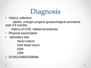
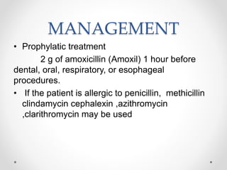
ENDOCARDITS
Endocarditis is an inflammation of the inner layer
of the heart, the endocardium. It usually involves
the heart valves (native or prosthetic valves).
TYPES
1. Infective endocarditis
2. Non-infective endocarditis
Infective endocarditis
• Previously known as bacterial endocarditis
• Infectious endocarditis involves the heart valves
and is most commonly found in people who
have underlying heart disease
CLASSIFICATION
1.Sub-acute bacterial endocarditis
2.Acute bacterial endocarditis
Subacute bacterial
endocarditis (SBE)
• usually develops insidiously and progresses
slowly (i.e, over weeks to months
• Typically affect those with preexisting valve
damage
Acute bacterial
endocarditis (ABE)
• usually develops abruptly and progresses
rapidly (ie, over days)
• A source of infection or portal of entry is often
evident.
Etiologic factors
• Causes by gram- ve, gram+ve bact & fungi.
• Most often caused by Staphylococcus aureus and
streptococcus viridans
• Fungi
candida albicans
• Viruses
coxsackie B virus
Predisposing factors
• Cardiac conditions
prosthetic valves
congenital heart diseases
pacemakers
cardiomyopathy
• Non cardiac conditions
IV Drug abuse
intravascular devices
procedures like dental extraction, tonsillectomy etc..
Clinical manifestations
• Low grade fever
• Chills
• Weakness
• Malaise
• Fatigue
• Anorexia
• clubbing
Vascular manifestations
splinter haemorrhages- black longitudinal streaks in
the nail beds
Oslers nodes- painful, tender, red , pea shaped
lesions found on the fingertips or toes
Janeway’s lesions- flat, painless, small, red spots
found on the palms and soles
Roth spots- haemorrhagic retinal lesions on
fundoscopic examination
Diagnosis
• History collection
dental, urologic,surgical gynaecological procedure
past 3-6 months
history of CVD, related procedures
• Physical examination
• laboratory test
blood culture
total blood count
ESR
CRP
• ECHOCARDIOGRAM
Duke Criteria
• Positive blood culture
presence of Cardiac murmer
intracardiac mass/vegetation on echo
• ECG- AV BLOCK
• XRAY- CARDIOMEGALY
MANAGEMENT
• Prophylatic treatment
2 g of amoxicillin (Amoxil) 1 hour before
dental, oral, respiratory, or esophageal
procedures.
• If the patient is allergic to penicillin, methicillin
clindamycin cephalexin ,azithromycin
,clarithromycin may be used
Pharmacologic
management
• Antibiotic therapy is usually administered
parenterally in a continuous intravenous infusion
for 2 to 6 weeks
eg- pencillin G
ceftriaxone
vancomycin
Surgical management
• After the patient recovers from the infectious process,
seriously damaged valves may need to be replaced
• Mitral valve replacement
• Aortic valve replacement
Nursing management
• Hyperthermia related to infection of cardiac tissue as
evidenced by elavated temp,chills,malaise, tachycardia
• Decreased cardiac output related to altered rhythm,
valvular insufficiency as evidenced by
tachycardia,murmer
• Deficient knowledge related to lack of experience and
exposure of information about disease and treatment
process

Endocardits

  • 1.
  • 2.
    Endocarditis is aninflammation of the inner layer of the heart, the endocardium. It usually involves the heart valves (native or prosthetic valves).
  • 3.
    TYPES 1. Infective endocarditis 2.Non-infective endocarditis
  • 4.
    Infective endocarditis • Previouslyknown as bacterial endocarditis • Infectious endocarditis involves the heart valves and is most commonly found in people who have underlying heart disease
  • 5.
  • 6.
    Subacute bacterial endocarditis (SBE) •usually develops insidiously and progresses slowly (i.e, over weeks to months • Typically affect those with preexisting valve damage
  • 7.
    Acute bacterial endocarditis (ABE) •usually develops abruptly and progresses rapidly (ie, over days) • A source of infection or portal of entry is often evident.
  • 8.
    Etiologic factors • Causesby gram- ve, gram+ve bact & fungi. • Most often caused by Staphylococcus aureus and streptococcus viridans • Fungi candida albicans • Viruses coxsackie B virus
  • 9.
    Predisposing factors • Cardiacconditions prosthetic valves congenital heart diseases pacemakers cardiomyopathy • Non cardiac conditions IV Drug abuse intravascular devices procedures like dental extraction, tonsillectomy etc..
  • 12.
    Clinical manifestations • Lowgrade fever • Chills • Weakness • Malaise • Fatigue • Anorexia • clubbing
  • 13.
    Vascular manifestations splinter haemorrhages-black longitudinal streaks in the nail beds Oslers nodes- painful, tender, red , pea shaped lesions found on the fingertips or toes Janeway’s lesions- flat, painless, small, red spots found on the palms and soles Roth spots- haemorrhagic retinal lesions on fundoscopic examination
  • 18.
    Diagnosis • History collection dental,urologic,surgical gynaecological procedure past 3-6 months history of CVD, related procedures • Physical examination • laboratory test blood culture total blood count ESR CRP • ECHOCARDIOGRAM
  • 19.
    Duke Criteria • Positiveblood culture presence of Cardiac murmer intracardiac mass/vegetation on echo • ECG- AV BLOCK • XRAY- CARDIOMEGALY
  • 20.
    MANAGEMENT • Prophylatic treatment 2g of amoxicillin (Amoxil) 1 hour before dental, oral, respiratory, or esophageal procedures. • If the patient is allergic to penicillin, methicillin clindamycin cephalexin ,azithromycin ,clarithromycin may be used
  • 21.
    Pharmacologic management • Antibiotic therapyis usually administered parenterally in a continuous intravenous infusion for 2 to 6 weeks eg- pencillin G ceftriaxone vancomycin
  • 22.
    Surgical management • Afterthe patient recovers from the infectious process, seriously damaged valves may need to be replaced • Mitral valve replacement • Aortic valve replacement
  • 23.
    Nursing management • Hyperthermiarelated to infection of cardiac tissue as evidenced by elavated temp,chills,malaise, tachycardia • Decreased cardiac output related to altered rhythm, valvular insufficiency as evidenced by tachycardia,murmer • Deficient knowledge related to lack of experience and exposure of information about disease and treatment process