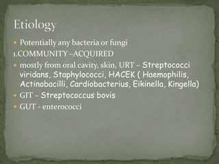
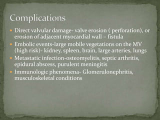
 This is an infection, usually bacterial, of the
endocardial surface of the heart
 Prototypic lesion – the vegetations( mass of platelets,
fibrin, microcolonies of microorganisms, scant
inflammatory cells
 Most commonly involves valves ( native and
prosthetic), may also affect mural endocardium,
intracardiac devices, A-V shunts, PDA, coarctation of
AO
 Acute febrile illness, rapid damage of cardiac
structures, hematogenically seeds extracardiac sites
resulting in death within weeks if untreated
 Subacute- cardiac damage is slow, gradual progression
 Potentially any bacteria or fungi
1.COMMUNITY –ACQUIRED
 mostly from oral cavity, skin, URT – Streptococci
viridans, Staphylococci, HACEK ( Haemophilis,
Actinobacilli, Cardiobacterius, Eikinella, Kingella)
 GIT – Streptococcus bovis
 GUT - enterococci
2. NOSOCOMIAL IE.
 Prosthetic valve endocarditis 2 months after surgery –
intraoperative contamination- usually coagulase-
negative Staphylococci, Gram(-) bacilli, diphteroids,
fungi
 Transvenous pacemaker leads or implanted
defibrillators – Staph.aureus and other Staph.
3. DRUG USERS
 Staph.aureus, Pseudomonas aeruginosa, Candida
 Fungi – difficult to diagnose and treat, mostly Candida
and Aspergilles
 5-15% of pts with IE have (-) blood culture. 1/3-1/2 of
them due to prior antibiotic exposure
 Normal endocardium is resistant to infection ( by
most bacteria) and to thrombus formation
 Endocardial injury (e.g. at the site of high velocity jets
or on the low pressure side of cardiac structure lesion)
– causes aberrant flow and allows direct infection by
virulent organism or the development of uninfected
platelet-fibrin thrombi – non-bacterial thrombotic
endocarditis (NBTE), subsequently site of bacterial
attachment
 Organisms enter the bloodstream from mucosa, skin,
sites of focal infection
 Some of them adhere directly to intact endocardium or
injured endothelium
 Others – adhere to thrombi, where they proliferate and
form vegetations
 Damage to cardiac structures causes hemodynamic
changes
 Embolization – infarction of other organs
 Infection of remote tissues due to dissemination of
infection, mycotic aneurysms
 Tissue injury due to deposition of circulating immune
complexes or immune response to deposited bacterial
antigens
 Cytokines release – constitutional symptoms
 Very variable
 Complete history including travel Hx and review of
systems, Hx of URTI, dental, surgical and other
procedures
 Fever, constitutional symptoms ( fatigue, malaise,
weight loss)
 Myalgias, arthritis, low back pain, pleuritic chest pain
 Thorough examination , search for stigmata of IE
 Fever
 Splinter hemorrhages on nail beds
 Osler’s nodes – painful nodes on palmar surfaces of toes
and fingers
 Janeway lesions –hemorrhagic papules on the palm and
soles
 Roth’s spots on fundoscopy
 IV injection marks
 Anemia
 Heart – cardiac murmurs, feature of CHF ( due to
valvular lesions, myocarditis or intracardiac fistulas),
various heart blocks (infectious process affecting
conduction system)
 Embolic events – extremities, spleen, kidneys, bowel,
brain
 Neurological complications – meningitis, intracranial
hemorrhages due to hemorrhagic infarcts, or ruptured
mycotic aneurysms, seizure, encepalopathy,
microabscesses
 Immune complex deposition on glomerular basement
membrane – diffuse glomerulonephritis and renal
dysfunction, emboli - hematuria
 Drug users – infection of Tricuspid V, high fever, faint
murmur, cough, chest pain, pulmonary infiltrates,
pyopneumothorax
 Prosthetic valves – CHF, arrhythmias, new murmurs
 Blood culture (+) for above mentioned organisms, at
least initially 3 sets
 FBC- Anemia of chronic illness
 WBC – normal or increased
 Urinalysis- Hematuria, proteinuria
 Abnormal CXR
 ECHO- Valvular Vegetations
 ECG – conduction blocks, MI
 Elevated ESR, (+) Rheumatoid factor
 Direct valvular damage- valve erosion ( perforation), or
erosion of adjacent myocardial wall – fistula
 Embolic events-large mobile vegetations on the MV
(high risk)- kidney, spleen, brain, large arteries, lungs
 Metastatic infection-osteomyelitis, septic arthritis,
epidural abscess, purulent meningitis
 Immunologic phenomena- Glomerulonephritis,
musculoskeletal conditions
 Duke criteria remain the clinical gold standard for
diagnosis
 2 major
 1 major + 3 minor
 5 minor
 Major : 1- +ve Blood culture in 2 separate
cultures;atleast 24 hrs apart
2- Endocardial involvement, +ve Echo.by
doing an echo e.g vulvular functions.
 Minor : 1- predisposition.e.g people whoz had
extracted a tooth and
2- fever > 38°c
3- vascular/immunological signs.
(roth’s spots, splinter hemorrhages, osler’s
nodes & janeway lesions)
4- +ve Blood Culture not meeting major
criteria.
5- +ve Echocardiogram.e.g affected heart
function .
 Antibiotic for 4-6 weeks
 Streptococci – Penicillin, Ceftriaxone, Vancomycin,
Gentamycin
 Enterococci – penicillin + gentamycin, ampicillin +
gentamycin, vancomycin
 Staphylococci- nafcillin, oxacillin, cefazolin,
gentamycin, vancomycin. If MRSA – vancomycin
 HACEK – ceftriaxone, ampicillin
 Candida – amphotericin B + flucytosin
 CHF
 Affected prosthetic valves
 Persistent bacteremia
 Lack of effective microbicide therapy ( Brucella, fungi)
 Staph.aureus prosthetic valve IE
 Relapse of prosthetic valve IE with optimal a/b
treatment
 Large > 10 mm vegetations
 Prognosis is poor :
 Older age
 Severe co morbidities.combination of different
conditions e.g cch and id and menngitis
 Delayed diagnosis
 Involvement of prosthetic valve or Aortic valve.e.g with
history of surgical RHD
 S.aureus or Pseudomonas, yeasts
 Intracardiac complications
 Neurologic complications
 Dental procedures: extraction, periodontal
procedures, implants, root canal, surgery beyond apex;
 Respiratory procedures: operations involving mucosa,
bronchoscopy with rigid bronchoscope
 GIT – esophageal stricture dilatation, sclerotherapy of
varices, endoscopic retrgrade cholangiography, biliary
tract surgery, surgery involving mucosa
 GUT – urethral dilation, prostate and urethral surgery,
cystoscopy
 CVS- prosthetic valves, prior IE, complex cyanotic
CHD, PDA, coarctation of Ao, surgical shunts,
Hypertrophic CMP, MVP, RHD
 Use of antibiotics before procedure:
 Amoxyl 2 g PO 1 hour before or ampicillin 2 g IV 30
min before proceure, Cefazolin 1 g IV 30 min before.
 If allergic to penicillin – clarythromycin 500 mg PO 1
hour before, Clindamycin 600 mg PO before
 Ampicillin + Gentamycin
 Vancomycin + Gentamycin
 Maintain good dental hygiene
 Aggressive treatment of local infections
Infective endocarditis.ppt infective endocarditis

Infective endocarditis.ppt infective endocarditis

  • 2.
     This isan infection, usually bacterial, of the endocardial surface of the heart  Prototypic lesion – the vegetations( mass of platelets, fibrin, microcolonies of microorganisms, scant inflammatory cells  Most commonly involves valves ( native and prosthetic), may also affect mural endocardium, intracardiac devices, A-V shunts, PDA, coarctation of AO
  • 3.
     Acute febrileillness, rapid damage of cardiac structures, hematogenically seeds extracardiac sites resulting in death within weeks if untreated  Subacute- cardiac damage is slow, gradual progression
  • 4.
     Potentially anybacteria or fungi 1.COMMUNITY –ACQUIRED  mostly from oral cavity, skin, URT – Streptococci viridans, Staphylococci, HACEK ( Haemophilis, Actinobacilli, Cardiobacterius, Eikinella, Kingella)  GIT – Streptococcus bovis  GUT - enterococci
  • 5.
    2. NOSOCOMIAL IE. Prosthetic valve endocarditis 2 months after surgery – intraoperative contamination- usually coagulase- negative Staphylococci, Gram(-) bacilli, diphteroids, fungi  Transvenous pacemaker leads or implanted defibrillators – Staph.aureus and other Staph.
  • 6.
    3. DRUG USERS Staph.aureus, Pseudomonas aeruginosa, Candida  Fungi – difficult to diagnose and treat, mostly Candida and Aspergilles  5-15% of pts with IE have (-) blood culture. 1/3-1/2 of them due to prior antibiotic exposure
  • 7.
     Normal endocardiumis resistant to infection ( by most bacteria) and to thrombus formation  Endocardial injury (e.g. at the site of high velocity jets or on the low pressure side of cardiac structure lesion) – causes aberrant flow and allows direct infection by virulent organism or the development of uninfected platelet-fibrin thrombi – non-bacterial thrombotic endocarditis (NBTE), subsequently site of bacterial attachment
  • 8.
     Organisms enterthe bloodstream from mucosa, skin, sites of focal infection  Some of them adhere directly to intact endocardium or injured endothelium  Others – adhere to thrombi, where they proliferate and form vegetations
  • 9.
     Damage tocardiac structures causes hemodynamic changes  Embolization – infarction of other organs  Infection of remote tissues due to dissemination of infection, mycotic aneurysms  Tissue injury due to deposition of circulating immune complexes or immune response to deposited bacterial antigens  Cytokines release – constitutional symptoms
  • 10.
     Very variable Complete history including travel Hx and review of systems, Hx of URTI, dental, surgical and other procedures  Fever, constitutional symptoms ( fatigue, malaise, weight loss)  Myalgias, arthritis, low back pain, pleuritic chest pain
  • 11.
     Thorough examination, search for stigmata of IE  Fever  Splinter hemorrhages on nail beds  Osler’s nodes – painful nodes on palmar surfaces of toes and fingers  Janeway lesions –hemorrhagic papules on the palm and soles  Roth’s spots on fundoscopy
  • 12.
     IV injectionmarks  Anemia  Heart – cardiac murmurs, feature of CHF ( due to valvular lesions, myocarditis or intracardiac fistulas), various heart blocks (infectious process affecting conduction system)
  • 13.
     Embolic events– extremities, spleen, kidneys, bowel, brain  Neurological complications – meningitis, intracranial hemorrhages due to hemorrhagic infarcts, or ruptured mycotic aneurysms, seizure, encepalopathy, microabscesses  Immune complex deposition on glomerular basement membrane – diffuse glomerulonephritis and renal dysfunction, emboli - hematuria
  • 14.
     Drug users– infection of Tricuspid V, high fever, faint murmur, cough, chest pain, pulmonary infiltrates, pyopneumothorax  Prosthetic valves – CHF, arrhythmias, new murmurs
  • 15.
     Blood culture(+) for above mentioned organisms, at least initially 3 sets  FBC- Anemia of chronic illness  WBC – normal or increased  Urinalysis- Hematuria, proteinuria  Abnormal CXR  ECHO- Valvular Vegetations  ECG – conduction blocks, MI  Elevated ESR, (+) Rheumatoid factor
  • 16.
     Direct valvulardamage- valve erosion ( perforation), or erosion of adjacent myocardial wall – fistula  Embolic events-large mobile vegetations on the MV (high risk)- kidney, spleen, brain, large arteries, lungs  Metastatic infection-osteomyelitis, septic arthritis, epidural abscess, purulent meningitis  Immunologic phenomena- Glomerulonephritis, musculoskeletal conditions
  • 17.
     Duke criteriaremain the clinical gold standard for diagnosis  2 major  1 major + 3 minor  5 minor  Major : 1- +ve Blood culture in 2 separate cultures;atleast 24 hrs apart 2- Endocardial involvement, +ve Echo.by doing an echo e.g vulvular functions.
  • 18.
     Minor :1- predisposition.e.g people whoz had extracted a tooth and 2- fever > 38°c 3- vascular/immunological signs. (roth’s spots, splinter hemorrhages, osler’s nodes & janeway lesions) 4- +ve Blood Culture not meeting major criteria. 5- +ve Echocardiogram.e.g affected heart function .
  • 19.
     Antibiotic for4-6 weeks  Streptococci – Penicillin, Ceftriaxone, Vancomycin, Gentamycin  Enterococci – penicillin + gentamycin, ampicillin + gentamycin, vancomycin  Staphylococci- nafcillin, oxacillin, cefazolin, gentamycin, vancomycin. If MRSA – vancomycin  HACEK – ceftriaxone, ampicillin  Candida – amphotericin B + flucytosin
  • 20.
     CHF  Affectedprosthetic valves  Persistent bacteremia  Lack of effective microbicide therapy ( Brucella, fungi)  Staph.aureus prosthetic valve IE  Relapse of prosthetic valve IE with optimal a/b treatment  Large > 10 mm vegetations
  • 21.
     Prognosis ispoor :  Older age  Severe co morbidities.combination of different conditions e.g cch and id and menngitis  Delayed diagnosis  Involvement of prosthetic valve or Aortic valve.e.g with history of surgical RHD  S.aureus or Pseudomonas, yeasts  Intracardiac complications  Neurologic complications
  • 22.
     Dental procedures:extraction, periodontal procedures, implants, root canal, surgery beyond apex;  Respiratory procedures: operations involving mucosa, bronchoscopy with rigid bronchoscope  GIT – esophageal stricture dilatation, sclerotherapy of varices, endoscopic retrgrade cholangiography, biliary tract surgery, surgery involving mucosa
  • 23.
     GUT –urethral dilation, prostate and urethral surgery, cystoscopy  CVS- prosthetic valves, prior IE, complex cyanotic CHD, PDA, coarctation of Ao, surgical shunts, Hypertrophic CMP, MVP, RHD
  • 24.
     Use ofantibiotics before procedure:  Amoxyl 2 g PO 1 hour before or ampicillin 2 g IV 30 min before proceure, Cefazolin 1 g IV 30 min before.  If allergic to penicillin – clarythromycin 500 mg PO 1 hour before, Clindamycin 600 mg PO before  Ampicillin + Gentamycin  Vancomycin + Gentamycin
  • 25.
     Maintain gooddental hygiene  Aggressive treatment of local infections