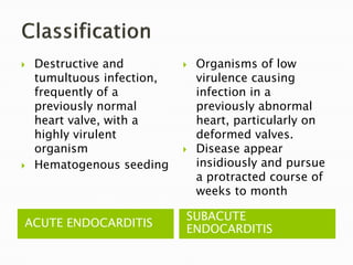
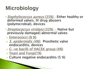
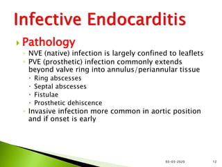
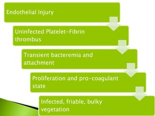
PROF DR FARHAT BASHIR
UNITED MEDICAL & DENTAL COLLEGE
 Infection of the endocardial surface of heart
characterized by
- Colonization or invasion of the heart valves
(native or prosthetic) or the mural endocardium
by a microbe,
- leading to formation of bulky, friable
vegetation composed of thrombotic debris and
organisms
- often associated with destruction of
underlying cardiac tissue.
• Heart valves
• Ventricular septum defects
• Mural endocardium
• Intracardiac devices
• INFECTIVE ENDARTERITIS – analogous
 Native valve endocarditis
 Prosthetic valve endocarditis
 Intravenous drug abuser infective
endocarditis
 Nosocomial endocarditis
ACUTE ENDOCARDITIS
SUBACUTE
ENDOCARDITIS
 Destructive and
tumultuous infection,
frequently of a
previously normal
heart valve, with a
highly virulent
organism
 Hematogenous seeding
 Organisms of low
virulence causing
infection in a
previously abnormal
heart, particularly on
deformed valves.
 Disease appear
insidiously and pursue
a protracted course of
weeks to month
Cardiac and vascular
abnormalities Host factors
 RHD
 Congenital heart
disease
◦ Myxomatous mitral valve
◦ Degenerative calcific
valvular stenosis
◦ Bicuspid aortic valves
 Prosthetic valves
 Neutropenia
 Immunodeficiency
 Malignancy
 Therapeutic
immunosuppression
 Diabetes mellitus
 Alcohol
 IV drug abuse
 Dental manipulation, dental
caries, dental abscess
 Extracardiac infections
 Instrumentation(urinary
tract, GIT, IV line)
 Cardiac surgery
 Staphylococcus aureus (35%) : Either healthy or
deformed valves, IV drug abusers
(polymicrobial), devices
 Streptococcus viridans (32%) : Native but
previously damaged/abnormal valves
 Enterococci (8 %)
 S. epidermidis (4%): Prosthetic valve
endocarditis, devices
 G –ve bacilli of HACEK group (4%)
 Yeast and Fungi(1%)
 Culture negative endocarditis (5 %)
 Pathology
◦ NVE (native) infection is largely confined to leaflets
◦ PVE (prosthetic) infection commonly extends
beyond valve ring into annulus/periannular tissue
 Ring abscesses
 Septal abscesses
 Fistulae
 Prosthetic dehiscence
◦ Invasive infection more common in aortic position
and if onset is early
03-03-2020 12
 Portal of entry:
◦ Dental / Surgical Procedures
◦ Contamination by IV drug use
◦ Obvious infections (RS/Skin)
◦ Occult source from gut, oral cavity
◦ Trivial injuries.
◦ Intravascular catheter infection
◦ Nosocomial wounds
◦ Invasive procedures
Turbulent blood flow within the heart -
most often (but not always) – patient has
risk factors for this
Turbulent blood flow disrupts valve surface
(endocardium) to produce suitable (sticky)
site for bacterial attachment
Platelet deposition + fibrin may lead to
non-bacterial thrombus or vegetation
Bacteraemia – delivers organisms to the
damaged (sticky) endocardial surface
resulting in adherence & colonisation
Eventual invasion of valve leaflets results in
infected vegetation (sheath of fibrin &
platelets, ideal conditions for further
bacterial multiplications, protection from
polymorphs) 03-03-2020 14
Endothelial Injury
Uninfected Platelet-Fibrin
thrombus
Transient bacteremia and
attachment
Proliferation and pro-coagulant
state
Infected, friable, bulky
vegetation
Feature Acute Subacute
Underlying Heart
Disease
Heart may be normal RHD,CHD, etc.
Organism S. aureus, Pneumococcus
S. pyogenes,
Enterococcus
viridans
Streptococci,
Entercoccus
03-03-2020 16
 Constitutional
symptoms
 --- Cytokine
release ?
 Symptoms
 - Damage to
intracardiac
structures
 - Embolization of
vegetation
fragments
 - Hematogenous
infection
 - Immune complex
1. Fever.
2. Murmurs
3. Petechial and cutaneous manifestations. Roth spots,
conjunctival and mucosal petechiae, splinter
hemorrhages, Osler nodes and Janway lesions.
4. Splenomegaly
5. Embolism. Septic or sterile. CNS, spleen, lung, retinal
vessels, coronary artery, large vessels.
6. Renal disease, infarction. Glomerulonephritis and
uremia
7. CHF
8. General. Weight loss, anorexia, debilitation.
03-03-2020 18
Endocarditis
• Persistent
fever
• Constitutional
symptoms
• New signs of
valve
dysfunction
• Heart failure
• Embolic Stroke
• Peripheral
arterial
embolism
03-03-2020Dr.T.V.Rao MD 21
03-03-2020 22
1. More specific
2. Erythematous, blanching macules
3. Nonpainful
4. Located on palms and soles
03-03-2020Dr.T.V.Rao MD 23
03-03-2020 24
1. Nonspecific
2. Nonblanching
3. Linear reddish-brown lesions found under the nail bed
4. Usually do NOT extend the entire length of the nail
 CBC
 ESR
 CRP
 Urine DR
 ECG
 CXR-PA view
 Serological tests
 BLOOD CULTURE
 ECHOCARDIOGRAPHY
Blood cultures: Echocardiography
 Key diagnostic investigation
in infective endocarditis.
 Isolation of microorganism
from culture is important for
diagnosis and also for
treatment.
 At least 3 sets of samples
should be taken from
different venepuncture sites
over 24 hours.
 It can identify the
 presence and size of vegetations,
 detect intracardiac complications
and
 assess cardiac function.
 Transthoracic
echocardiography is
noninvasive and has high
specificity for visualising
vegetations.
 Transesophageal
echocardiography is more
sensitive than TTE. It can
detect small vegetations,
prosthetic endocarditis and
intra cardiac complications.
 Definitive Endocarditis if,
- Two major or,
- One major and three
minor or,
- five minor
 Possible Endocarditis if,
- One major and one minor
or,
- Three minor
 Positive blood culture
◦ Typical organism from two cultures
◦ Persistent positive blood cultures taken >
12 hours apart
◦ Three or more positive cultures taken over
more than 1 hour.
 Endocardial involvement
◦ Positive echocardiographic findings of
vegetations
◦ New valvular regurgitation
◦ Myocardial abscess
◦ New partial dehiscence of prosthetic valve
1. Predisposition: Predisposing
valvular or cardiac abnormality
2. Intravenous drug misuse
3. Pyrexia ≥38°C (≥100.4°F)
4. Embolic phenomenon
5. Vasculitic/ immunologic
phenomenon
6. Blood cultures suggestive: -
organism grown but not achieving
major criteria
7. Suggestive echocardiographic
findings
Antimicrobial Therapy
 Therapy requires identification
of specific pathogen and its
susceptibility to antimicrobials.
 Empirical therapy should be
started as soon as possible
targeting most likely pathogens.
 Bactericidal drugs should be
used.
 Resolution of fever occurs in 5
to 7 days. If fever persists
patient should be evaluated for
complications like paravalvular
abscess and extracardiac
abscess.
 Serologic abnormalities resolve
slowly and do not reflect
response to treatment.
Antibiotic regimen for infective endocarditis
 Streptococci
Benzyl penicillin (2-3 million units 4 hourly) 4-6 weeks
Gentamicin (1mg/kg 8-12 hourly) 4-6 weeks
(ceftriaxone & vancomycin)
 Enterococci
o Ampicillin sensitive
Ampicillin (2 g 4 hourly) 4-6 weeks, and
Gentamicin (1mg/kg 8-12 hourly)
o Ampicillin resistant
Vancomycin(1g 12hourly) 4-6 weeks, and
Gentamicin (1mg/kg 8-12 hourly)
 Staphylococci
o Penicillin sensitive
Benzyl penicillin I.V(2-3 Munits 4 hourly)
+ gentamicin
o Penicillin resistant but methicillin sensitive
Flucloxacillin I.V (2g 4 hourly )
o Both penicillin and methicillin resistant
Vancomycin I.V (1g 12 hourly) and
Gentamicin
Surgery
 Indications
 patients with direct extension of infection to
myocardial structures.
 Prosthetic valve dysfunction.
 Congestive heart failure.
 Badly damaged valves.
 IE caused by fungi or gram-ve or resistant
organisms.
 Large vegetations on echocardiography
 Recurrent embolic attacks.
 Prophylaxis
High risk category
 prosthetic cardiac valves
 Previous bacterial endocarditis, even in
absence of heart disease.
 Complex cyanotic congenital heart
disease(TGA,TOF)
 Surgically constructed systemic pulmonary
shunts.
Moderate risk category
 Rheumatic and other valvular dysfunction
 Congenital cardiac malformations
 Hypertrophic cardiomyopathy
 Mitral valve prolapse with valvular
regurgitation
 High risk lesions
◦ Prosthetic valves
◦ Prior IE
◦ Cyanotic congenital heart
disease
◦ PDA
◦ AR, AS, MR,MS with MR
◦ VSD
◦ Coarctation
◦ Surgical systemic-pulmonary
shunts
 Intermediate risk
◦ MVP with murmur
◦ Pure MS
◦ Tricuspid disease
◦ Pulmonary stenosis
◦ ASH
◦ Bicuspid Ao valve with no
hemodynamic
significance
03-03-2020 42
Lesions at highest risk
Regimen for IE prophylaxis
 Standard oral regime
Amoxicillin 2 g 1hr before procedure
 Inability to take oral medication
Ampicillin 2g IV or IM 1hr before procedure
 Penicillin allergy
Clindamycin 600 mg
Clarithromycin 500 mg
Cephalexin 2 g.
Thank you.

Infective endocarditis

  • 1.
    PROF DR FARHATBASHIR UNITED MEDICAL & DENTAL COLLEGE
  • 3.
     Infection ofthe endocardial surface of heart characterized by - Colonization or invasion of the heart valves (native or prosthetic) or the mural endocardium by a microbe, - leading to formation of bulky, friable vegetation composed of thrombotic debris and organisms - often associated with destruction of underlying cardiac tissue.
  • 4.
    • Heart valves •Ventricular septum defects • Mural endocardium • Intracardiac devices • INFECTIVE ENDARTERITIS – analogous
  • 5.
     Native valveendocarditis  Prosthetic valve endocarditis  Intravenous drug abuser infective endocarditis  Nosocomial endocarditis
  • 7.
    ACUTE ENDOCARDITIS SUBACUTE ENDOCARDITIS  Destructiveand tumultuous infection, frequently of a previously normal heart valve, with a highly virulent organism  Hematogenous seeding  Organisms of low virulence causing infection in a previously abnormal heart, particularly on deformed valves.  Disease appear insidiously and pursue a protracted course of weeks to month
  • 8.
    Cardiac and vascular abnormalitiesHost factors  RHD  Congenital heart disease ◦ Myxomatous mitral valve ◦ Degenerative calcific valvular stenosis ◦ Bicuspid aortic valves  Prosthetic valves  Neutropenia  Immunodeficiency  Malignancy  Therapeutic immunosuppression  Diabetes mellitus  Alcohol  IV drug abuse  Dental manipulation, dental caries, dental abscess  Extracardiac infections  Instrumentation(urinary tract, GIT, IV line)  Cardiac surgery
  • 9.
     Staphylococcus aureus(35%) : Either healthy or deformed valves, IV drug abusers (polymicrobial), devices  Streptococcus viridans (32%) : Native but previously damaged/abnormal valves  Enterococci (8 %)  S. epidermidis (4%): Prosthetic valve endocarditis, devices  G –ve bacilli of HACEK group (4%)  Yeast and Fungi(1%)  Culture negative endocarditis (5 %)
  • 10.
     Pathology ◦ NVE(native) infection is largely confined to leaflets ◦ PVE (prosthetic) infection commonly extends beyond valve ring into annulus/periannular tissue  Ring abscesses  Septal abscesses  Fistulae  Prosthetic dehiscence ◦ Invasive infection more common in aortic position and if onset is early 03-03-2020 12
  • 11.
     Portal ofentry: ◦ Dental / Surgical Procedures ◦ Contamination by IV drug use ◦ Obvious infections (RS/Skin) ◦ Occult source from gut, oral cavity ◦ Trivial injuries. ◦ Intravascular catheter infection ◦ Nosocomial wounds ◦ Invasive procedures
  • 12.
    Turbulent blood flowwithin the heart - most often (but not always) – patient has risk factors for this Turbulent blood flow disrupts valve surface (endocardium) to produce suitable (sticky) site for bacterial attachment Platelet deposition + fibrin may lead to non-bacterial thrombus or vegetation Bacteraemia – delivers organisms to the damaged (sticky) endocardial surface resulting in adherence & colonisation Eventual invasion of valve leaflets results in infected vegetation (sheath of fibrin & platelets, ideal conditions for further bacterial multiplications, protection from polymorphs) 03-03-2020 14
  • 13.
    Endothelial Injury Uninfected Platelet-Fibrin thrombus Transientbacteremia and attachment Proliferation and pro-coagulant state Infected, friable, bulky vegetation
  • 14.
    Feature Acute Subacute UnderlyingHeart Disease Heart may be normal RHD,CHD, etc. Organism S. aureus, Pneumococcus S. pyogenes, Enterococcus viridans Streptococci, Entercoccus 03-03-2020 16
  • 15.
     Constitutional symptoms  ---Cytokine release ?  Symptoms  - Damage to intracardiac structures  - Embolization of vegetation fragments  - Hematogenous infection  - Immune complex
  • 16.
    1. Fever. 2. Murmurs 3.Petechial and cutaneous manifestations. Roth spots, conjunctival and mucosal petechiae, splinter hemorrhages, Osler nodes and Janway lesions. 4. Splenomegaly 5. Embolism. Septic or sterile. CNS, spleen, lung, retinal vessels, coronary artery, large vessels. 6. Renal disease, infarction. Glomerulonephritis and uremia 7. CHF 8. General. Weight loss, anorexia, debilitation. 03-03-2020 18
  • 17.
    Endocarditis • Persistent fever • Constitutional symptoms •New signs of valve dysfunction • Heart failure • Embolic Stroke • Peripheral arterial embolism
  • 19.
  • 20.
    03-03-2020 22 1. Morespecific 2. Erythematous, blanching macules 3. Nonpainful 4. Located on palms and soles
  • 21.
  • 22.
    03-03-2020 24 1. Nonspecific 2.Nonblanching 3. Linear reddish-brown lesions found under the nail bed 4. Usually do NOT extend the entire length of the nail
  • 23.
     CBC  ESR CRP  Urine DR  ECG  CXR-PA view  Serological tests  BLOOD CULTURE  ECHOCARDIOGRAPHY
  • 24.
    Blood cultures: Echocardiography Key diagnostic investigation in infective endocarditis.  Isolation of microorganism from culture is important for diagnosis and also for treatment.  At least 3 sets of samples should be taken from different venepuncture sites over 24 hours.  It can identify the  presence and size of vegetations,  detect intracardiac complications and  assess cardiac function.  Transthoracic echocardiography is noninvasive and has high specificity for visualising vegetations.  Transesophageal echocardiography is more sensitive than TTE. It can detect small vegetations, prosthetic endocarditis and intra cardiac complications.
  • 27.
     Definitive Endocarditisif, - Two major or, - One major and three minor or, - five minor  Possible Endocarditis if, - One major and one minor or, - Three minor
  • 28.
     Positive bloodculture ◦ Typical organism from two cultures ◦ Persistent positive blood cultures taken > 12 hours apart ◦ Three or more positive cultures taken over more than 1 hour.  Endocardial involvement ◦ Positive echocardiographic findings of vegetations ◦ New valvular regurgitation ◦ Myocardial abscess ◦ New partial dehiscence of prosthetic valve
  • 29.
    1. Predisposition: Predisposing valvularor cardiac abnormality 2. Intravenous drug misuse 3. Pyrexia ≥38°C (≥100.4°F) 4. Embolic phenomenon 5. Vasculitic/ immunologic phenomenon 6. Blood cultures suggestive: - organism grown but not achieving major criteria 7. Suggestive echocardiographic findings
  • 30.
    Antimicrobial Therapy  Therapyrequires identification of specific pathogen and its susceptibility to antimicrobials.  Empirical therapy should be started as soon as possible targeting most likely pathogens.  Bactericidal drugs should be used.
  • 31.
     Resolution offever occurs in 5 to 7 days. If fever persists patient should be evaluated for complications like paravalvular abscess and extracardiac abscess.  Serologic abnormalities resolve slowly and do not reflect response to treatment.
  • 32.
    Antibiotic regimen forinfective endocarditis  Streptococci Benzyl penicillin (2-3 million units 4 hourly) 4-6 weeks Gentamicin (1mg/kg 8-12 hourly) 4-6 weeks (ceftriaxone & vancomycin)  Enterococci o Ampicillin sensitive Ampicillin (2 g 4 hourly) 4-6 weeks, and Gentamicin (1mg/kg 8-12 hourly) o Ampicillin resistant Vancomycin(1g 12hourly) 4-6 weeks, and Gentamicin (1mg/kg 8-12 hourly)
  • 33.
     Staphylococci o Penicillinsensitive Benzyl penicillin I.V(2-3 Munits 4 hourly) + gentamicin o Penicillin resistant but methicillin sensitive Flucloxacillin I.V (2g 4 hourly ) o Both penicillin and methicillin resistant Vancomycin I.V (1g 12 hourly) and Gentamicin
  • 34.
    Surgery  Indications  patientswith direct extension of infection to myocardial structures.  Prosthetic valve dysfunction.  Congestive heart failure.  Badly damaged valves.  IE caused by fungi or gram-ve or resistant organisms.  Large vegetations on echocardiography  Recurrent embolic attacks.
  • 35.
     Prophylaxis High riskcategory  prosthetic cardiac valves  Previous bacterial endocarditis, even in absence of heart disease.  Complex cyanotic congenital heart disease(TGA,TOF)  Surgically constructed systemic pulmonary shunts.
  • 36.
    Moderate risk category Rheumatic and other valvular dysfunction  Congenital cardiac malformations  Hypertrophic cardiomyopathy  Mitral valve prolapse with valvular regurgitation
  • 37.
     High risklesions ◦ Prosthetic valves ◦ Prior IE ◦ Cyanotic congenital heart disease ◦ PDA ◦ AR, AS, MR,MS with MR ◦ VSD ◦ Coarctation ◦ Surgical systemic-pulmonary shunts  Intermediate risk ◦ MVP with murmur ◦ Pure MS ◦ Tricuspid disease ◦ Pulmonary stenosis ◦ ASH ◦ Bicuspid Ao valve with no hemodynamic significance 03-03-2020 42 Lesions at highest risk
  • 38.
    Regimen for IEprophylaxis  Standard oral regime Amoxicillin 2 g 1hr before procedure  Inability to take oral medication Ampicillin 2g IV or IM 1hr before procedure  Penicillin allergy Clindamycin 600 mg Clarithromycin 500 mg Cephalexin 2 g.
  • 39.